

Follistatin Effects in Migration, Vascularization, and Osteogenesis in vitro and Bone Repair in vivo
- PMID: 30881954
- PMCID: PMC6405513
- DOI: 10.3389/fbioe.2019.00038
Follistatin Effects in Migration, Vascularization, and Osteogenesis in vitro and Bone Repair in vivo
Abstract
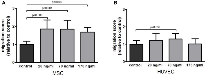
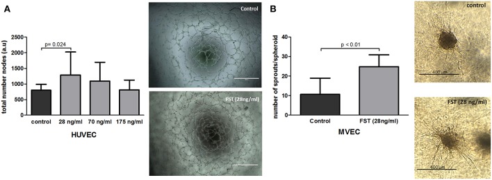
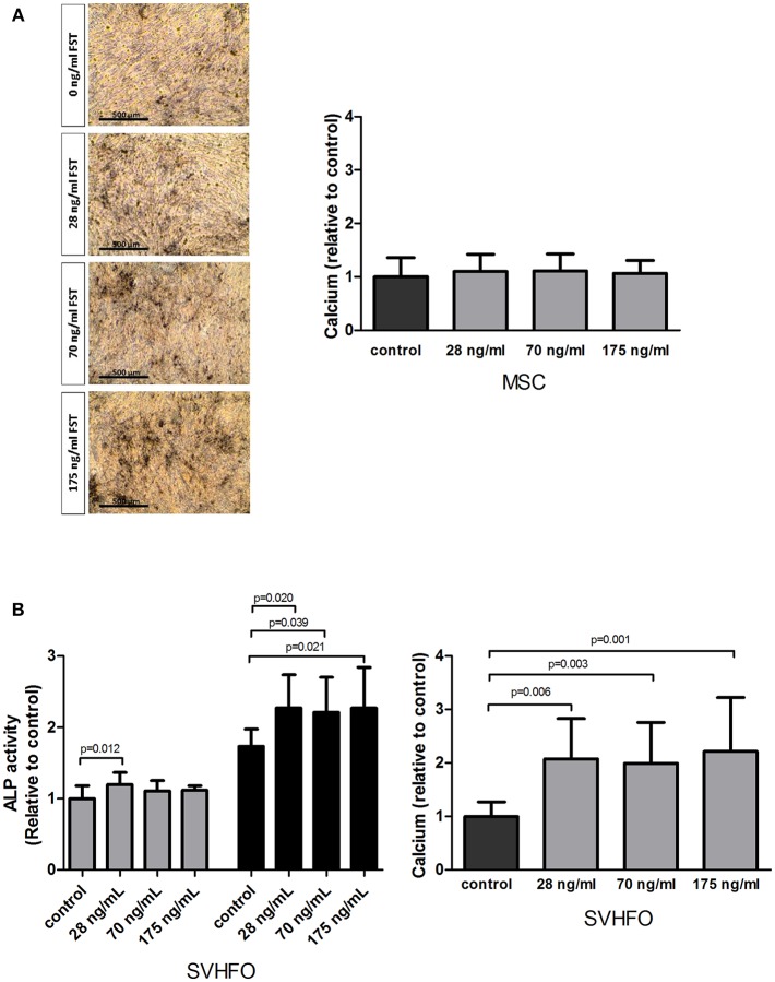
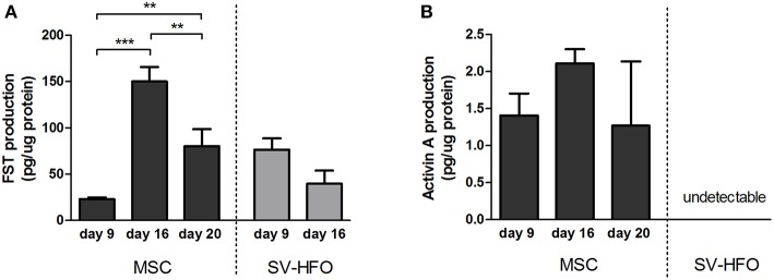
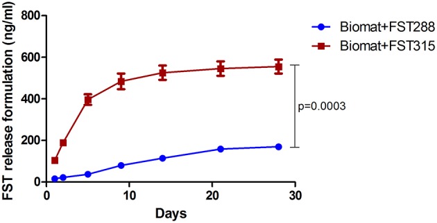
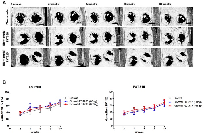
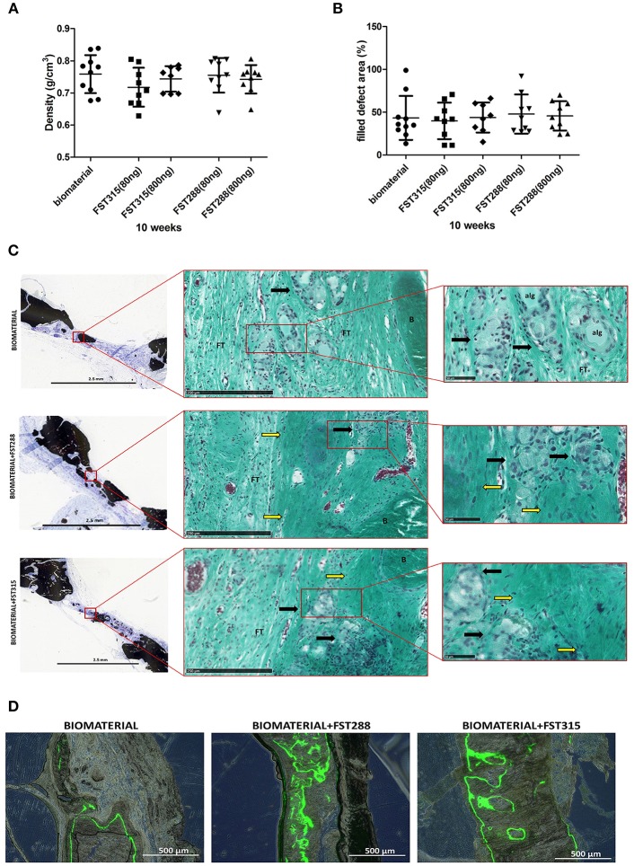
The use of biomaterials and signaling molecules to induce bone formation is a promising approach in the field of bone tissue engineering. Follistatin (FST) is a glycoprotein able to bind irreversibly to activin A, a protein that has been reported to inhibit bone formation. We investigated the effect of FST in critical processes for bone repair, such as cell recruitment, osteogenesis and vascularization, and ultimately its use for bone tissue engineering. In vitro, FST promoted mesenchymal stem cell (MSC) and endothelial cell (EC) migration as well as essential steps in the formation and expansion of the vasculature such as EC tube-formation and sprouting. FST did not enhance osteogenic differentiation of MSCs, but increased committed osteoblast mineralization. In vivo, FST was loaded in an in situ gelling formulation made by alginate and recombinant collagen-based peptide microspheres and implanted in a rat calvarial defect model. Two FST variants (FST288 and FST315) with major differences in their affinity to cell-surface proteoglycans, which may influence their effect upon in vivo bone repair, were tested. In vitro, most of the loaded FST315 was released over 4 weeks, contrary to FST288, which was mostly retained in the biomaterial. However, none of the FST variants improved in vivo bone healing compared to control. These results demonstrate that FST enhances crucial processes needed for bone repair. Further studies need to investigate the optimal FST carrier for bone regeneration.
Keywords: bone tissue engineering; follistatin 288 (FST288); follistatin 315 (FST315); injectable in situ gelling slow release system; migration; osteogenesis; regenerative medicine; vascularization.
Figures
Similar articles
-
Biological activity of follistatin isoforms and follistatin-like-3 is dependent on differential cell surface binding and specificity for activin, myostatin, and bone morphogenetic proteins.Endocrinology. 2006 Jul;147(7):3586-97. doi: 10.1210/en.2006-0089. Epub 2006 Apr 20. Endocrinology. 2006. PMID: 16627583
-
Progesterone stimulates expression of follistatin splice variants Fst288 and Fst315 in the mouse uterus.Reprod Biomed Online. 2012 Mar;24(3):364-74. doi: 10.1016/j.rbmo.2011.12.004. Epub 2011 Dec 20. Reprod Biomed Online. 2012. PMID: 22285243
-
The follistatin-288 isoform alone is sufficient for survival but not for normal fertility in mice.Endocrinology. 2010 Mar;151(3):1310-9. doi: 10.1210/en.2009-1176. Epub 2009 Dec 23. Endocrinology. 2010. PMID: 20032047 Free PMC article.
-
Clinical and Therapeutic Implications of Follistatin in Solid Tumours.Cancer Genomics Proteomics. 2016 11-12;13(6):425-435. doi: 10.21873/cgp.20005. Cancer Genomics Proteomics. 2016. PMID: 27807065 Free PMC article. Review.
-
Osteoblast recruitment to sites of bone formation in skeletal development, homeostasis, and regeneration.Birth Defects Res C Embryo Today. 2013 Sep;99(3):170-91. doi: 10.1002/bdrc.21047. Birth Defects Res C Embryo Today. 2013. PMID: 24078495 Review.
Cited by
-
Role of fibro-adipogenic progenitor cells in muscle atrophy and musculoskeletal diseases.Curr Opin Pharmacol. 2021 Jun;58:1-7. doi: 10.1016/j.coph.2021.03.003. Epub 2021 Apr 8. Curr Opin Pharmacol. 2021. PMID: 33839480 Free PMC article. Review.
-
A 1, 4-benzoquinone derivative isolated from Ardisia crispa (Thunb.) A. DC. root suppresses angiogenesis via its angiogenic signaling cascades.Saudi Pharm J. 2024 Jan;32(1):101891. doi: 10.1016/j.jsps.2023.101891. Epub 2023 Dec 1. Saudi Pharm J. 2024. PMID: 38111673 Free PMC article.
-
Endogenous Follistatin-like 1 guarantees the immunomodulatory properties of mesenchymal stem cells during liver fibrotic therapy.Stem Cell Res Ther. 2022 Aug 5;13(1):403. doi: 10.1186/s13287-022-03042-4. Stem Cell Res Ther. 2022. PMID: 35932064 Free PMC article.
-
CD41-deficient exosomes from non-traumatic femoral head necrosis tissues impair osteogenic differentiation and migration of mesenchymal stem cells.Cell Death Dis. 2020 Apr 27;11(4):293. doi: 10.1038/s41419-020-2496-y. Cell Death Dis. 2020. PMID: 32341357 Free PMC article.
-
Dynamic proteomic profiling of human periodontal ligament stem cells during osteogenic differentiation.Stem Cell Res Ther. 2021 Feb 3;12(1):98. doi: 10.1186/s13287-020-02123-6. Stem Cell Res Ther. 2021. PMID: 33536073 Free PMC article.
References
-
- Alves R. D., Eijken M., Bezstarosti K., Demmers J. A., van Leeuwen J. P. (2013). Activin A suppresses osteoblast mineralization capacity by altering extracellular matrix (ECM) composition and impairing matrix vesicle (MV) production. Mol. Cell Proteomics 12, 2890–2900. 10.1074/mcp.M112.024927 - DOI - PMC - PubMed
LinkOut - more resources
Full Text Sources
Medical
Miscellaneous