

MuSK myasthenia gravis monoclonal antibodies: Valency dictates pathogenicity
- PMID: 30882021
- PMCID: PMC6410930
- DOI: 10.1212/NXI.0000000000000547
MuSK myasthenia gravis monoclonal antibodies: Valency dictates pathogenicity
Abstract
Objective: To isolate and characterize muscle-specific kinase (MuSK) monoclonal antibodies from patients with MuSK myasthenia gravis (MG) on a genetic and functional level.
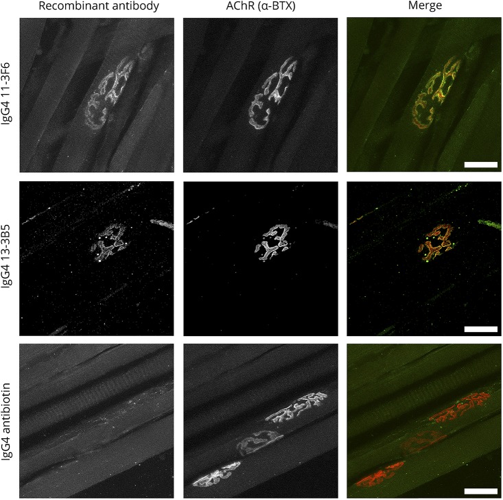
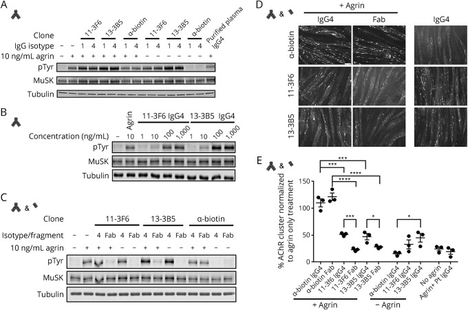
Methods: We generated recombinant MuSK antibodies from patient-derived clonal MuSK-specific B cells and produced monovalent Fab fragments from them. Both the antibodies and Fab fragments were tested for their effects on neural agrin-induced MuSK phosphorylation and acetylcholine receptor (AChR) clustering in myotube cultures.
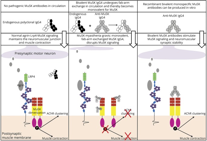
Results: The isolated MuSK monoclonal antibody sequences included IgG1, IgG3, and IgG4 that had undergone high levels of affinity maturation, consistent with antigenic selection. We confirmed their specificity for the MuSK Ig-like 1 domain and binding to neuromuscular junctions. Monovalent MuSK Fab, mimicking functionally monovalent MuSK MG patient Fab-arm exchanged serum IgG4, abolished agrin-induced MuSK phosphorylation and AChR clustering. Surprisingly, bivalent monospecific MuSK antibodies instead activated MuSK phosphorylation and partially induced AChR clustering, independent of agrin.
Conclusions: Patient-derived MuSK antibodies can act either as MuSK agonist or MuSK antagonist, depending on the number of MuSK binding sites. Functional monovalency, induced by Fab-arm exchange in patient serum, makes MuSK IgG4 antibodies pathogenic.
Figures
Similar articles
-
IgG4 autoantibodies against muscle-specific kinase undergo Fab-arm exchange in myasthenia gravis patients.J Autoimmun. 2017 Feb;77:104-115. doi: 10.1016/j.jaut.2016.11.005. Epub 2016 Dec 10. J Autoimmun. 2017. PMID: 27965060
-
Characterization of pathogenic monoclonal autoantibodies derived from muscle-specific kinase myasthenia gravis patients.JCI Insight. 2019 Jun 20;4(12):e127167. doi: 10.1172/jci.insight.127167. eCollection 2019 Jun 20. JCI Insight. 2019. PMID: 31217355 Free PMC article.
-
MuSK myasthenia gravis IgG4 disrupts the interaction of LRP4 with MuSK but both IgG4 and IgG1-3 can disperse preformed agrin-independent AChR clusters.PLoS One. 2013 Nov 7;8(11):e80695. doi: 10.1371/journal.pone.0080695. eCollection 2013. PLoS One. 2013. PMID: 24244707 Free PMC article.
-
Passive transfer models of myasthenia gravis with muscle-specific kinase antibodies.Ann N Y Acad Sci. 2018 Feb;1413(1):111-118. doi: 10.1111/nyas.13543. Epub 2018 Jan 21. Ann N Y Acad Sci. 2018. PMID: 29356029 Review.
-
MuSK antibodies, lessons learned from poly- and monoclonality.J Autoimmun. 2020 Aug;112:102488. doi: 10.1016/j.jaut.2020.102488. Epub 2020 Jun 4. J Autoimmun. 2020. PMID: 32505442 Review.
Cited by
-
Reemergence of pathogenic, autoantibody-producing B cell clones in myasthenia gravis following B cell depletion therapy.Acta Neuropathol Commun. 2022 Oct 28;10(1):154. doi: 10.1186/s40478-022-01454-0. Acta Neuropathol Commun. 2022. PMID: 36307868 Free PMC article.
-
Defective autophagy and autophagy activators in myasthenia gravis: a rare entity and unusual scenario.Autophagy. 2024 Jul;20(7):1473-1482. doi: 10.1080/15548627.2024.2315893. Epub 2024 Mar 6. Autophagy. 2024. PMID: 38346408 Free PMC article. Review.
-
MuSK cysteine-rich domain antibodies are pathogenic in a mouse model of autoimmune myasthenia gravis.J Clin Invest. 2025 Jun 12;135(15):e173308. doi: 10.1172/JCI173308. eCollection 2025 Aug 1. J Clin Invest. 2025. PMID: 40504622 Free PMC article.
-
MuSK-Associated Myasthenia Gravis: Clinical Features and Management.Front Neurol. 2020 Jul 23;11:660. doi: 10.3389/fneur.2020.00660. eCollection 2020. Front Neurol. 2020. PMID: 32793097 Free PMC article. Review.
-
N2 year in review.Neurol Neuroimmunol Neuroinflamm. 2019 Dec 12;7(1):e644. doi: 10.1212/NXI.0000000000000644. Print 2020 Jan. Neurol Neuroimmunol Neuroinflamm. 2019. PMID: 31831570 Free PMC article. No abstract available.
References
-
- Hoch W, McConville J, Helms S, Newsom-Davis J, Melms A, Vincent A. Auto-antibodies to the receptor tyrosine kinase MuSK in patients with myasthenia gravis without acetylcholine receptor antibodies. Nat Med 2001;7:365–368. - PubMed
-
- McConville J, Farrugia ME, Beeson D, et al. . Detection and characterization of MuSK antibodies in seronegative myasthenia gravis. Ann Neurol 2004;55:580–584. - PubMed
-
- Lighaam LC, Rispens T. The immunobiology of immunoglobulin G4. Semin Liver Dis 2016;36:200–215. - PubMed
-
- van der Neut Kolfschoten M, Schuurman J, Losen M, et al. . Anti-inflammatory activity of human IgG4 antibodies by dynamic Fab arm exchange. Science 2007;317:1554–1557. - PubMed
-
- Shiraishi H, Motomura M, Yoshimura T, et al. . Acetylcholine receptors loss and postsynaptic damage in MuSK antibody-positive myasthenia gravis. Ann Neurol 2005;57:289–293. - PubMed
Publication types
MeSH terms
Substances
LinkOut - more resources
Full Text Sources
Other Literature Sources
Medical