Review
. 2019 Apr 17;30(4):1006-1027.
doi: 10.1021/acs.bioconjchem.9b00112. Epub 2019 Apr 5.

Trapped! A Critical Evaluation of Methods for Measuring Total Cellular Uptake versus Cytosolic Localization

Affiliations
Review

Trapped! A Critical Evaluation of Methods for Measuring Total Cellular Uptake versus Cytosolic Localization

Kirsten Deprey et al. Bioconjug Chem. .

Abstract

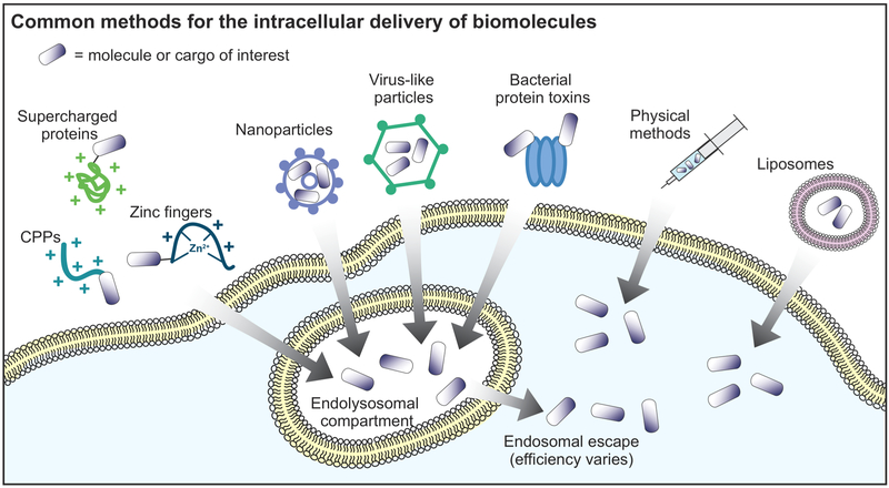
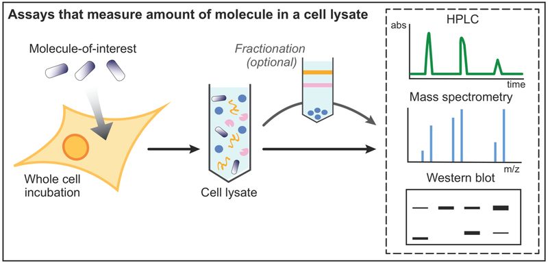
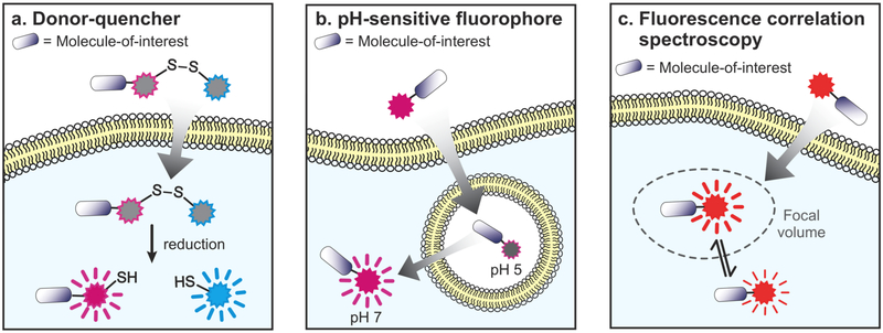
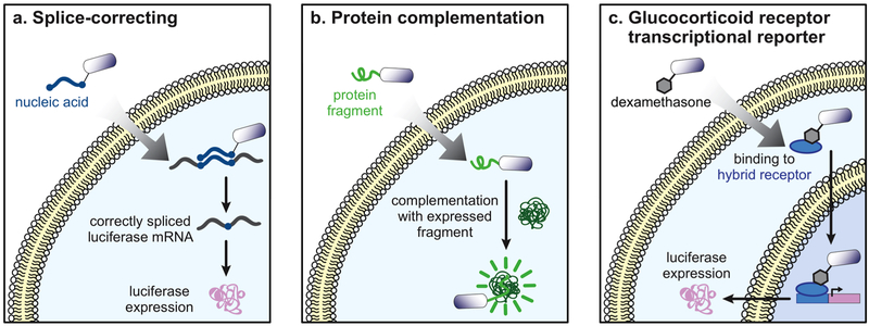
Biomolecules have many properties that make them promising for intracellular therapeutic applications, but delivery remains a key challenge because large biomolecules cannot easily enter the cytosol. Furthermore, quantification of total intracellular versus cytosolic concentrations remains demanding, and the determination of delivery efficiency is thus not straightforward. In this review, we discuss strategies for delivering biomolecules into the cytosol and briefly summarize the mechanisms of uptake for these systems. We then describe commonly used methods to measure total cellular uptake and, more selectively, cytosolic localization, and discuss the major advantages and drawbacks of each method. We critically evaluate methods of measuring "cell penetration" that do not adequately distinguish total cellular uptake and cytosolic localization, which often lead to inaccurate interpretations of a molecule's cytosolic localization. Finally, we summarize the properties and components of each method, including the main caveats of each, to allow for informed decisions about method selection for specific applications. When applied correctly and interpreted carefully, methods for quantifying cytosolic localization offer valuable insight into the bioactivity of biomolecules and potentially the prospects for their eventual development into therapeutics.


Figures

Figure 1.
Figure 1.
Common methods for the intracellular delivery of biomolecules. Cargo molecules can be fused to positively charged molecules, fused to bacterial protein toxins, or packed in nanoparticles or virus-like particles for cellular uptake and eventual endosomal escape. Physical methods and liposomes can directly deliver cargo molecules to the cytosol.
Figure 2.
Figure 2.
Assays that measure the amount of molecule in a cell lysate. The cells are incubated with the molecule or cargo being investigated, followed by cell lysis. The efficient removal (or their exclusion from the assay) of molecules still bound to the surface is a critical step. Subcellular fractionation is an optional step that can be used to isolate specific subcellular compartments, requiring additional controls for excluding postlysis redistribution. The cell lysate (or fraction of the cell lysate) can then be used in several different assays to quantitate internalized molecule, including HPLC, mass spectrometry, and quantitative Western blotting.
Figure 3.
Figure 3.
Assays that distinguish cytosolic fluorescence from fluorescence in endosomes or other compartments. (a) If a molecule-of-interest or CPP cargo is labeled with a fluorophore (magenta), and also linked to a fluorescence quencher (blue) via a disulfide bond, then cytosolic localization can be inferred from dequenching of the fluorophore following reduction of the disulfide bond. (b) The pH-sensitive dye naphthofluorescein has low fluorescence in acidic environments such as endosomes and higher fluorescence in the cytosol where pH is close to 7. (c) Fluorescence correlation spectroscopy uses a diffusion model to quantitate absolute concentrations of a fluorescent dye within a focal volume chosen to exclude endosomes and other subcellular compartments. Caveats for each of these methods are summarized in Table 2.
Figure 4.
Figure 4.
Assays that measure expression of a reporter protein. (a) In the splice-correcting assay, molecules-of-interest are labeled with a nucleic acid sequence that is complementary to an interrupted luciferase mRNA transcript. When the molecule reaches the cytosol, the nucleic acid label corrects the aberrantly spliced transcript, resulting in luciferase expression that can be detected using standard luminescence methods. (b) Molecule-of-interest labeled with dexamethasone can bind to the cytosolic glucocorticoid receptor. This leads to translocation to the nucleus and expression of a reporter gene (luciferase or GFP). (c) A molecule-of-interest labeled with a GFP-derived peptide can reconstitute GFP upon cytosolic localization by complementing a genetically expressed, larger fragment. Caveats for each of these methods are summarized in Table 2.
Figure 5.
Figure 5.
Assays that measure direct interactions with cytosolic enzymes. (a) The farnesylation assay involves the transfer of a farnesyl group to a CaaX motif appended to the molecule-of-interest. The extent of farnesylation can be monitored by SDS-PAGE or Western blot. (b) In the deubiquitination assay, a dye-labeled ubiquitin is released from the molecule-of-interest by cytosolic deubiquitinases. This change is also measured by SDS-PAGE or Western blot. (c) Using a molecule-of-interest tagged with a diglycosylated fluorescein, cytosolic localization is quantitated based on extent of galactopyranoside cleavage as measured by fluorescent signal. This signal can be detected by flow cytometry since it can only originate from the cytosol. (d) When it enters the cytosol, molecule-of-interest is biotinylated by cytosolic bacterial biotin ligase. The extent of biotinylation can be measured using a quantitative Western blot. (e) Chloroalkane-labeled molecule-of-interest irreversibly labels HaloTag protein expressed in the cytosol. The relative amount of unreacted HaloTag is measured by chasing with a chloroalkane dye and measured by flow cytometry. Caveats for each of these methods are summarized in Table 2.

Similar articles

Cited by

References

    1. Mayor S, and Pagano RE (2007) Pathways of clathrin-independent endocytosis. Nat. Rev. Mol. Cell Biol 8, 603–612. - PMC - PubMed
    1. Kaksonen M, and Roux A (2018) Mechanisms of clathrin-mediated endocytosis. Nat. Rev. Mol. Cell Biol 19, 313–326. - PubMed
    1. Pei D, and Buyanova M (2019) Overcoming Endosomal Entrapment in Drug Delivery. Bioconjugate Chem 30, 273–283. - PMC - PubMed
    1. Stewart MP, Langer R, and Jensen KF (2018) Intracellular delivery by membrane disruption: Mechanisms, strategies, and concepts. Chem. Rev 118, 7409–7531. - PMC - PubMed
    1. Bechara C, and Sagan S (2013) Cell-penetrating peptides: 20 years later, where do we stand? FEBS Lett 587, 1693–1702. - PubMed

Publication types