

Hepatoid adenocarcinoma of fallopian tube: A case report and review of the literature
- PMID: 30882620
- PMCID: PMC6426549
- DOI: 10.1097/MD.0000000000014534
Hepatoid adenocarcinoma of fallopian tube: A case report and review of the literature
Abstract
Rationale: Hepatoid adenocarcinoma (HAC) of the fallopian tubes is a rare malignant tumor in the female reproductive system.
Patient concerns: An 81-year-old Chinese woman presented with an elevated serum alpha-fetoprotein (AFP) level.
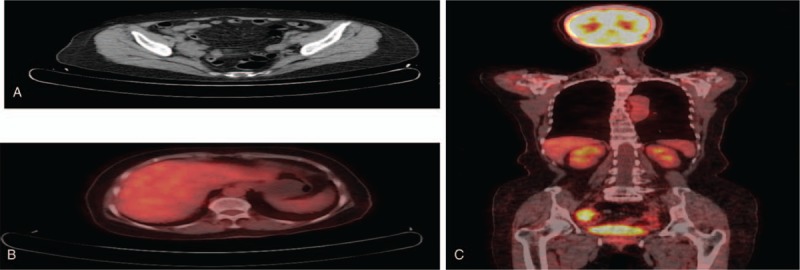
Diagnosis: Positron emission tomography-computed tomography (PET-CT) scan revealed a mass of approximately 47 × 27 mm located in the right adnexa. The tumor was diagnosed as a HAC arising from fallopian tube by immunohistochemical and histochemical technique.
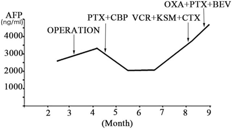
Interventions: This patient underwent surgical treatment including a bilateral adnexectomy and appendectomy. In addition, the patient underwent 5 cycles of postoperative chemotherapy.
Outcomes: The disease has recurred approximately six months after surgery and therefore, this patient will continue to be observed.
Lessons: Up to this point, only 4 known cases of HAC originating in fallopian tube have been published in the English literature. Further studies are needed to better understand the clinical characteristics, the prognosis, and the pathological mechanism of HAC development in the fallopian tubes.
Conflict of interest statement
The authors have no conflicts of interest.
Figures

Similar articles
-
alpha-Fetoprotein-producing (hepatoid) carcinoma of the fallopian tube.Gynecol Oncol. 1996 Nov;63(2):261-6. doi: 10.1006/gyno.1996.0317. Gynecol Oncol. 1996. PMID: 8910638
-
AFP-producing hepatoid adenocarcinoma of appendix: a case report of 18F-FDG PET/CT.Clin Imaging. 2014 Jul-Aug;38(4):526-528. doi: 10.1016/j.clinimag.2014.02.019. Epub 2014 Mar 10. Clin Imaging. 2014. PMID: 24721022
-
Primary malignant mixed Müllerian tumors of the fallopian tube with cervix metastasis: A rare case report and literature review.Medicine (Baltimore). 2018 Jul;97(28):e11311. doi: 10.1097/MD.0000000000011311. Medicine (Baltimore). 2018. PMID: 29995765 Free PMC article.
-
[Primary uterine hepatoid adenocarcinoma: Clinicopathological analysis of 2 cases and literature review].Beijing Da Xue Xue Bao Yi Xue Ban. 2024 Dec 18;56(6):1126-1131. doi: 10.19723/j.issn.1671-167X.2024.06.030. Beijing Da Xue Xue Bao Yi Xue Ban. 2024. PMID: 39690782 Free PMC article. Review. Chinese.
-
[Primary metastasizing fallopian tube carcinoma. Case report and overview of current therapy].Zentralbl Gynakol. 1990;112(23):1477-80. Zentralbl Gynakol. 1990. PMID: 2291372 Review. German.
Cited by
-
Primary hepatoid carcinoma of the ovary: A case report and review of the literature.Medicine (Baltimore). 2020 May;99(19):e20051. doi: 10.1097/MD.0000000000020051. Medicine (Baltimore). 2020. PMID: 32384467 Free PMC article. Review.
References
-
- Dogeas E, Peng L, Choti MA. Hepatoid adenocarcinoma of unknown primary masquerading as a pancreatic tumor. J Gastrointest Surg 2017;21:2132–4. - PubMed
-
- Ishibashi K, Kishimoto T, Yonemori Y, et al. Primary hepatoid adenocarcinoma of the uterine corpus: a case report with immunohistochemical study for expression of liver-enriched nuclear factors. Pathol Res Pract 2011;207:332–6. - PubMed
Publication types
MeSH terms
Substances
LinkOut - more resources
Full Text Sources
Medical
