

Primary adrenal extranasal NK/T cell lymphoma with subcutaneous involvement demonstrated on FDG PET/CT: A clinical case report
- PMID: 30882662
- PMCID: PMC6426610
- DOI: 10.1097/MD.0000000000014818
Primary adrenal extranasal NK/T cell lymphoma with subcutaneous involvement demonstrated on FDG PET/CT: A clinical case report
Abstract
Rationale: Primary adrenal non-Hodgkin lymphomas are predominant diffuse large B cell lymphoma with frequently bilateral adrenal involvement, but the occurrence of nasal type extranodal NK/T cell lymphoma is relatively rare.
Patient concerns: A 40-year-old woman complaining of left back pain for 2-month was admitted to our department.
Diagnosis: Based on the feature of enhanced computed tomography (CT) images which showed huge bilateral well-defined adrenal masses with heterogeneous enhancement, she was tentatively diagnosed as having primary adrenal malignancy. Postoperative pathology revealed the diagnosis of primary adrenal Epstein-Barr virus-associated nasal type extranodal NK/T-cell lymphoma.
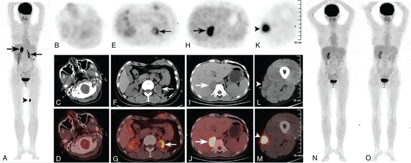
Interventions: Then, she underwent F-fluoro-2-deoxy-D-glucose (F-FDG) positron emission tomography (PET)/CT examination for staging, which showed homogeneously increased FDG uptake in the right adrenal gland and left thigh subcutaneous lesion, as well as heterogeneous increased FDG uptake in the left adrenal gland region with no abnormal uptake in the nasal cavity. Subsequently, the patient has performed 7 cycles of gemcitabine, L-asparaginase, ifosfamide, dexamethasone, etoposide (GLIDE) regimen and autologous stem cell transplantation.
Outcomes: Fortunately, the subsequent 2 follow-up FDG PET/CT scans within 1 year revealed complete resolution with no abnormal FDG uptake in the initially involved sites after 7 cycles of GLIDE chemotherapy and autologous stem cell transplantation.
Lessons: The enhanced CT and FDG PET/CT features of primary adrenal extranasal NK/T cell lymphoma are huge bilateral well-defined adrenal masses with heterogeneous enhancement, high FDG uptake, especially with subcutaneous involvement. And the awareness of this entity may help clinicians to differentiate it from other primary adrenal tumors and make reasonable therapeutic strategies. Besides, FDG PET/CT scan is very useful for the treatment follow-up of the primary adrenal extranasal NK/T cell lymphoma.
Conflict of interest statement
The authors declare that there is no conflict of interests.
Figures

Similar articles
-
Primary Adrenal Lymphoma: Two Case Series From China.Front Endocrinol (Lausanne). 2022 Jan 28;12:778984. doi: 10.3389/fendo.2021.778984. eCollection 2021. Front Endocrinol (Lausanne). 2022. PMID: 35154000 Free PMC article.
-
Extranodal NK/T-cell lymphoma with localized relapse in bone marrow of lower leg detected using PET-CT.J Clin Exp Hematop. 2024;64(1):45-51. doi: 10.3960/jslrt.23046. J Clin Exp Hematop. 2024. PMID: 38538318 Free PMC article.
-
Disseminated extranodal NK/T-cell lymphoma, nasal type, with multiple subcutaneous nodules: utility of 18F-FDG PET in staging.Clin Nucl Med. 2008 May;33(5):365-6. doi: 10.1097/RLU.0b013e31816a7a23. Clin Nucl Med. 2008. PMID: 18431160
-
Recurrence of nasal type NK/T cell lymphoma presenting as neurolymphomatosis on 18F-FDG PET/CT: A case report and literature review.Medicine (Baltimore). 2020 Jan;99(1):e18640. doi: 10.1097/MD.0000000000018640. Medicine (Baltimore). 2020. PMID: 31895825 Free PMC article. Review.
-
Beyond the lymph nodes: FDG-PET/CT in primary extranodal lymphoma.Clin Imaging. 2017 Mar-Apr;42:25-33. doi: 10.1016/j.clinimag.2016.11.006. Epub 2016 Nov 16. Clin Imaging. 2017. PMID: 27875758 Review.
Cited by
-
Primary Adrenal Lymphoma: Two Case Series From China.Front Endocrinol (Lausanne). 2022 Jan 28;12:778984. doi: 10.3389/fendo.2021.778984. eCollection 2021. Front Endocrinol (Lausanne). 2022. PMID: 35154000 Free PMC article.
-
Primary adrenal CD56-negative extranodal NK/T-cell lymphoma: Case report and literature review.Sci Prog. 2025 Jul-Sep;108(3):368504251368731. doi: 10.1177/00368504251368731. Epub 2025 Aug 17. Sci Prog. 2025. PMID: 40820315 Free PMC article.
-
Primary adrenal extranodal NK/T-cell lymphoma: A case report and literature review.Leuk Res Rep. 2020 Sep 28;14:100223. doi: 10.1016/j.lrr.2020.100223. eCollection 2020. Leuk Res Rep. 2020. PMID: 33024692 Free PMC article.
References
-
- Li YX, Fang H, Liu QF, et al. Clinical features and treatment outcome of nasal-type NK/T-cell lymphoma of Waldeyer ring. Blood 2008;112:3057–64. - PubMed
-
- AlShemmari SH, Ameen RM, Sajnani KP. Extranodal lymphoma: a comparative study. Hematology 2008;13:163–9. - PubMed
-
- Chim CS, Au WY, Shek TW, et al. Primary CD56 positive lymphomas of the gastrointestinal tract. Cancer 2001;91:525–33. - PubMed
Publication types
MeSH terms
Substances
LinkOut - more resources
Full Text Sources
Medical
