. 2019 Apr 11;62(7):3722-3740.
doi: 10.1021/acs.jmedchem.9b00231. Epub 2019 Apr 1.

Dopamine D4 Receptor-Selective Compounds Reveal Structure-Activity Relationships that Engender Agonist Efficacy

Affiliations

Dopamine D4 Receptor-Selective Compounds Reveal Structure-Activity Relationships that Engender Agonist Efficacy

Thomas M Keck et al. J Med Chem. .

Abstract

The dopamine D4 receptor (D4R) plays important roles in cognition, attention, and decision making. Novel D4R-selective ligands have promise in medication development for neuropsychiatric conditions, including Alzheimer's disease and substance use disorders. To identify new D4R-selective ligands, and to understand the molecular determinants of agonist efficacy at D4R, we report a series of eighteen novel ligands based on the classical D4R agonist A-412997 (1, 2-(4-(pyridin-2-yl)piperidin-1-yl)- N-( m-tolyl)acetamide). Compounds were profiled using radioligand binding displacement assays, β-arrestin recruitment assays, cyclic AMP inhibition assays, and molecular dynamics computational modeling. We identified several novel D4R-selective ( Ki ≤ 4.3 nM and >100-fold vs other D2-like receptors) compounds with diverse partial agonist and antagonist profiles, falling into three structural groups. These compounds highlight receptor-ligand interactions that control efficacy at D2-like receptors and may provide insights into targeted drug discovery, leading to a better understanding of the role of D4Rs in neuropsychiatric disorders.


Conflict of interest statement

The authors declare no competing financial interest.

Figures

Figure 1
Figure 1
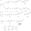
Three classic D4R-selective partial agonists.
Scheme 1
Scheme 1. Synthesis of 2-(4-(Pyridin-2-yl)piperidin-1-yl)-N-(m-tolyl)acetamide Analogues
Reagents and conditions: (a) triethylamine, EtOAc, RT; (b) CH3CN, K2CO3, reflux, appropriate arylpiperazine or arylpiperidine.
Figure 2
Figure 2
Three classes of modifications to the structure of 1 resulting in differing binding and efficacy profiles at D2-like receptors. (A) Substitution of the piperidine ring for piperazine induced a gain of efficacy at D2R and D3R with insubstantial changes to D4R efficacy. (B) Substitution of the pyridine ring with a phenyl or napthyl moiety produced modest D4R subtype selectivity improvements and lowered partial agonist efficacy at D4R with no agonist activity at D2R or D3R. (C) Para-substituted pyridine rings produced highly D4R-selective antagonists.
Figure 3
Figure 3
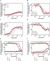
Compounds 10 (red) and 21 (gray) show similar pharmacology to parent compound 1 (black). D4R-expressing stable cells lines were plated and compounds were assayed for agonist (A) and antagonist (B) activity on β-arrestin recruitment. Similarly, D4R-mediated inhibition of cAMP accumulation was also examined in both agonist (C), and antagonist (D) modes, as indicated. Assays were conducted as described in the Experimental Methods; briefly, agonist assays were conducted by incubating the cells with the indicated concentration of test compound and measuring luminescence. Antagonist assays were conducted by incubating the compound with an EC80 concentration of dopamine (1 μM for β-arrestin and 10 nM in cAMP) and the indicated concentration of the test compound. For cAMP assays, cells were first stimulated with 10 μM forskolin. Agonist mode assays are expressed as a percentage of the maximum dopamine response, whereas antagonist mode assays are expressed as a percentage of dopamine’s EC80 response. Emax and EC50 values are shown in Tables 2 and 3. Data were fit using nonlinear regression of individual experiments performed in triplicate and are shown as means ± SEM; n = 3. Dopamine and sulpiride were run during each assay as positive controls for a full agonist and full antagonist respectively (data not shown). Compounds were also tested for both agonist and antagonist activity on cells stably expressing the closely related D2R (E) or D3R (F). Assays were conducted as described in the Experimental Methods. Agonist mode assays (open symbols) are expressed as a percentage of the maximum dopamine response observed for each receptor, whereas antagonist mode assays (solid symbols) are expressed as a percentage of dopamine’s EC80 response. Emax and EC50 values are shown in Tables 2 and 3. Data were fit using nonlinear regression of individual experiments performed in triplicate and are shown as means ± SEM; n = 3.
Figure 4
Figure 4
Compounds 13 (yellow) and 6 (blue) show diminished agonist activity at the D4R compared to parent compound 1 (black). D4R-expressing stable cells lines were plated and compounds were assayed for agonist (A) and antagonist (B) activity on β-arrestin recruitment. Similarly, D4R-mediated inhibition of cAMP accumulation was also examined in both agonist (C), and antagonist (D) modes, as indicated. Assays were conducted as described in the Experimental Methods; briefly, agonist assays were conducted by incubating the cells with the indicated concentration of test compound and measuring luminescence. Antagonist assays were conducted by incubating the compound with an EC80 concentration of dopamine (1 μM for β-arrestin and 10 nM in cAMP) and the indicated concentration of test compound. For cAMP assays, cells were first stimulated with 10 μM forskolin. Agonist mode assays are expressed as a percentage of the maximum dopamine response, whereas antagonist mode assays are expressed as a percentage of dopamine’s EC80 response. Emax and EC50 values are shown in Tables 2 and 3. Dopamine and sulpiride were run during each assay as positive controls for a full agonist and full antagonist, respectively (data not shown). Data were fit using nonlinear regression of individual experiments performed in triplicate and are shown as means ± SEM; n = 3.
Figure 5
Figure 5
Compounds 12 (green) and 9 (purple) are full antagonists at the D4R. D4R-expressing stable cells lines were plated and compounds were assayed for agonist (A) and antagonist (B) activity on β-arrestin recruitment. Similarly, D4R-mediated inhibition of cAMP accumulation was also examined in both agonist (C), and antagonist (D) modes, as indicated. Assays were conducted as described in the Experimental Methods; briefly, agonist assays were conducted by incubating the cells with the indicated concentration of test compound and measuring luminescence. Antagonist assays were conducted by incubating the compound with an EC80 concentration of dopamine (1 μM for β-arrestin and 10 nM in cAMP) and the indicated concentration of test compound. For cAMP assays, cells were first stimulated with 10 μM forskolin. Assays were conducted as described in the Experimental Methods. Agonist mode assays are expressed as a percentage of the maximum dopamine response, whereas antagonist mode assays are expressed as a percentage of dopamine’s EC80 (1 μM in β-arrestin and 10 nM in cAMP) response. Emax and EC50 values are shown in Tables 2 and 3. Dopamine and sulpiride were run during each assay as positive controls for a full agonist and full antagonist respectively (data not shown). Data were fit using nonlinear regression of individual experiments performed in triplicate and are shown as means ± SEM; n = 3.
Figure 6
Figure 6
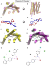
1 and 10 docked at D2R. (A–D) Comparative alignment of 1 (red ligand, yellow TM domains) and 10 (blue ligand, purple TM domains) following MD simulations of the D2R (PDB: 6CM4(26)). (E–H) Analysis of ligand interactions with specific side chains of 1 (E,G) and 10 (F,H). Although the structural difference between 1 and 10 is only a piperidine vs a piperazine ring, this drives a dramatic shift in ligand orientation in which 10 is “flipped” and rotated by 180° about its longitudinal axis, with its pyridine ring deepest in the binding pocket. This allows the basic nitrogen of the neighboring piperazine ring to engage the conserved aspartate in TM3, a common feature of dopaminergic agonists.
Figure 7
Figure 7
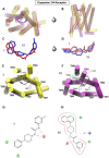
1 and 10 docked at D3R. (A–D) Comparative alignment of 1 (red ligand, yellow TM domains) and 10 (blue ligand, purple TM domains) following MD simulations of the D3R (PDB: 3PBL(25)). (E–H) Analysis of ligand interactions with specific side chains of 1 (E,G) and 10 (F,H). As seen in the D2R model, 10 is also “flipped” and rotated by 180° about its longitudinal axis in the binding pocket at D3R compared to 1. This allows for a different set of hydrophobic interactions and the engagement of the basic nitrogen of the piperazine ring to with the conserved aspartate in TM3.
Figure 8
Figure 8
1 and 13 docked at D4R. (A–D) Comparative alignment of 1 (red ligand, yellow TM domains) and 13 (blue ligand, purple TM domains) following MD simulations of the D4R (PDB: 5WIU(24)). (E–H) Analysis of ligand interactions with specific side chains of 1 (E,G) and 13 (F,H). The bulky napthyl ring of 13 shifts the overall fit within the extended binding pocket, partially disrupting the engagement of the basic nitrogen of the piperazine ring to with the conserved aspartate in TM3.
Figure 9
Figure 9
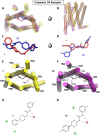
1 and 9 docked at D4R. (A–D) Comparative alignment of 1 (red ligand, yellow TM domains) and 9 (blue ligand, purple TM domains) following MD simulations of the D4R (PDB: 5WIU(24)). (E–H) Analysis of ligand interactions with specific side chains of 1 (E,G) and 9 (F,H). The inclusion of a single para substitution on the pyridine ring of 9 induces a “flipped” orientation of the ligand, in which the binding pose is rotated by 180° about its longitudinal axis, with its pyridine ring deepest in the binding pocket driving the arylamide into a deeper binding position.

References

    1. Beaulieu J.-M.; Gainetdinov R. R. The physiology, signaling, and pharmacology of dopamine receptors. Pharmacol. Rev. 2011, 63, 182–217. 10.1124/pr.110.002642. - DOI - PubMed
    1. González S.; Rangel-Barajas C.; Peper M.; Lorenzo R.; Moreno E.; Ciruela F.; Borycz J.; Ortiz J.; Lluís C.; Franco R.; McCormick P. J.; Volkow N. D.; Rubinstein M.; Floran B.; Ferré S. Dopamine D4 receptor, but not the ADHD-associated D4.7 variant, forms functional heteromers with the dopamine D2S receptor in the brain. Mol. Psychiatr. 2012, 17, 650–662. 10.1038/mp.2011.93. - DOI - PMC - PubMed
    1. Oak J. N.; Oldenhof J.; Van Tol H. H. M. The dopamine D4 receptor: one decade of research. Eur. J. Pharmacol. 2000, 405, 303–327. 10.1016/s0014-2999(00)00562-8. - DOI - PubMed
    1. Powell S.; Paulus M. P.; Hartman D. S.; Godel T.; Geyer M. A. RO-10-5824 is a selective dopamine D4 receptor agonist that increases novel object exploration in C57 mice. Neuropharmacology 2003, 44, 473–481. 10.1016/s0028-3908(02)00412-4. - DOI - PubMed
    1. Woolley M. L.; Waters K. A.; Reavill C.; Bull S.; Lacroix L. P.; Martyn A. J.; Hutcheson D. M.; Valerio E.; Bate S.; Jones D. N. C.; Dawson L. A. Selective dopamine D4 receptor agonist (A-412997) improves cognitive performance and stimulates motor activity without influencing reward-related behaviour in rat. Behav. Pharmacol. 2008, 19, 765–776. 10.1097/fbp.0b013e32831c3b06. - DOI - PubMed

Publication types

LinkOut - more resources