. 2019 Sep;89(5):758-767.
doi: 10.2319/082018-612.1. Epub 2019 Mar 18.

Reliability of 3D dental and skeletal landmarks on CBCT images

Reliability of 3D dental and skeletal landmarks on CBCT images

Joorok Park et al. Angle Orthod. 2019 Sep.

Abstract

Objectives: To quantify reliability of three-dimensional skeletal landmarks and a comprehensive set of dental landmarks in cone-beam computed tomography (CBCT) and to determine the shapes of envelope of error.

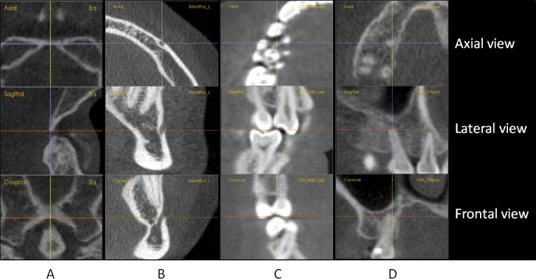
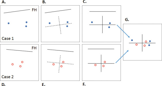
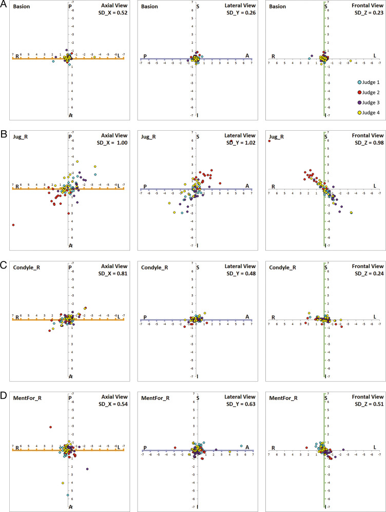
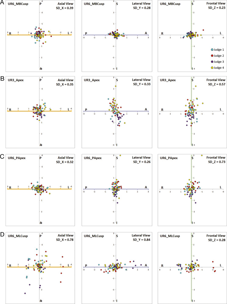
Materials and methods: Three judges located 31 skeletal landmarks and 60 dental landmarks on the pre- and posttreatment CBCT images of 22 patients. Landmark error was determined by calculating the distance of deviation of landmark locations around their average. Standard deviation and mean radial spherical error were calculated. Scatterplots were constructed to characterize envelope of error.

Results: The midline landmarks of the cranial base were highly reliable. Bilateral skeletal landmarks tended to have larger error than midline landmarks. Among the nonconventional landmarks, fronto-zygomatic suture, condyle, and mental foramen showed relatively high reliability. However, foramen spinosum and temporal fossa showed larger errors. Gonion was the least reliable landmark. Most dental landmarks were located more reliably than skeletal landmarks. The highest reliability was found at incisal edges. Mesiobuccal cusp of first molars also showed high reliability.

Conclusions: There were differences in the size and shape of the distributions of errors of different landmarks. Most landmarks showed elongated envelopes. Bilateral structures tended to show greater errors than midline structures. Most dental landmarks were more reliable than skeletal landmarks.

Keywords: Cone-beam computed tomography; Dental landmark; Reliability; Skeletal landmark.


Figures

Figure 1.
Figure 1.
Axial, sagittal, and coronal sectional views used for locating landmarks. (A) Basion. (B) Mental foramen_left. (C) Lower right first molar mesiobuccal cusp. (D) Upper right first molar palatal root apex
Figure 2.
Figure 2.
The method for constructing scatterplots. (A) Each judge located the landmark independently. The individual images of three judges were registered with their coordinates expressed in terms of the image AFOR. (B) The estimates of the three judges were averaged. The average was taken as the origin of a local AFOR for that landmark on that image with coordinates parallel and perpendicular to the image's Frankfort plane. (C) The local landmark's data were rotated and translated such that the axes became horizontal and vertical. (D–F) For the same landmark from another case, Steps A through C were repeated. (G) The plot of the second case was translated and registered on the plot of the first case. In a similar manner, the estimates for the same landmark on the remaining 20 cases were registered on the plot from G, yielding the final plot for all 22 cases.
Figure 3.
Figure 3.
Scatterplots of skeletal landmarks. (A) Basion. (B) Jugale_R. (C) Condyle_R. (D) Mental foramen_R
Figure 4.
Figure 4.
Scatterplots of dental landmarks. (A) UR6_MBCusp. (B) UR3_Apex. (C) UR6_PApex. (D) UR6_MLCusp.
Figure 5.
Figure 5.
Molar rotation can be measured by the angle formed by the distobuccal cusp, mesiolingual cusp, and midline of the palate (the line connecting ANS and PNS).

References

    1. Baumrind S. The road to three-dimensional imaging in orthodontics. Semin Orthod. 2011;17:2–12.
    1. Kapila S, Conley RS, Harrell WE., Jr The current status of cone beam computed tomography imaging in orthodontics. Dentomaxillofac Radiol. 2011;40:2–34. - PMC - PubMed
    1. Cevidanes LH, Heymann G, Cornelis MA, DeClerck HJ, Tulloch JF. Superimposition of 3-dimensional cone-beam computed tomography models of growing patients. Am J Orthod Dentofacial Orthop. 2009;136:94–99. - PMC - PubMed
    1. Weissheimer A, Menezes LM, Koerich L, Pham J, Cevidanes LH. Fast three-dimensional superimposition of cone beam computed tomography for orthopaedics and orthognathic surgery evaluation. Int J Oral Maxillofac Surg. 2015;44:1188–1196. - PMC - PubMed
    1. Baumrind S, Frantz RC. The reliability of head film measurements: 1. Landmark identification. Am J Orthod. 1971;60:111–127. - PubMed

LinkOut - more resources