. 2019 Mar 18;14(3):e0214053.
doi: 10.1371/journal.pone.0214053. eCollection 2019.

The TRPP2-dependent channel of renal primary cilia also requires TRPM3

Affiliations

The TRPP2-dependent channel of renal primary cilia also requires TRPM3

Steven J Kleene et al. PLoS One. .

Abstract

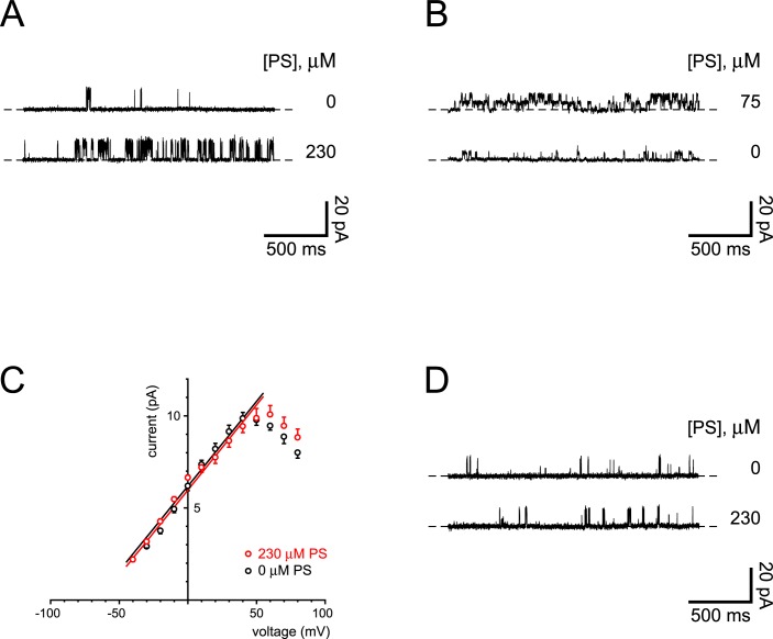
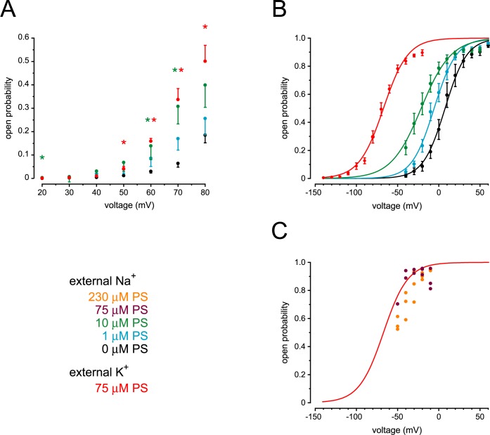
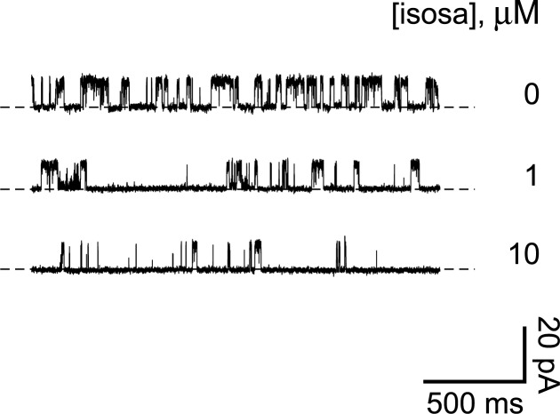
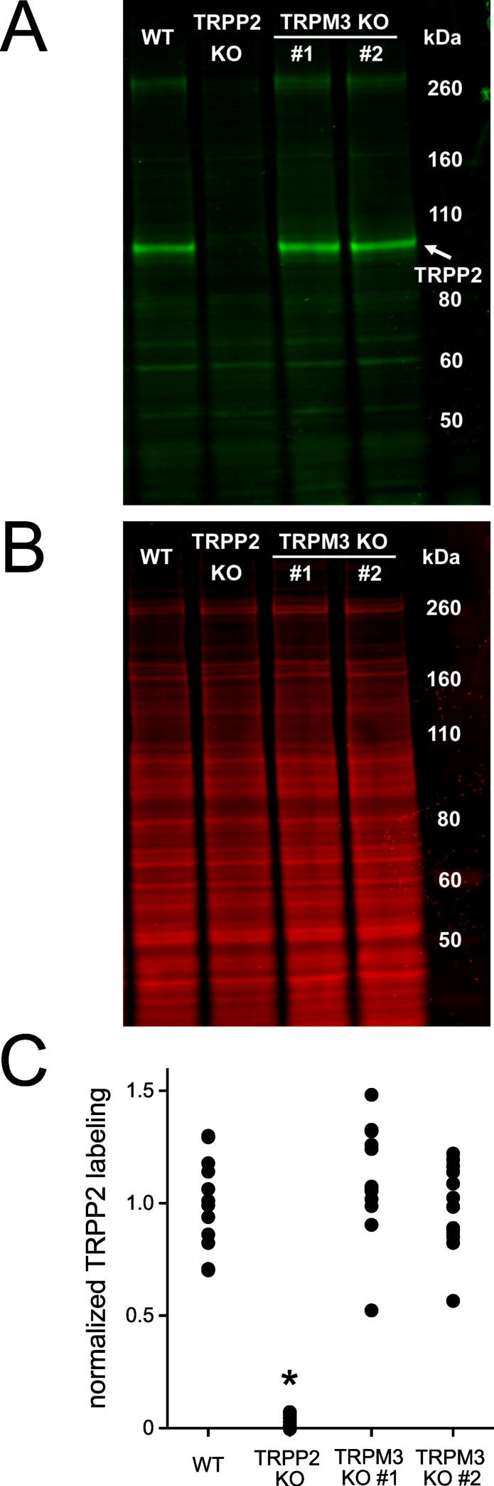
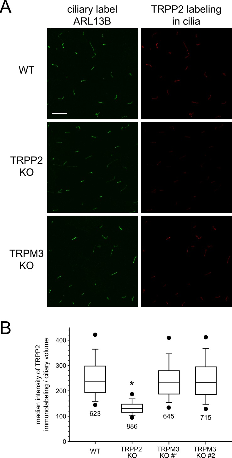
Primary cilia of renal epithelial cells express several members of the transient receptor potential (TRP) class of cation-conducting channel, including TRPC1, TRPM3, TRPM4, TRPP2, and TRPV4. Some cases of autosomal dominant polycystic kidney disease (ADPKD) are caused by defects in TRPP2 (also called polycystin-2, PC2, or PKD2). A large-conductance, TRPP2-dependent channel in renal cilia has been well described, but it is not known whether this channel includes any other protein subunits. To study this question, we investigated the pharmacology of the TRPP2-dependent channel through electrical recordings from the cilia of mIMCD-3 cells, a murine cell line of renal epithelial origin. The pharmacology was found to match that of TRPM3 channels. The ciliary TRPP2-dependent channel is known to be activated by depolarization and by increasing cytoplasmic Ca2+. This activation was greatly enhanced by external pregnenolone sulfate, an agonist of TRPM3 channels. Pregnenolone sulfate did not change the single-channel current-voltage relation. The channels were effectively blocked by isosakuranetin, a specific inhibitor of TRPM3 channels. Both pregnenolone sulfate and isosakuranetin were effective at concentrations as low as 1 μM. Knocking out TRPM3 by CRISPR/Cas9 genome editing eliminated the ciliary channel. Thus the channel is both TRPM3-dependent and TRPP2-dependent, suggesting that it may include both types of subunit. Knocking out TRPM3 did not change the level of TRPP2 protein in the cilia, so it is unlikely that the absence of functional ciliary channels results from a failure of trafficking.


Conflict of interest statement

The authors have declared that no competing interests exist.

Figures

Fig 1
Fig 1. Activation of ciliary channels by pregnenolone sulfate.
(A) The top recording shows the activity of a single ciliary channel observed while recording in the standard solutions (with 0.1 μM cytoplasmic free Ca2+). The bottom recording shows increased channel activity in the same cilium 5 min after perfusion of the pipette with standard external solution to which 230 μM pregnenolone sulfate had been added. The holding potential was +60 mV for both recordings. The dashed lines indicate the current level when the channel was closed. (B) The top recording shows the activity of two ciliary channels observed with the standard pipette (external) solution to which 75 μM pregnenolone sulfate had been added. The bottom recording shows decreased channel activity in the same cilium 4 min after perfusion of the pipette with the standard external solution, which lacks pregnenolone sulfate. For both recordings, free Ca2+ was 3 μM and the holding potential was −10 mV. (C) Single-channel current-voltage relation measured in standard external solution without (black) or with (red) 230 μM pregnenolone sulfate. For all recordings, cytoplasmic free Ca2+ was either 0.1 or 3 μM. Each point shown is the mean of measurements in 3 to 5 cilia (without pregnenolone sulfate) or 7 to 10 cilia (with pregnenolone sulfate). Each straight line is fit to the linear portion of its relation (−40 to +50 mV; R2 0.972, P < 0.001 without pregnenolone sulfate; R2 0.992, P < 0.001 with pregnenolone sulfate). (D) The top recording shows the activity of a single ciliary channel observed while recording in the standard solutions. The bottom recording shows channel activity in the same cilium 5 min after replacing the cytoplasmic solution with one containing 230 μM pregnenolone sulfate. Both cytoplasmic solutions contained 0.1 μM free Ca2+. The holding potential was +60 mV for both recordings.
Fig 2
Fig 2. Dependence of channel activation on voltage and pregnenolone sulfate concentration.
(A) Channel open probability was measured as a function of membrane potential. Cytoplasmic solutions contained 0.1 μM free Ca2+. The pipette contained pregnenolone sulfate (0 μM to 75 μM as indicated) dissolved in an external solution that also contained 0.3% (w/v) DMSO. Each point shown is the mean of measurements in 4 to 8 cilia. * Significantly different from 0 μM pregnenolone sulfate for the given voltage, P < 0.001–0.014, t-tests and Mann-Whitney rank sum tests controlled by false discovery rate correction for 21 comparisons. (B) The same procedures as in A but with 3 μM cytoplasmic free Ca2+. The external (pipette) solution included pregnenolone sulfate (0 μM to 75 μM as indicated). Each point shown is the mean of measurements in 4 to 11 cilia. The solid smooth curves represent best fits of the open probability-voltage relations to Boltzmann functions as described previously [19]. Constants and strength and significance of fit for the best-fitting Boltzmann functions are given in S1 Table. Representative single-channel recordings are shown in S2 Fig. (C) Responses in standard external solution to which was added 75 μM (purple circles) or 230 μM (orange circles) pregnenolone sulfate. As in (B), cytoplasmic solutions contained 3 μM free Ca2+. The red Boltzmann function is copied from (B) (response to 75 μM pregnenolone sulfate with a KCl-based external solution).
Fig 3
Fig 3. Isosakuranetin reduces the activity of a ciliary channel.
The top recording shows the activity of a single ciliary channel in the absence of isosakuranetin. The middle and bottom recordings show reduced channel activity in the same cilium following transfer to solutions containing 1 μM (5 min) or 10 μM (2 min) isosakuranetin, respectively. In all cases cytoplasmic free Ca2+ was 3 μM and the holding potential was +30 mV. Isosa, isosakuranetin.
Fig 4
Fig 4. Knockout of TRPM3 does not change TRPP2 membrane protein levels.
(A,B) Representative western blot of membrane proteins labeled for TRPP2 (A) or total protein (B). TRPP2 is present in wild-type (WT) and TRPM3-knockout (TRPM3 KO) cell lines but absent from the TRPP2-knockout (TRPP2 KO) cell line. Each blot had a duplicate set of lanes also used for analysis but not shown in the figure. (C) Quantification of TRPP2 labeling. Membrane protein labeling for TRPP2 in two TRPM3 knockout cell lines is not significantly different from that in the wild type (P = 0.75 and P ≥ 0.99, one-way ANOVA with Tukey's all pairwise comparison). The TRPP2 knockout has less labeling than the other lines (* P < 0.001). Six cell passages of the four cell lines were tested with one blot per passage and two lanes per cell line on each blot. The fluorescent signal of each lane’s TRPP2 band was normalized by dividing by a lane normalization factor, which was the Revert total protein stain’s fluorescent signal for that entire lane divided by the Revert total protein stain’s fluorescent signal for the brightest lane on the blot. To obtain the normalized TRPP2 labeling value (y-axis), each lane's ratio of TRPP2 signal/lane normalization factor was divided by the average of the TRPP2 signal/lane normalization factor for the two lanes containing the wild-type samples for that blot.
Fig 5
Fig 5. Ciliary TRPP2 immunolabeling is not affected by knocking out TRPM3.
(A) For each of the wild-type (WT), TRPP2-knockout (TRPP2 KO), and TRPM3-knockout (TRPM3 KO) cell lines, representative maximum intensity projections through a Z stack of images are shown. The cells were immunolabeled for ARL13B, a ciliary protein [34] (first column). The ARL13B labeling was used to define ciliary volumes in the 3D data set as described in Materials and Methods. The maximum intensity projection for the TRPP2 labeling in those ciliary volumes (second column) shows that knocking out TRPM3 does not alter the TRPP2 ciliary labeling. Knocking out TRPP2 reduces the TRPP2 ciliary fluorescence but non-specific labeling remains. Bar = 10 μm. All images had contrast enhanced in the same manner. A few brightly labeled pixels were saturated so that lightly labeled pixels would be visible while keeping gamma at 1. No pixels were saturated for the quantification of intensity used to make the graph in (B). (B) A box plot of the median intensity of TRPP2 immunolabeling/ciliary volume for each cell line. Knocking out TRPM3 does not alter the TRPP2 ciliary labeling (P = 0.62 and P ≥ 0.99, Kruskal-Wallis one-way ANOVA on ranks with Dunn’s all pairwise comparison). Knocking out TRPP2 reduces the TRPP2 ciliary fluorescence by about half compared to the other lines (* P < 0.001). The remaining fluorescence in the TRPP2 knockout is considered non-specific labeling by the non-affinity purified, polyclonal TRPP2 antibody, which had several non-specific bands in a western blot of whole-cell lysate from these same wild-type and TRPP2-knockout cell lines (see Fig 9A in [19]). The box plot's top, middle, and bottom lines indicate 75th, 50th, and 25th percentile values, respectively. Whiskers indicate 90% and 10% values; circles indicate the 95% and 5% values. The numbers below each box plot indicate the number of ciliary volumes followed. Four cell passages were used for each cell line.

Similar articles

Cited by

References

    1. Goetz SC, Anderson KV. The primary cilium: a signalling centre during vertebrate development. Nat Rev Genet. 2010;11: 331–344. 10.1038/nrg2774 . - DOI - PMC - PubMed
    1. Hildebrandt F, Benzing T, Katsanis N. Ciliopathies. N Engl J Med. 2011;364: 1533–1543. 10.1056/NEJMra1010172 . - DOI - PMC - PubMed
    1. Waters AM, Beales PL. Ciliopathies: an expanding disease spectrum. Pediatr Nephrol. 2011;26: 1039–1056. 10.1007/s00467-010-1731-7 . - DOI - PMC - PubMed
    1. Reiter JF, Leroux MR. Genes and molecular pathways underpinning ciliopathies. Nat Rev Mol Cell Biol. 2017;18: 533–547. 10.1038/nrm.2017.60 . - DOI - PMC - PubMed
    1. Delling M, DeCaen PG, Doerner JF, Febvay S, Clapham DE. Primary cilia are specialized calcium signalling organelles. Nature. 2013;504: 311–314. 10.1038/nature12833 . - DOI - PMC - PubMed

Publication types

MeSH terms