. 2019 Jul;67(7):1344-1358.
doi: 10.1002/glia.23609. Epub 2019 Mar 18.

Vascular endothelial growth factor increases the function of calcium-impermeable AMPA receptor GluA2 subunit in astrocytes via activation of protein kinase C signaling pathway

Affiliations

Vascular endothelial growth factor increases the function of calcium-impermeable AMPA receptor GluA2 subunit in astrocytes via activation of protein kinase C signaling pathway

Zeng-Wei Kou et al. Glia. 2019 Jul.

Abstract

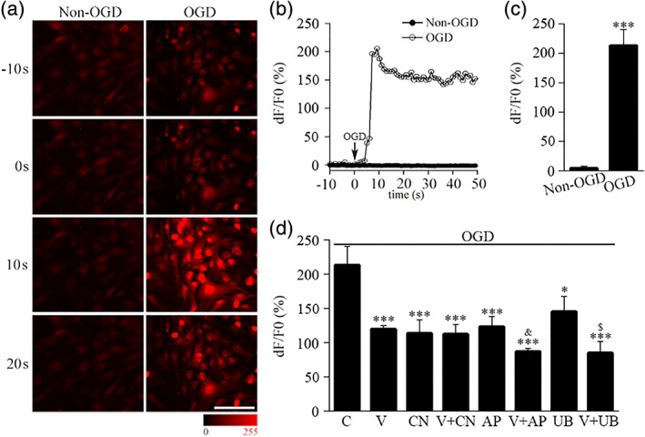
Astrocytic calcium signaling plays pivotal roles in the maintenance of neural functions and neurovascular coupling in the brain. Vascular endothelial growth factor (VEGF), an original biological substance of vessels, regulates the movement of calcium and potassium ions across neuronal membrane. In this study, we investigated whether and how VEGF regulates glutamate-induced calcium influx in astrocytes. We used cultured astrocytes combined with living cell imaging to detect the calcium influx induced by glutamate. We found that VEGF quickly inhibited the glutamate/hypoxia-induced calcium influx, which was blocked by an AMPA receptor antagonist CNQX, but not D-AP5 or UBP310, NMDA and kainate receptor antagonist, respectively. VEGF increased phosphorylation of PKCα and AMPA receptor subunit GluA2 in astrocytes, and these effects were diminished by SU1498 or calphostin C, a PKC inhibitor. With the pHluorin assay, we observed that VEGF significantly increased membrane insertion and expression of GluA2, but not GluA1, in astrocytes. Moreover, siRNA-produced knockdown of GluA2 expression in astrocytes reversed the inhibitory effect of VEGF on glutamate-induced calcium influx. Together, our results suggest that VEGF reduces glutamate-induced calcium influx in astrocytes via enhancing PKCα-mediated GluA2 phosphorylation, which in turn promotes the membrane insertion and expression of GluA2 and causes AMPA receptors to switch from calcium-permeable to calcium-impermeable receptors, thereby inhibiting astrocytic calcium influx. The present study reveals that excitatory neurotransmitter glutamate-mediated astrocytic calcium influx can be regulated by vascular biological factor via activation of AMPA receptor GluA2 subunit and uncovers a novel coupling mechanism between astrocytes and endothelial cells within the neurovascular unit.

Keywords: AMPA receptors; calcium imaging; neurovascular unit; siRNA; vascular endothelial growth factor.


Conflict of interest statement

The authors declare no conflicts of interest.

Figures

Figure 1
Figure 1
VEGF increases serine phosphorylation of AMPA receptor GluA2 subunit in astrocytes via activation of Flk1 receptor. (a–d) Total cellular proteins from cultured astrocytes with different VEGF treatment times were precipitated with antibodies against Flk1, Flt1, GluA1 or GluA2. The immunoprecipitates were examined by Western blot with antibodies against phosphotyrosine (PY20) or phosphoserine (pSer) to detect tyrosine phosphorylated Flk1 (a, p‐Flk1) or Flt1 (b, p‐Flt1), and serine phosphorylated GluA1 (c, p‐GluA1) or GluA2 (d, p‐GluA2). Then the blots were reprobed with antibodies against Flk1, Flt1, GluA1 or GluA2 to detect the total protein loading (a, t‐Flk1, b, t‐Flt1, c, t‐GluA1 d, t‐GluA2). The relative quantity of phosphorylated Flk1, Flt1, GluA1 or GluA2 was calculated as p‐Flk1/t‐Flk1, p‐Flt1/t‐Flt1, p‐GluA1/t‐GluA1 or p‐GluA2/t‐GluA2. (e, f) Total cellular proteins from cultured astrocytes with 20 min different treatments were precipitated with an antibody against GluA2. The immunoprecipitates were examined by Western blot with an antibody against phosphoserine to detect phosphorylated GluA2. Then the blots were reprobed with antibody against GluA2 to detect the total loading of GluA2 protein. The relative quantity of phosphorylated GluA2 was calculated as p‐GluA2/t‐GluA2. C: Vehicle; V: VEGF; V + S: VEGF + SU1498; S: SU1498. To make sure equal proteins were used for immunoprecipitation, 10% amount of proteins (input) for immunoprecipitation from each group were electrophoresed and immunoblotted with antibodies against Flk1, Flt1, GluA1 or GluA2. N = 4 in (a, b, d, f). N = 3 in (c). * p < 0.05 vs. vehicle group; *** p < 0.01 vs. vehicle group; ### p < 0.01 vs. VEGF group
Figure 2
Figure 2
VEGF increases membrane insertion and expression of AMPA receptor GluA2 subunit in astrocytes. (a–e) Recording of the GluA1 and GluA2 insertion events by the pHluorin assay. (aI) the representative image showed the whole cell fluorescence from the pH‐GluA2 plasmid transfected astrocyte. Bar = 10 μm. (aII) series of images showed the time course of one pH‐GluA2 insertion event. (aIII) Y‐t projection image showed the insertion event that shown in (aII). (b, c) Representative images and quantification of the insertion events from the pH‐GluA2 plasmid transfected astrocytes with different treatments. C: Vehicle; V: VEGF; V + S: VEGF + SU1498; S: SU1498. (d, e) representative images and quantification of the insertion events from the pH‐GluA1 plasmid transfected astrocytes with vehicle or VEGF treatment. (f, g) representative blots for GluA2 (mGluA2) and pan‐cadherin in membrane proteins from cultured astrocytes with 20 min different treatments. The relative quantity of mGluA2 was calculated as mGluA2/pan‐cadherin. (h, i) representative blots for GluA1 (mGluA1), GluA2 (mGluA2) and pan‐cadherin in membrane proteins from cultured astrocytes with 20 min vehicle or VEGF treatment. The ratio of (mGluA2/mGluA1) was compared between vehicle and VEGF treatment group. N = 4 in (c, e, g). N = 3 in (i). *** p < 0.01 vs. vehicle group; ### p < 0.01 vs. VEGF group
Figure 3
Figure 3
VEGF increases serine phosphorylation of AMPA receptor GluA2 subunit in astrocytes via activation of PKCα signaling pathway. (a) Representative images of triple stained Flk1, GluA2, and GFAP in cultured astrocytes. Bar = 100 μm. (b) Total cellular proteins from cultured astrocytes were precipitated with anti‐GluA2 antibody or IgG. The immunoprecipitates were examined by Western blot with antibodies against Flk1 and GluA2. (c) Total cellular proteins from cultured astrocytes were precipitated with anti‐PKCα antibody or IgG. The immunoprecipitates were examined by Western blot with antibodies against Flk1, GluA2, and PKCα. (d) Representative blots for phosphorylated PKCα (p‐PKCα) and total PKC (t‐PKCα) in total cellular proteins from cultured astrocytes with 20 min different treatments. The relative quantity of phosphorylated PKCα was calculated as p‐PKCα/t‐PKCα. C: Vehicle; V: VEGF; V + S: VEGF + SU1498; S: SU1498. (e) Total cellular proteins from cultured astrocytes with 20 min different treatments were precipitated with an antibody against GluA2. The immunoprecipitates were examined by Western blot with an antibody against phosphoserine (pSer) to detect phosphorylated GluA2 (p‐GluA2). Then the blots were reprobed with antibody against GluA2 to detect the total loading of GluA2 protein (t‐GluA2). The relative quantity of phosphorylated GluA2 was calculated as p‐GluA2/t‐GluA2. V + Ca: VEGF + calphostin C; Ca: Calphostin C. N = 4 in (d, e). *** p < 0.01 vs. vehicle group; ### p < 0.01 vs. VEGF group [Color figure can be viewed at wileyonlinelibrary.com]
Figure 4
Figure 4
VEGF inhibits glutamate‐induced calcium influx in astrocytes via activation of AMPA receptor GluA2 subunit and PKC signaling. (a) Series of images showed the glutamate‐induced Fluo‐4 fluorescence changes in cultured astrocytes with vehicle (C) or VEGF (V) treatment. Bar = 200 μm. (b) Representative curves showed the glutamate‐induced changes of calcium in cultured astrocytes with different treatments. V + S: VEGF + SU1498; S: SU1498. (c) Quantification of the glutamate‐induced peak calcium response representatively shown in (b). (d) Representative curves showed the glutamate‐induced changes of calcium in cultured astrocytes with vehicle or VEGF treatment in Ca2+‐free recording buffer. (e) Quantification of the glutamate‐induced peak calcium response representatively shown in (d). (f) Quantification of the glutamate‐induced peak calcium response in cultured astrocytes with different treatments. CN: CNQX, V + CN: VEGF + CNQX; AP: D‐AP5; V + AP: VEGF +D‐AP5; UB: UBP310; V + UB: VEGF + UBP310. (g) Representative curves showed the glutamate‐induced changes of calcium in cultured astrocytes with different treatments. V + Ca: VEGF + calphostin C; Ca: Calphostin C. (h) Quantification of the glutamate‐induced peak calcium response representatively shown in (g). N = 4 in (c, e, h); in (f), n = 6 for C, V, CN, V + CN, AP, V + AP. N = 3 for UB, V + UB; *** p < 0.01 vs. vehicle group; ### p < 0.01 vs. V group; &&& p < 0.01 vs. AP group; $ p < 0.05 vs. UB group [Color figure can be viewed at wileyonlinelibrary.com]
Figure 5
Figure 5
Knockdown of AMPA receptor GluA2 subunit reverses the inhibitory effect of VEGF on glutamate‐induced calcium influx in astrocytes. (a) Representative blots for GluA2 and β‐actin in total cellular proteins from astrocytes transfected with scramble non‐target siRNA or GluA2 siRNA. The relative quantity of GluA2 was calculated as GluA2/β‐actin. (b) Curves showed the glutamate‐induced changes of calcium in siRNA transfected astrocytes with vehicle (C) or VEGF treatment (V). (c) Representative in cell‐Western with antibodies against GluA2 and β‐actin in the same wells performed calcium imaging. (d) Quantification of in cell‐Western results representatively shown in (c). N = 3 in (a, b, d). In (a) *** p < 0.01 vs. scramble siRNA group. In (b) * p < 0.05 vs. scramble siRNA + C group *** p < 0.01 vs. scramble siRNA + C group. In (d), ### p < 0.01 vs. scramble siRNA + V group
Figure 6
Figure 6
VEGF inhibits OGD‐induced calcium influx via AMPA receptor in astrocytes. (a) Series of images showed the Fluo‐4 fluorescence changes in cultured astrocytes with (OGD) or without OGD (non‐OGD) treatment. Bar = 100 μm. (b) Representative curves showed the changes of Fluo‐4 fluorescence in cultured astrocytes with or without OGD treatment. (c) Quantification of the peak calcium response representatively shown in (b). (d) Quantification of the OGD‐induced peak calcium response in cultured astrocytes with different treatments. C: vehicle; V: VEGF; CN: CNQX; V + CN: VEGF + CNQX; AP: D‐AP5; V + AP: VEGF + D‐AP5; UB: UBP310; V + UB: VEGF + UBP310. N = 4 in (c, d). In (c), ***p < 0.01 vs. non‐OGD group. In (d), ***p < 0.01 vs. C group; & p < 0.05 vs. AP group; $ p < 0.05 vs. UB group [Color figure can be viewed at wileyonlinelibrary.com]
Figure 7
Figure 7
Schematic of VEGF increases the function of calcium‐impermeable AMPA receptor GluA2 subunit in astrocytes via activation of protein kinase C signaling pathway. A, VEGF binds to FLK1 receptors and induces phosphorylation at tyrosine residues. B, The p‐Flk1 induced PKCα phosphorylated at threonine 638. C, p‐PKCα increased GluA2 phosphorylation at serine residues. D, Phosphorylated GluA2 inserts into cell membrane and inhibits AMPA receptor‐calcium influx in glutamate treatment [Color figure can be viewed at wileyonlinelibrary.com]

Similar articles

Cited by

References

    1. Allen, N. J. , & Barres, B. A. (2009). Neuroscience: Glia—More than just brain glue. Nature, 457(7230), 675–677. 10.1038/457675a - DOI - PubMed
    1. Ahn, S. M. , & Choe, E. S. (2010). Alterations in GluR2 AMPA receptor phosphorylation at serine 880 following group I metabotropic glutamate receptor stimulation in the rat dorsal striatum. Journal of Neuroscience Research, 88(5), 992–999. 10.1002/jnr.22275 - DOI - PubMed
    1. Anthony, T. E. , Klein, C. , Fishell, G. , & Heintz, N. (2004). Radial glia serve as neuronal progenitors in all regions of the central nervous system. Neuron, 41(6), 881–890. - PubMed
    1. Araki, Y. , Lin, D. T. , & Huganir, R. L. (2010). Plasma membrane insertion of the AMPA receptor GluA2 subunit is regulated by NSF binding and Q/R editing of the ion pore. Proceedings of the National Academy of Sciences of the United States of America, 107(24), 11080–11085. 10.1073/pnas.1006584107 - DOI - PMC - PubMed
    1. Avraham‐Lubin, B. C. , Goldenberg‐Cohen, N. , Sadikov, T. , & Askenasy, N. (2012). VEGF induces neuroglial differentiation in bone marrow‐derived stem cells and promotes microglia conversion following mobilization with GM‐CSF. Stem Cell Reviews, 8(4), 1199–1210. 10.1007/s12015-012-9396-1 - DOI - PubMed

Publication types

MeSH terms

Substances