. 2019 Jun;111(6):1652-1670.
doi: 10.1111/mmi.14243. Epub 2019 Apr 26.

Analysis of a flagellar filament cap mutant reveals that HtrA serine protease degrades unfolded flagellin protein in the periplasm of Borrelia burgdorferi

Affiliations

Analysis of a flagellar filament cap mutant reveals that HtrA serine protease degrades unfolded flagellin protein in the periplasm of Borrelia burgdorferi

Kai Zhang et al. Mol Microbiol. 2019 Jun.

Abstract

Unlike external flagellated bacteria, spirochetes have periplasmic flagella (PF). Very little is known about how PF are assembled within the periplasm of spirochaetal cells. Herein, we report that FliD (BB0149), a flagellar cap protein (also named hook-associated protein 2), controls flagellin stability and flagellar filament assembly in the Lyme disease spirochete Borrelia burgdorferi. Deletion of fliD leads to non-motile mutant cells that are unable to assemble flagellar filaments and pentagon-shaped caps (10 nm in diameter, 12 nm in length). Interestingly, FlaB, a major flagellin protein of B. burgdorferi, is degraded in the fliD mutant but not in other flagella-deficient mutants (i.e., in the hook, rod, or MS-ring). Biochemical and genetic studies reveal that HtrA, a serine protease of B. burgdorferi, controls FlaB turnover. Specifically, HtrA degrades unfolded but not polymerized FlaB, and deletion of htrA increases the level of FlaB in the fliD mutant. Collectively, we propose that the flagellar cap protein FliD promotes flagellin polymerization and filament growth in the periplasm. Deletion of fliD abolishes this process, which leads to leakage of unfolded FlaB proteins into the periplasm where they are degraded by HtrA, a protease that prevents accumulation of toxic products in the periplasm.


Figures

FIG. 1.
FIG. 1.. Transcriptional analyses of fliDBb gene.
(A) Genes adjacent to fliDBb. Black arrows represent three pairs of primers for co-RT-PCR. Vertical arrows denote promoters of flaB (pflaB) and fliDBb (pfliD). (B) fliDBb is monocistronic. DNA gel electrophoresis analysis of RT-PCR (top panel) and PCR (bottom panel) products. The lane numbers correspond to the primers labeled in panel A. (C) Upstream DNA sequence of fliDBb. Underlined sequences are the predicted −12 and −35 regions of pfliD. Asterisk denotes the previously mapped transcription start site (TSS) (Adams et al., 2017). (D) Comparison of pfliD with the consensus sequences of σ70 promoters identified in E. coli and B. burgdorferi. Asterisk represents TSS. (E) Transcriptional analysis of pfliD using lacZ as a reporter. For this assay, pfliD or pflaB was fused to the promoterless lacZ gene in the pRS414 plasmid. The empty pRS414 was used as a negative control, and the plasmid containing pflaB used as a positive control. β-galactosidase activity was measured and expressed as the average Miller units of triplicate samples from two independent experiments, as previously described (Bian et al., 2011). (F) & (G) GFP reporter assay. For this assay, pfliD-gfp (F) and gfp alone (G) were cloned into pJSB275, respectively. The resultant vectors were transformed into B31A strain. The expression of GFP was visualized with fluorescence microcopy (Axiostar Plus, Ziess). All of the primers used here are listed in Table S1.
FIG. 2.
FIG. 2.. Domain organization and overall structure of FliDBb.
(A) The domain organization of FliDBb. Domains were determined by multiple sequence alignments of FliD proteins (Fig. S2), including E. coli and S. typhymurium FliD (ecFliD and stFliD). (B) Monomeric structure of FliDBb. An all-atom model of FliDBb was generated using the crystal structure of ecFliD (PDB ID 5H5V) (Song et al., 2017) as the template. Despite the low sequence homology (16% identity), the model has ~72% coverage (480 of 665 aa). Due to a lack of sequence similarity, the variable region (VR) of 185 residues (dotted lines) was unable to be modeled. (C) Hexamer structure of FliDBb. The homology model was generated using the hexamer of ecFliD as the template. The domains that resemble the cap plate and leg of FliD are labeled.
FIG. 3.
FIG. 3.. Construction of a fliDBb deletion mutant and its complemented strain.
(A) Construction of fliD::kan for targeted mutagenesis of fliDBb. To construct this vector, a kanamycin-resistance marker (kan) was inserted into fliDBb at a HindIII cut site. (B) Construction of cis-complementation vector of fliD/cisCom by inserting fliDBb into an intergenic region between bb0445 and bb0446 on the chromosome of B. burgdorferi, as previously reported (Sze et al., 2013, Li et al., 2007). (C) Characterization of WT, a fliDBb mutant (∆fliD), and its complemented strain (∆fliDcom) by immunoblotting with a specific antibody against FliD (αFliD). DnaK was used as an internal control, as previously described (Sze et al., 2013).
FIG. 4.
FIG. 4.. Deletion of fliDBb leads to mutant cells that are non-motile and rod-shaped.
(A-D) Swimming plate assays. These assays were conducted by inoculating 5 μl of B. burgdorferi cells into 0.35% agar plates, as previously described (Li et al., 2002). ΔflaB, a previously constructed non-motile mutant (Motaleb et al., 2000), was used as a control to determine the initial inoculum size. At least four plates were included for each strain and the data are expressed as averaged diameters of the rings ± standard deviations (n=4): WT (12 ± 0.4 mm), ΔfliD (7.25 ± 0.6 mm), ΔfliDcom (11.75 ± 0.3 mm), and ΔflaB (7.5 ± 0.5 mm). (E-F) Dark-field microscopy analysis of WT and ∆fliD.
FIG. 5.
FIG. 5.. Visualization of ∆fliD and ΔflaB mutant cells by cryo-ET.
(A, B) Representative tomograms of ∆fliD and ΔflaB mutant cells. The insertion images show enlarged views of the flagellar hook structure and length. (C, D) Three-dimensional surface views of the cells shown in (A) and (B). The insertion images show the enlarged views of the hooks and hook-associated caps.
FIG. 6.
FIG. 6.. Three-dimensional averaged structures of ΔflaB and ∆fliD show difference in the FliD cap region.
(A) A central section of the averaged structure from the ΔflaB mutant. The averaged structure was generated from 1492 sub-tomograms. (B) A cross section on the hook region from (A) shows six subunits with a diameter of 16 nm. (C) A cross section on the cap region from (A) shows five subunits with a diameter of 10 nm. (D) Two different views of solid-surface rendering map on the structure in (A) show the symmetry mismatching between hook and cap. The hook region has helical packing at about 11 subunits per two turns. The cap region has an asymmetric structure that is close to 5-fold symmetry. The top part of the cap that interacts with the hook is not symmetric. (E) A central section of the averaged structure from ∆fliD. The averaged structure was generated from 599 sub-tomograms. (F) A cross section on the hook region from (E) also shows six subunits with a diameter of 16 nm. (G) Two different views of the surface rendering of the hook structure from (A). It has the same helical symmetry as the hook region in (D).
FIG. 7.
FIG. 7.. Deletion of fliDBb impairs the level of flagellar filament proteins.
(A) Immunoblotting analyses to measure the levels of five flagellar proteins that represent three parts of the PF –the filament (FlaA and FlaB), the hook (FlgE), and the rod (FlgG) – as well as the MS-ring (FliF). For this experiment, similar amounts of whole cell lysates of WT, ∆fliD, and ∆fliDcom were analyzed by SDS-PAGE and then probed against different antibodies as indicated. α- Abbreviation of antibody. DnaK was used as a loading control. Immunoblots were developed using horseradish peroxidase secondary antibody with an ECL luminol assay, as previously described (Sze et al., 2013). (B) Detection of flaB transcripts by qRT-PCR. This experiment was conducted, as previously described (Sze et al., 2013). The transcript of the enolase gene (eno) was used as an internal control to normalize the qPCR data. The results were expressed as the mean threshold cycles (CT) of triplicate samples. (C, D) Measuring FlaB stability by protein turnover assays. To arrest protein translation, 100 µg/ml spectinomycin was added to the WT and ∆fliD mutant cultures. Samples were collected at the indicated time points for immunoblotting analyses. DnaK was used as a sample loading control. The levels of FlaB were measured by densitometry and expressed as the percent protein density at zero time compared to subsequent time points.
FIG. 8.
FIG. 8.. Detection of FlaB in different flagellar mutants by immunoblotting.
(A) Overall structure of PF. Red arrows denote five flagellar genes that were inactivated by targeted mutagenesis. (B) Detection of FlaB in WT and five flagellar mutants by immunoblotting. Similar amounts of whole cell lysates were subjected to SDS-PAGE, followed by immunoblotting using antibodies against FlaB (αFlaB) and DnaK (αDnaK). DnaK was used as an internal control. (C) Measuring the stability of FlaB in different flagellar mutants by protein turnover assays.
FIG. 9.
FIG. 9.. Deletion of htrABb or csrABb partially restores the level of FlaB in the ∆fliD mutant.
(A) Characterization of WT, ∆fliD, ∆htrA (a single-deletion mutant of htrABb, kanamycin resistance), and ∆fliD-htrA (a double mutant of fliDBb and htrABb). (B) Detection of FlaB in ∆fliD, ∆fliD-htrA, and ∆fliD-csrA (a double mutant of fliDBb-csrABb) by immunoblotting. DnaK was used as a sample loading control.
FIG. 10.
FIG. 10.. Proteolytic assay of HtrABb against FlaB proteins.
2 μg of rHtrABb (wild type recombinant HtrABb) or rHtrABbS226A (a site-directed mutant of rHtrABb in which Ser226 was replaced by Ala) was incubated with equal amounts of polymerized FlaB (pFlaB) or denatured FlaB (dFlaB) for 12 hrs. The resultant samples were subjected to SDS-PAGE, followed by Coomassie blue staining. pFlaB, flagellar filaments isolated from ΔflaA; dFlaB, heat-inactivated pFlaB. The flagellar filaments of B. burgdorferi are composed of a major flagellin protein, FlaB, and a minor sheath protein, FlaA. The filaments isolated from the ΔflaA mutant only contain FlaB, as shown in lanes 2, 4 and 7. Some minor bands were observed below rHtrABb (lanes 1, 3, and 5) but not rHtrABbS226A (lanes 6 and 8), indicative of autolysis of rHtrABb, as previously reported (Coleman et al., 2013).
FIG. 11.
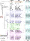
FIG. 11.. Phylogenetic analysis of FliD proteins.
Protein sequences were aligned using MUSCLE (Edgar, 2004). A maximum likelihood tree was inferred using FastTree (Price et al., 2010). The tree was subsequently re-rooted at the mid-point, and branches with lower than 90% bootstrap support were removed using the BIOTREE utility of the BpWrapper package (Hernandez et al., 2018). The tree was visualized and graphed using the APE package in R (Paradis et al., 2004). To reconstruct the evolutionary origin of the central domain, we inferred its gains or losses based on the principle of maximum parsimony. FliD homologs were identified using BLASTp with FliDBb (BB0149) as a query, and protein sequences were downloaded from GenBank. Note: Borrelia burgdorferi was recently named Borreliella burgdorferi.
FIG. 12.
FIG. 12.. Model illustrating how flagellar assembly is regulated in B. burgdorferi.
In this model, the FlaB-FliW-CsrABb interaction regulates flagellin homeostasis in the cytoplasm by a partner-switching mechanism, and HtrABb functions as a house cleaner by removal of unpolymerized FlaB in the periplasm. Upon completion of the hook assembly, FlaB monomers are transported from the cytoplasm into the periplasm via fT3SS, reducing the cytoplasmic level of FlaB. When FlaB falls below a threshold level, FliW is released and binds to CsrABb, thereby relieving the translational repression such that more FlaB proteins are synthesized and exported to assemble nascent filaments. When the filament assembly is completed or fT3SS is disrupted, as occurs in the ∆flgE mutant, the secretion of FlaB is blocked. This leads to FlaB accumulation in the cytoplasm. When FlaB rises above a threshold level, it binds to FliW, releasing CsrABb, which in turn binds to the SD sequence of flaB mRNA, blocking translation. Deletion of fliDBb leads to FlaB monomers leaking into the periplasm, where they are degraded by HtrABb. As a result, the homeostatic equilibrium of CsrABb-FliW-FlaB in the cytoplasm is perturbed, which further leads to restriction of flaB translation.

Similar articles

Cited by

References

    1. Adams PP, Flores Avile C., Popitsch N, Bilusic I, Schroeder R, Lybecker M & Jewett MW, (2017) In vivo expression technology and 5’ end mapping of the Borrelia burgdorferi transcriptome identify novel RNAs expressed during mammalian infection. Nucleic Acids Res 45: 775–792. - PMC - PubMed
    1. Agulleiro JI & Fernandez JJ, (2011) Fast tomographic reconstruction on multicore computers. Bioinformatics 27: 582–583. - PubMed
    1. Aldridge P & Hughes KT, (2002) Regulation of flagellar assembly. Curr Opin Microbiol 5: 160–165. - PubMed
    1. Aldridge PD, Karlinsey JE, Aldridge C, Birchall C, Thompson D, Yagasaki J & Hughes KT, (2006) The flagellar-specific transcription factor, sigma28, is the Type III secretion chaperone for the flagellar-specific anti-sigma28 factor FlgM. Genes Dev 20: 2315–2326. - PMC - PubMed
    1. Arora SK, Ritchings BW, Almira EC, Lory S & Ramphal R, (1998) The Pseudomonas aeruginosa flagellar cap protein, FliD, is responsible for mucin adhesion. Infect Immun 66: 1000–1007. - PMC - PubMed

Publication types