

Vascular smooth muscle cell-specific progerin expression in a mouse model of Hutchinson-Gilford progeria syndrome promotes arterial stiffness: Therapeutic effect of dietary nitrite
- PMID: 30884114
- PMCID: PMC6516150
- DOI: 10.1111/acel.12936
Vascular smooth muscle cell-specific progerin expression in a mouse model of Hutchinson-Gilford progeria syndrome promotes arterial stiffness: Therapeutic effect of dietary nitrite
Abstract
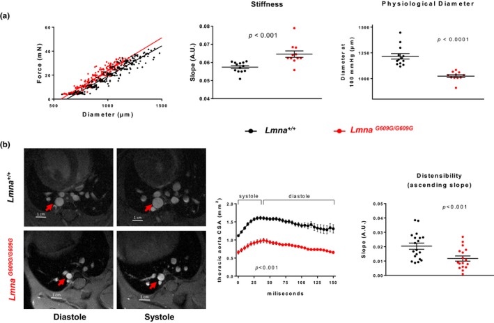
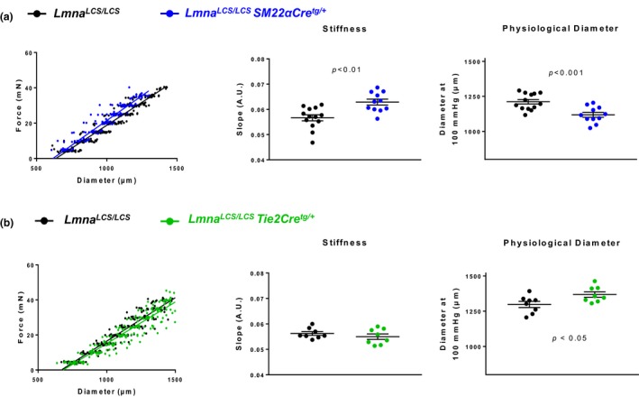
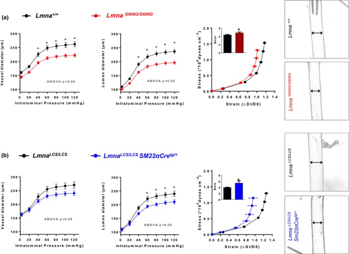
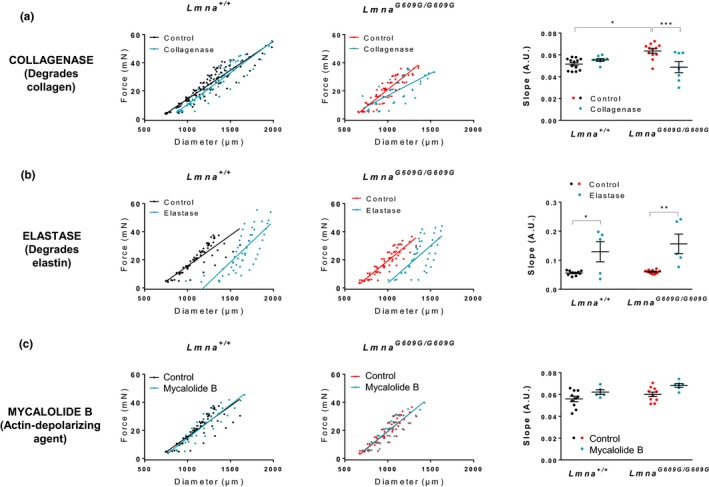
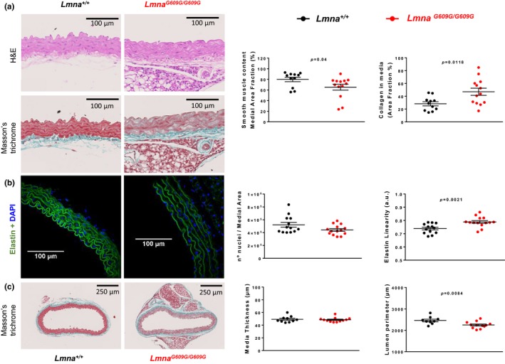
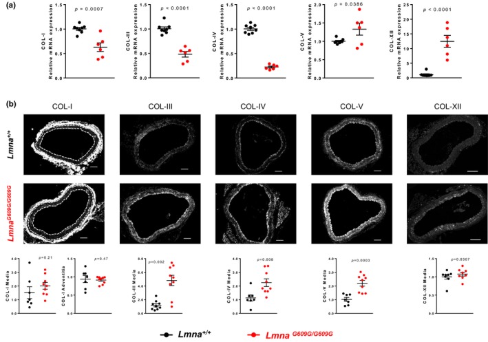
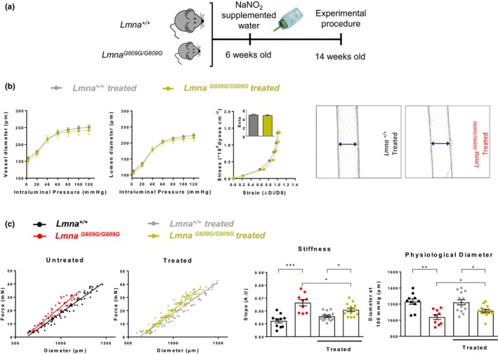
Vascular stiffness is a major cause of cardiovascular disease during normal aging and in Hutchinson-Gilford progeria syndrome (HGPS), a rare genetic disorder caused by ubiquitous progerin expression. This mutant form of lamin A causes premature aging associated with cardiovascular alterations that lead to death at an average age of 14.6 years. We investigated the mechanisms underlying vessel stiffness in LmnaG609G/G609G mice with ubiquitous progerin expression, and tested the effect of treatment with nitrites. We also bred LmnaLCS/LCS Tie2Cre+/tg and LmnaLCS/LCS SM22αCre+/tg mice, which express progerin specifically in endothelial cells (ECs) and in vascular smooth muscle cells (VSMCs), respectively, to determine the specific contribution of each cell type to vascular pathology. We found vessel stiffness and inward remodeling in arteries of LmnaG609G/G609G and LmnaLCS/LCS SM22αCre+/tg , but not in those from LmnaLCS/LCS Tie2Cre+/tg mice. Structural alterations in aortas of progeroid mice were associated with decreased smooth muscle tissue content, increased collagen deposition, and decreased transverse waving of elastin layers in the media. Functional studies identified collagen (unlike elastin and the cytoskeleton) as an underlying cause of aortic stiffness in progeroid mice. Consistent with this, we found increased deposition of collagens III, IV, V, and XII in the media of progeroid aortas. Vessel stiffness and inward remodeling in progeroid mice were prevented by adding sodium nitrite in drinking water. In conclusion, LmnaG609G/G609G arteries exhibit stiffness and inward remodeling, mainly due to progerin-induced damage to VSMCs, which causes increased deposition of medial collagen and a secondary alteration in elastin structure. Treatment with nitrites prevents vascular stiffness in progeria.
Keywords: aging; dietary nitrite; progeria; smooth muscle cells; vascular stiffness.
© 2019 The Authors. Aging Cell published by the Anatomical Society and John Wiley & Sons Ltd.
Conflict of interest statement
None declared.
Figures
Similar articles
-
Vascular Smooth Muscle Cell-Specific Progerin Expression Provokes Contractile Impairment in a Mouse Model of Hutchinson-Gilford Progeria Syndrome that Is Ameliorated by Nitrite Treatment.Cells. 2020 Mar 8;9(3):656. doi: 10.3390/cells9030656. Cells. 2020. PMID: 32182706 Free PMC article.
-
Vascular Smooth Muscle-Specific Progerin Expression Accelerates Atherosclerosis and Death in a Mouse Model of Hutchinson-Gilford Progeria Syndrome.Circulation. 2018 Jul 17;138(3):266-282. doi: 10.1161/CIRCULATIONAHA.117.030856. Epub 2018 Feb 28. Circulation. 2018. PMID: 29490993 Free PMC article.
-
Defective extracellular pyrophosphate metabolism promotes vascular calcification in a mouse model of Hutchinson-Gilford progeria syndrome that is ameliorated on pyrophosphate treatment.Circulation. 2013 Jun 18;127(24):2442-51. doi: 10.1161/CIRCULATIONAHA.112.000571. Epub 2013 May 20. Circulation. 2013. PMID: 23690466
-
Vascular smooth muscle cell loss underpins the accelerated atherosclerosis in Hutchinson-Gilford progeria syndrome.Nucleus. 2019 Dec;10(1):28-34. doi: 10.1080/19491034.2019.1589359. Nucleus. 2019. PMID: 30900948 Free PMC article. Review.
-
Molecular and Cellular Mechanisms Driving Cardiovascular Disease in Hutchinson-Gilford Progeria Syndrome: Lessons Learned from Animal Models.Cells. 2021 May 11;10(5):1157. doi: 10.3390/cells10051157. Cells. 2021. PMID: 34064612 Free PMC article. Review.
Cited by
-
Aortic Stiffness in L-NAME Treated C57Bl/6 Mice Displays a Shift From Early Endothelial Dysfunction to Late-Term Vascular Smooth Muscle Cell Dysfunction.Front Physiol. 2022 Jun 16;13:874015. doi: 10.3389/fphys.2022.874015. eCollection 2022. Front Physiol. 2022. PMID: 35800344 Free PMC article.
-
Progerin, an Aberrant Spliced Form of Lamin A, Is a Potential Therapeutic Target for HGPS.Cells. 2023 Sep 18;12(18):2299. doi: 10.3390/cells12182299. Cells. 2023. PMID: 37759521 Free PMC article. Review.
-
Vascular Smooth Muscle Cell-Specific Progerin Expression Provokes Contractile Impairment in a Mouse Model of Hutchinson-Gilford Progeria Syndrome that Is Ameliorated by Nitrite Treatment.Cells. 2020 Mar 8;9(3):656. doi: 10.3390/cells9030656. Cells. 2020. PMID: 32182706 Free PMC article.
-
Endothelial YAP/TAZ activation promotes atherosclerosis in a mouse model of Hutchinson-Gilford progeria syndrome.J Clin Invest. 2024 Oct 1;134(22):e173448. doi: 10.1172/JCI173448. J Clin Invest. 2024. PMID: 39352768 Free PMC article.
-
From Cell Architecture to Mitochondrial Signaling: Role of Intermediate Filaments in Health, Aging, and Disease.Int J Mol Sci. 2025 Jan 27;26(3):1100. doi: 10.3390/ijms26031100. Int J Mol Sci. 2025. PMID: 39940869 Free PMC article. Review.
References
-
- Briones, A. M. , Rodriguez‐Criado, N. , Hernanz, R. , Garcia‐Redondo, A. B. , Rodrigues‐Diez, R. R. , Alonso, M. J. , … Salaices, M. (2009). Atorvastatin prevents angiotensin II‐induced vascular remodeling and oxidative stress. Hypertension, 54(1), 142–149. 10.1161/hypertensionaha.109.133710 - DOI - PubMed
-
- Chen, C. , Yun, X. J. , Liu, L. Z. , Guo, H. , Liu, L. F. , & Chen, X. L. (2017). Exogenous nitric oxide enhances the prophylactic effect of aminoguanidine, a preferred iNOS inhibitor, on bleomycin‐induced fibrosis in the lung: Implications for the direct roles of the NO molecule in vivo. Nitric Oxide, 70, 31–41. 10.1016/j.niox.2017.07.005 - DOI - PubMed
Publication types
MeSH terms
Substances
Grants and funding
LinkOut - more resources
Full Text Sources
Miscellaneous
