Review
. 2019 Aug:57:84-92.
doi: 10.1016/j.sbi.2019.01.017. Epub 2019 Mar 16.

Optically inducible membrane recruitment and signaling systems

Affiliations
Review

Optically inducible membrane recruitment and signaling systems

Pimkhuan Hannanta-Anan et al. Curr Opin Struct Biol. 2019 Aug.

Abstract

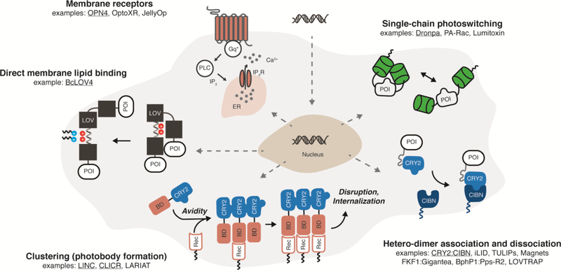
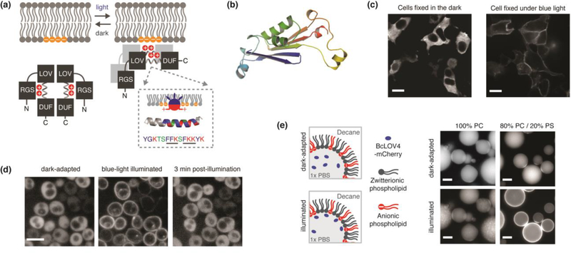
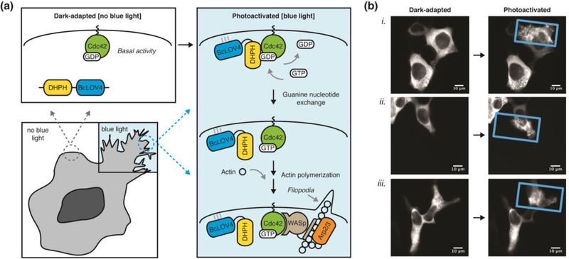
Optical induction of intracellular signaling by membrane-associated and integral membrane proteins allows spatiotemporally precise control over second messenger signaling and cytoskeletal rearrangements that are important to cell migration, development, and proliferation. Optogenetic membrane recruitment of a protein-of-interest to control its signaling by altering subcellular localization is a versatile means to these ends. Here, we summarize the signaling characteristics and underlying structure-function of RGS-LOV photoreceptors as single-component membrane recruitment tools that rapidly, reversibly, and efficiently carry protein cargo from the cytoplasm to the plasma membrane by a light-regulated electrostatic interaction with the membrane itself. We place the technology-relevant features of these recently described natural photosensory proteins in context of summarized protein engineering and design strategies for optically controlling membrane protein signaling.


Figures

Figure 1.
Figure 1.. Fully genetically encoded strategies for optically inducible intracellular signaling by integral membrane and membrane-associated proteins in mammalian cells.
Examples schematized are underlined. Abbreviations: POI = protein of interest. BD = binding domain, CIBN = cryptochrome interacting binding partner, CRY2 = cryptochrome, ER = endoplasmic reticulum, Gq* = activated Gαq, IP3 = inositol triphosphate, IP3R = IP3 receptor, LOV = light-oxygen-voltage, PLC = phospholipase C, Rec = membrane receptor.
Figure 2.
Figure 2.. BcLOV4, a membrane-interacting photoreceptor and single-component system for dynamic membrane recruitment across diverse model systems.
(a) Schematized signaltransmission mode of membrane recruitment by a directly light-regulated and high-affinity electrostatic interaction with anionic phospholipids, which are largely enriched in the plasma membrane in mammalian cells. The interaction is inhibited by an N-terminal RGS (Regulator of G-protein signaling). A critical membrane binding site exists in a polybasic amphipathic helix in the linker region between the LOV sensor and a C-terminal DUF (domain of unidentified function). Inset: Schematized helix-membrane interaction and amphipathic helix sequence from BcLOV4, with known membrane binding motifs underlined. Blue = hydrophobic. Red = Basic. Green = Polar. (b) Pymol rendered model of predicted DUF structure by de novo energy minimization modeling in Rosetta. The DUF has a PAS-like mixed α-helix / anti-parallel β-sheet topology, suggesting that the LOV-DUF interaction is an evolutionary conserved PAS-PAS interaction. (c-e) Fluorescence micrographs of light-activated membrane recruitment in diverse contexts: (c) Expressed as FLAG epitope-tagged protein in mammalian HEK cells, fixed in the dark/light, and visualized by immunocytochemistry with Alexa488-conjugated anti-FLAG antibody. Scale = 10um. (d) Expressed in fungal S. cerevisiae cells and visualized by mCherry-fusion tag. Scale = 5 um. (e) In vitro as purified recombinant mCherry-tagged protein that binds lipid-stabilized water-in-oil emulsion interfaces. BcLOV4 does not bind purely zwitterionic interfaces of phosphatidylcholine (PC), but binds anionic interfaces with 20% phosphatidylserine (PS) that emulates mammalian plasma membrane inner leaflets. Scale = 25 um. Images modified from Reference 7. Copyright 2018 National Academy of Sciences.
Figure 3.
Figure 3.. Spatially localized and optically induced cytoskeletal rearrangements by BcLOV4-derived opto-DHPH.
(a) Schematic overview of spatially precise opto-DHPH membrane recruitment and consequent activation of Cdc42 by the DHPH domain of the Intersectin GEF, which drives downstream actin polymerization. Cdc42 = Cell division control protein 42. DHPH = Diffuse B-cell lymphoma homology, Pleckstrin homology domain. GEF = Guanine exchange factor. Arp2/3 = Actin-related protein-2/3. WASp = Wiskott-Aldrich Syndrome protein. (b) Optically induced filopodia formation in HEK cells visualized by fluorescence imaging of a C-terminal mCherry tag. Only the blue light-illuminated (rectangle) regions show pronounced protusions, which are induced with very little stimulation (duty cycle = 0.8% = 0.5 sec per minute, λ = 450nm, 15 mW/cm2; spatially patterned by a digital micromirror device). Post-illumination times (i) 300 sec, (ii, iii) 500 sec Supplementary Movie 1 corresponds to cell (i).

References

    1. Losi A, Gardner KH, Möglich A: Blue-Light Receptors for Optogenetics. Chemical Reviews 2018, 118:10659–10709. - PMC - PubMed
    1. Rost BR, Schneider-Warme F, Schmitz D, Hegemann P: Optogenetic Tools for Subcellular Applications in Neuroscience. Neuron 2017, 96:572–603. - PubMed
    1. Ankenbruck N, Courtney T, Naro Y, Deiters A: Optochemical Control of Biological Processes in Cells and Animals. Angewandte Chemie International Edition 2018, 57:2768–2798. - PMC - PubMed
    1. Kienzler MA, Isacoff EY: Precise modulation of neuronal activity with synthetic photoswitchable ligands. Current Opinion in Neurobiology 2017, 45:202–209. - PMC - PubMed
    1. Caldwell RM, Bermudez JG, Thai D, Aonbangkhen C, Schuster BS, Courtney T, Deiters A, Hammer DA, Chenoweth DM, Good MC: Optochemical Control of Protein Localization and Activity within Cell-like Compartments. Biochemistry 2018, 57:2590–2596. - PMC - PubMed

Publication types

MeSH terms

LinkOut - more resources