

The Mite-Gallery Unit: A New Concept for Describing Scabies through Entodermoscopy
- PMID: 30884795
- PMCID: PMC6473440
- DOI: 10.3390/tropicalmed4010048
The Mite-Gallery Unit: A New Concept for Describing Scabies through Entodermoscopy
Abstract
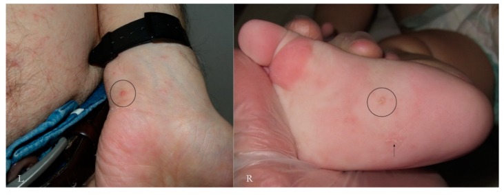
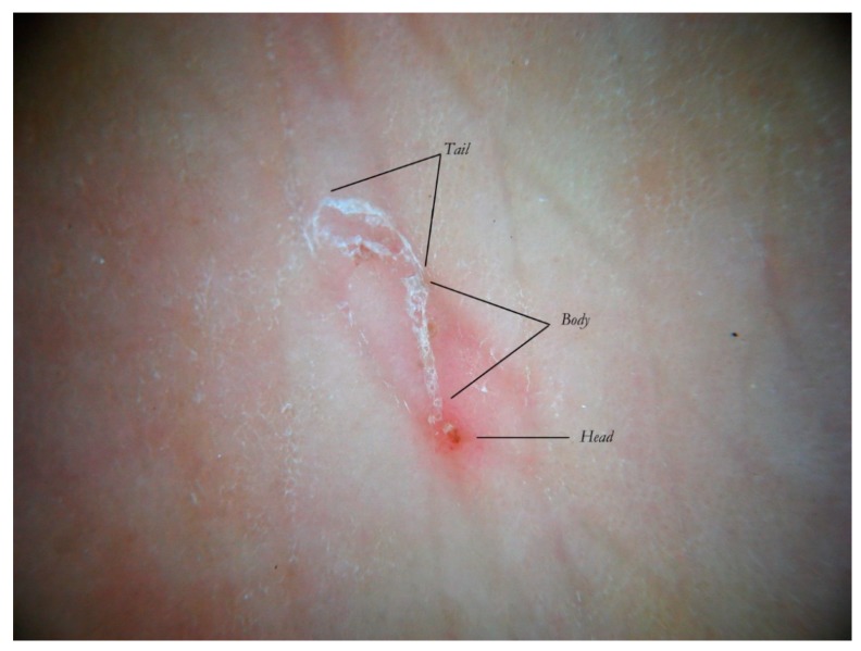
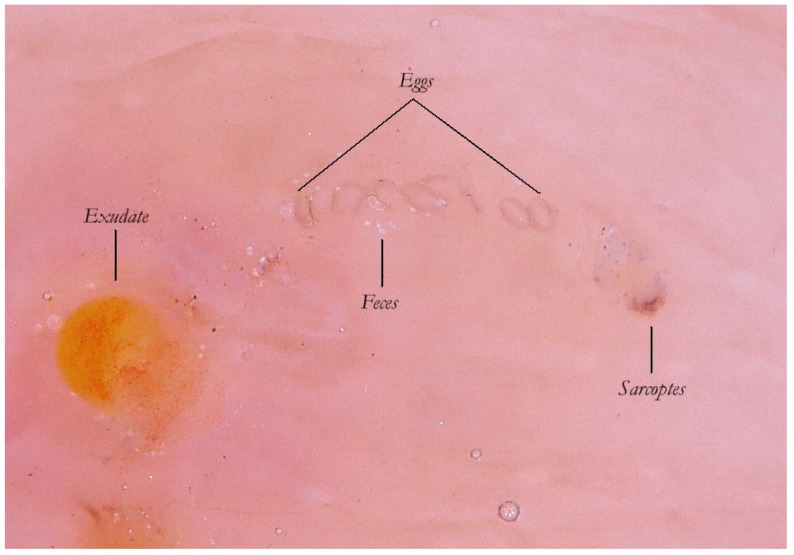
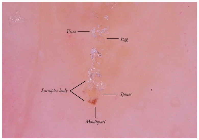
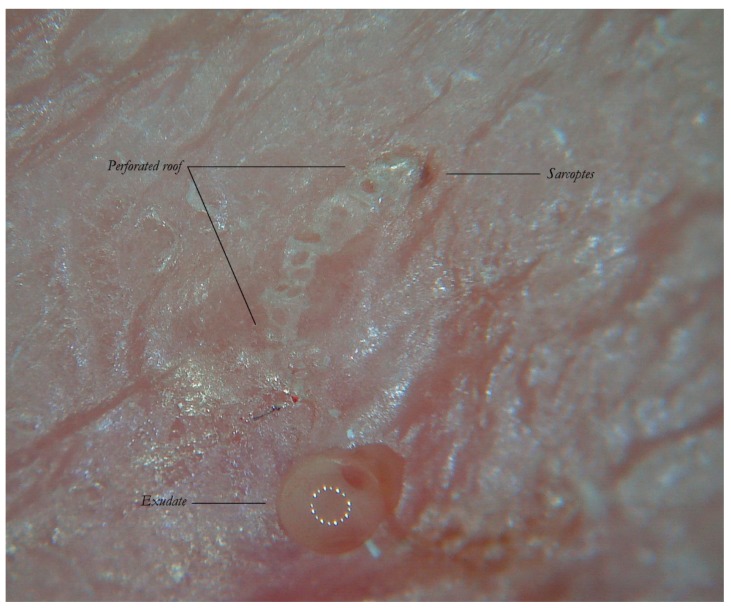
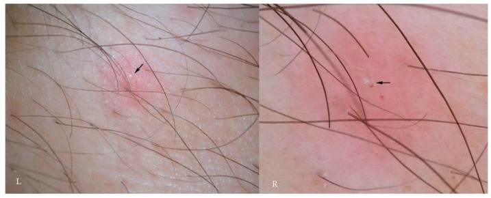
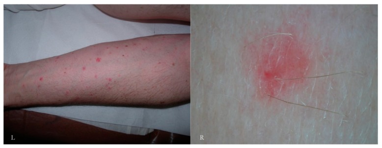
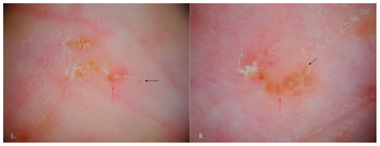
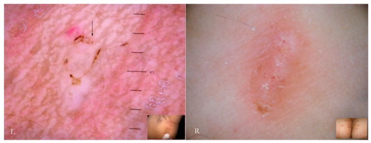
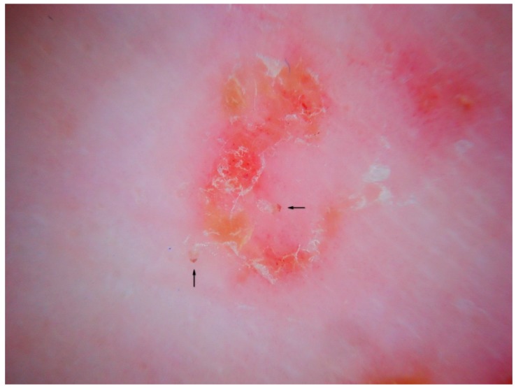
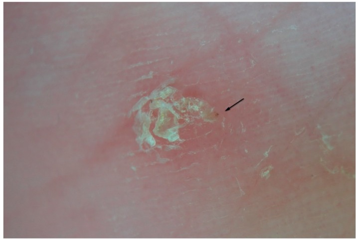
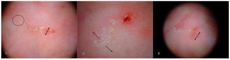
Scabies has always represented a diagnostic challenge for dermatologists, especially in subclinical cases or in atypical ones due to the coexistence of other diseases. Fortunately, dermatoscopy has enabled easier and faster in situ diagnosis. The aim of this study is to examine old and new dermatoscopic signs that Sarcoptes scabiei produces on the skin during its whole life cycle through entodermoscopy (dermatoscopy with an entomological focus) which, unlike traditional optical microscope examination, allows the local micro-environment to be preserved intact. Patients were enrolled during outbreaks of scabies from hospitals or nursing homes for the elderly in Bari (Italy). The study was performed applying both immersion and polarized dry dermatoscopy. The systematic use of dermatoscopy highlighted the morphological complexity of the Sarcoptes tunnel that had been described previously as a simple unitary structure. On the contrary, it is possible to distinguish three separate segments of the burrow that introduce a new anatomo-functional concept called the Mite-Gallery Unit (MGU). This approach, based on the mite life cycle and local skin turnover (the latter usually being ignored), allows the dermatologist to recognize not only Sarcoptes using the gallery, but also new descriptors including tunnels without Sarcoptes, those with acari alone, and those with associated signs of inflammation. The diagnosis of scabies using optical microscopy until recently has always involved demonstrating the mite and its products outside the human body (on a glass slide) without taking into account exactly what happens within the epidermis. Entodermoscopy is a term used to encapsulate both the presence of the parasite, the usual target of microscopy, and the changes produced in the superficial layers of the epidermis in situ. Thus, the scabies tunnel or burrow can be shown to be composed of three parts, the Head, Body, and Tail, in which different events affecting both mite and host develop. The Mite-Gallery Unit provides a new anatomical and functional explanation of scabies because it provides a more comprehensive in vivo and in situ dermatoscopic diagnosis. In this respect, dermatoscopy takes into account the behavior of the mite in addition to its interaction with its habitat, the human skin.
Keywords: Dry Dermatoscopy (d-DS); Enhanced Dermatoscopy (e-DS); Entodermoscopy (EDS); Mite-Gallery Unit (MGU); Wet Dermatoscopy (w-DS).
Conflict of interest statement
The author declares no conflict of interest.
Figures

References
-
- Scanni G., Bonifazi E. Viability of the head louse eggs in pediculosis capitis. A dermoscopy study. Eur. J. Pediat. Dermatol. 2006;16:201–204.
LinkOut - more resources
Full Text Sources
