Distribution of Gadolinium in Rat Heart Studied by Fast Field Cycling Relaxometry and Imaging SIMS
- PMID: 30884846
- PMCID: PMC6471734
- DOI: 10.3390/ijms20061339
Distribution of Gadolinium in Rat Heart Studied by Fast Field Cycling Relaxometry and Imaging SIMS
Abstract
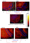
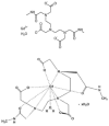
Research on microcirculatory alterations in human heart disease is essential to understand the genesis of myocardial contractile dysfunction and its evolution towards heart failure. The use of contrast agents in magnetic resonance imaging is an important tool in medical diagnostics related to this dysfunction. Contrast agents significantly improve the imaging by enhancing the nuclear magnetic relaxation rates of water protons in the tissues where they are distributed. Gadolinium complexes are widely employed in clinical practice due to their high magnetic moment and relatively long electronic relaxation time. In this study, the behavior of gadolinium ion as a contrast agent was investigated by two complementary methods, relaxometry and secondary ion mass spectrometry. The study examined the distribution of blood flow within the microvascular network in ex vivo Langendorff isolated rat heart models, perfused with Omniscan® contrast agent. The combined use of secondary ion mass spectrometry and relaxometry allowed for both a qualitative mapping of agent distribution as well as the quantification of gadolinium ion concentration and persistence. This combination of a chemical mapping and temporal analysis of the molar concentration of gadolinium ion in heart tissue allows for new insights on the biomolecular mechanisms underlying the microcirculatory alterations in heart disease.
Keywords: NMRD profiles; ToF-SIMS; gadolinium; tissue microimaging.
Conflict of interest statement
The authors declare no conflict of interest.
Figures


References
-
- Vickerman J.C., Briggs D. ToF-SIMS: Surface Analysis by mass Spectrometry. 2nd ed. IM Publications and Surface Spectra Limited; Chichester, UK: 2003.
-
- Kailas L., Audinot J.-N., Migeon H.-N., Bertrand P. ToF-SIMS molecular characterization and nano-SIMS imaging of submicron domain formation at the surface of PS/PMMA blend and copolymer thin films. Appl. Surf. Sci. 2004;231:289–295. doi: 10.1016/j.apsusc.2004.03.063. - DOI
-
- Piras F.M., Di Mundo R., Fracassi F., Magnani A. Silicon nitride and oxynitride films deposited from organosilicon plasmas: ToF-SIMS characterization with multivariate analysis. Surf. Coat. Technol. 2008;202:1606–1614. doi: 10.1016/j.surfcoat.2007.07.016. - DOI
-
- Leone G., Consumi M., Tognazzi A., Magnani A. Realisation and chemical characterisation of a model system for saccharide-based biosensor. Thin Solid Films. 2010;519:462–470. doi: 10.1016/j.tsf.2010.07.099. - DOI
MeSH terms
Substances
LinkOut - more resources
Full Text Sources
Medical

