Human bone marrow mesenchymal stem cells-derived exosomes alleviate liver fibrosis through the Wnt/β-catenin pathway
- PMID: 30885249
- PMCID: PMC6421647
- DOI: 10.1186/s13287-019-1204-2
Human bone marrow mesenchymal stem cells-derived exosomes alleviate liver fibrosis through the Wnt/β-catenin pathway
Abstract
Background: Mesenchymal stem cells (MSCs) are increasingly being applied as a therapy for liver fibrosis. Exosomes possess similar functions to their parent cells; however, they are safe and effective cell-free reagents with controllable and predictable outcomes. In this study, we investigated the therapeutic potential and underlying molecular mechanism for human bone mesenchymal stem cells-derived exosomes (hBM-MSCs-Ex) in the treatment of liver fibrosis.
Methods: We established an 8-week CCl4-induced rat liver fibrosis model, after which, we administered hBM-MSCs-Ex in vivo for 4 weeks. The resulting histopathology, liver function, and inflammatory cytokines were analyzed. In addition, we investigated the anti-fibrotic mechanism of hBM-MSCs-Ex in hepatic stellate cells (HSCs) and liver fibrosis tissue, by western blotting for the expression of Wnt/β-catenin signaling pathway-related genes.
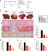
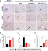
Results: In vivo administration of hBM-MSCs-Ex effectively alleviated liver fibrosis, including a reduction in collagen accumulation, enhanced liver functionality, inhibition of inflammation, and increased hepatocyte regeneration. Moreover, based on measurement of the collagen area, Ishak fibrosis score, MDA levels, IL-1, and IL-6, the therapeutic effect of hBM-MSCs-Ex against liver fibrosis was significantly greater than that of hBM-MSCs. In addition, we found that hBM-MSCs-Ex inhibited the expression of Wnt/β-catenin pathway components (PPARγ, Wnt3a, Wnt10b, β-catenin, WISP1, Cyclin D1), α-SMA, and Collagen I, in both HSCs and liver fibrosis tissue.
Conclusions: These results suggest that hBM-MSCs-Ex treatment could ameliorate CCl4-induced liver fibrosis via inhibition of HSC activation through the Wnt/β-catenin pathway.
Keywords: Exosomes; Liver fibrosis; Wnt/β-catenin; hBM-MSCs.
Conflict of interest statement
Ethics approval and consent to participate
All experiments were performed in accordance with the guidelines and study protocols of the Animal Experiment Ethic Committee of Jilin University (Approval No. 201802072).
Consent for publication
Not applicable.
Competing interests
The authors declare that they have no competing interests.
Publisher’s Note
Springer Nature remains neutral with regard to jurisdictional claims in published maps and institutional affiliations.
Figures


Similar articles
-
Exosomes derived from bone marrow mesenchymal stem cells reverse epithelial-mesenchymal transition potentially via attenuating Wnt/β-catenin signaling to alleviate silica-induced pulmonary fibrosis.Toxicol Mech Methods. 2021 Nov;31(9):655-666. doi: 10.1080/15376516.2021.1950250. Epub 2021 Aug 12. Toxicol Mech Methods. 2021. PMID: 34225584
-
Human amniotic mesenchymal stem cells-derived IGFBP-3, DKK-3, and DKK-1 attenuate liver fibrosis through inhibiting hepatic stellate cell activation by blocking Wnt/β-catenin signaling pathway in mice.Stem Cell Res Ther. 2022 Jun 3;13(1):224. doi: 10.1186/s13287-022-02906-z. Stem Cell Res Ther. 2022. PMID: 35659360 Free PMC article.
-
MSCs reduce airway remodeling in the lungs of asthmatic rats through the Wnt/β-catenin signaling pathway.Eur Rev Med Pharmacol Sci. 2020 Nov;24(21):11199-11211. doi: 10.26355/eurrev_202011_23608. Eur Rev Med Pharmacol Sci. 2020. PMID: 33215438
-
Progress of mesenchymal stem cells (MSCs) & MSC-Exosomes combined with drugs intervention in liver fibrosis.Biomed Pharmacother. 2024 Jul;176:116848. doi: 10.1016/j.biopha.2024.116848. Epub 2024 Jun 3. Biomed Pharmacother. 2024. PMID: 38834005 Review.
-
Wnt signaling in liver fibrosis: progress, challenges and potential directions.Biochimie. 2013 Dec;95(12):2326-35. doi: 10.1016/j.biochi.2013.09.003. Epub 2013 Sep 13. Biochimie. 2013. PMID: 24036368 Review.
Cited by
-
Effects of Lycopene Attenuating Injuries in Ischemia and Reperfusion.Oxid Med Cell Longev. 2022 Oct 7;2022:9309327. doi: 10.1155/2022/9309327. eCollection 2022. Oxid Med Cell Longev. 2022. PMID: 36246396 Free PMC article. Review.
-
Differential Wound Healing Capacity of Mesenchymal Stem Cell-Derived Exosomes Originated From Bone Marrow, Adipose Tissue and Umbilical Cord Under Serum- and Xeno-Free Condition.Front Mol Biosci. 2020 Jun 24;7:119. doi: 10.3389/fmolb.2020.00119. eCollection 2020. Front Mol Biosci. 2020. PMID: 32671095 Free PMC article.
-
Immunomodulatory role of mesenchymal stem cell therapy in liver fibrosis.Front Immunol. 2023 Jan 4;13:1096402. doi: 10.3389/fimmu.2022.1096402. eCollection 2022. Front Immunol. 2023. PMID: 36685534 Free PMC article. Review.
-
Stem cells for treatment of liver fibrosis/cirrhosis: clinical progress and therapeutic potential.Stem Cell Res Ther. 2022 Jul 26;13(1):356. doi: 10.1186/s13287-022-03041-5. Stem Cell Res Ther. 2022. PMID: 35883127 Free PMC article. Review.
-
Structural and Temporal Dynamics of Mesenchymal Stem Cells in Liver Diseases From 2001 to 2021: A Bibliometric Analysis.Front Immunol. 2022 May 19;13:859972. doi: 10.3389/fimmu.2022.859972. eCollection 2022. Front Immunol. 2022. PMID: 35663940 Free PMC article. Review.
References
Publication types
MeSH terms
Substances
LinkOut - more resources
Full Text Sources
Medical
Research Materials

