

HHLA2 in intrahepatic cholangiocarcinoma: an immune checkpoint with prognostic significance and wider expression compared with PD-L1
- PMID: 30885276
- PMCID: PMC6421676
- DOI: 10.1186/s40425-019-0554-8
HHLA2 in intrahepatic cholangiocarcinoma: an immune checkpoint with prognostic significance and wider expression compared with PD-L1
Abstract
Background: Intrahepatic cholangiocarcinoma (ICC) is a highly mortal malignancy with limited therapeutic options. Immunotherapies targeting PD-1/PD-L1 pathway represent a promising treatment for ICC. However, PD-L1 expression and microsatellite instability are not common in ICC. This study aimed to investigate whether HHLA2, a newly identified B7 family immune checkpoint for T cells, could be a therapeutic target next to PD-L1 in ICC.
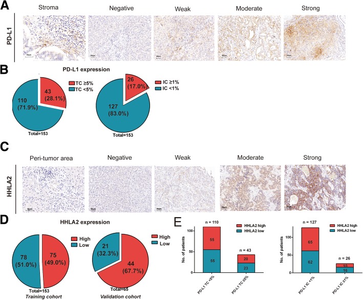
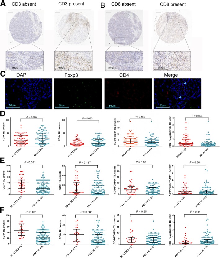
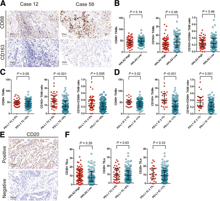
Methods: Expression levels of PD-L1 and HHLA2 as well as infiltrations of CD3+, CD8+, CD4 + Foxp3+, CD68+, CD163+ and CD20+ cells were evaluated by immunohistochemistry in 153 resected ICC samples. Comprehensive comparisons were made between PD-L1 and HHLA2 in terms of the expression rates, clinicopathological features and infiltrations of different immune cells. The expression level and prognostic significance of HHLA2 were further validated in an independent cohort.
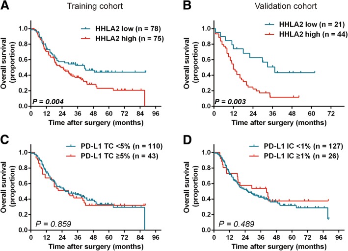
Results: Expression of HHLA2 is more frequent than PD-L1 in ICC (49.0% vs 28.1%). Co-expression of both immune checkpoints was infrequent (13.1%) and 50% PD-L1 negative cases were with elevated HHLA2. HHLA2 overexpression was associated with sparser CD3+ tumor infiltrating lymphocytes (TILs), CD8+ TILs and a higher CD4 + Foxp3+/CD8+ TIL ratio, whereas PD-L1 expression was associated with prominent T cells and CD163+ tumor associated macrophages infiltrations. PD-L1 failed to stratify overall survival (OS) but HHLA2 was identified as an independent prognostic indicator for OS in two independent cohorts.
Conclusions: Compared with PD-L1, HHLA2 is more prevalent and possesses more explicit prognostic significance, which confer the rationale for HHLA2 as a potential immunotherapeutic target next to PD-L1 for ICC patients.
Keywords: HHLA2; Immunotherapy; Intrahepatic cholangiocarcinoma; PD-L1; Prognosis; Tumor associated macrophages; Tumor infiltrating lymphocytes.
Conflict of interest statement
Authors’ information
Not applicable.
Ethics approval and consent to participate
All the patients signed informed consent before surgery that permitted the usage of resected tumors and clinical profiles in research under the condition of anonymity. The study was approved by the Clinical Research Ethic Committee of Zhongshan hospital.
Consent for publication
Not applicable.
Competing interests
The authors declare that they have no competing interests.
Publisher’s Note
Springer Nature remains neutral with regard to jurisdictional claims in published maps and institutional affiliations.
Figures
Similar articles
-
CTLA-4 Synergizes With PD1/PD-L1 in the Inhibitory Tumor Microenvironment of Intrahepatic Cholangiocarcinoma.Front Immunol. 2021 Aug 30;12:705378. doi: 10.3389/fimmu.2021.705378. eCollection 2021. Front Immunol. 2021. PMID: 34526987 Free PMC article.
-
Clinical prognosticators and targets in the immune microenvironment of intrahepatic cholangiocarcinoma.Oncoimmunology. 2024 Oct 1;13(1):2406052. doi: 10.1080/2162402X.2024.2406052. eCollection 2024. Oncoimmunology. 2024. PMID: 39359389 Free PMC article.
-
Cancer-associated fibroblast senescence and its relation with tumour-infiltrating lymphocytes and PD-L1 expressions in intrahepatic cholangiocarcinoma.Br J Cancer. 2022 Feb;126(2):219-227. doi: 10.1038/s41416-021-01569-6. Epub 2021 Oct 6. Br J Cancer. 2022. PMID: 34616011 Free PMC article.
-
The role of tumor-infiltrating lymphocytes in cholangiocarcinoma.J Exp Clin Cancer Res. 2022 Apr 7;41(1):127. doi: 10.1186/s13046-022-02340-2. J Exp Clin Cancer Res. 2022. PMID: 35392957 Free PMC article. Review.
-
The prognostic role of PD-L1 expression for survival in head and neck squamous cell carcinoma: A systematic review and meta-analysis.Oral Oncol. 2018 Nov;86:81-90. doi: 10.1016/j.oraloncology.2018.09.016. Epub 2018 Sep 17. Oral Oncol. 2018. PMID: 30409325
Cited by
-
Cell Division Cycle-Associated Genes Are Potential Immune Regulators in Nasopharyngeal Carcinoma.Front Oncol. 2022 Feb 14;12:779175. doi: 10.3389/fonc.2022.779175. eCollection 2022. Front Oncol. 2022. PMID: 35237510 Free PMC article.
-
Crosstalk between the B7/CD28 and EGFR pathways: Mechanisms and therapeutic opportunities.Genes Dis. 2021 Sep 20;9(5):1181-1193. doi: 10.1016/j.gendis.2021.08.009. eCollection 2022 Sep. Genes Dis. 2021. PMID: 35873032 Free PMC article. Review.
-
Long Non-Coding RNA GRIK1-AS1 Inhibits the Proliferation and Invasion of Gastric Cancer Cells by Regulating the miR-375/IFIT2 Axis.Front Oncol. 2021 Sep 30;11:754834. doi: 10.3389/fonc.2021.754834. eCollection 2021. Front Oncol. 2021. PMID: 34660323 Free PMC article.
-
Rethinking Immune Check Point Inhibitors Use in Liver Transplantation: Implications and Resistance.Cell Mol Gastroenterol Hepatol. 2025;19(1):101407. doi: 10.1016/j.jcmgh.2024.101407. Epub 2024 Sep 24. Cell Mol Gastroenterol Hepatol. 2025. PMID: 39326581 Free PMC article. Review.
-
B7-H7 is a prognostic biomarker in epithelial ovarian cancer.Transl Cancer Res. 2020 Sep;9(9):5360-5370. doi: 10.21037/tcr-20-697. Transl Cancer Res. 2020. PMID: 35117901 Free PMC article.
References
-
- Capecitabine Extends Survival for Biliary Tract Cancer. Cancer Discov. 2017;7(7):OF1. - PubMed
Publication types
MeSH terms
Substances
LinkOut - more resources
Full Text Sources
Other Literature Sources
Medical
Molecular Biology Databases
Research Materials