. 2019 May 3;431(10):1940-1955.
doi: 10.1016/j.jmb.2019.03.010. Epub 2019 Mar 15.

G-Quadruplexes in Human Ribosomal RNA

Affiliations

G-Quadruplexes in Human Ribosomal RNA

Santi Mestre-Fos et al. J Mol Biol. .

Abstract

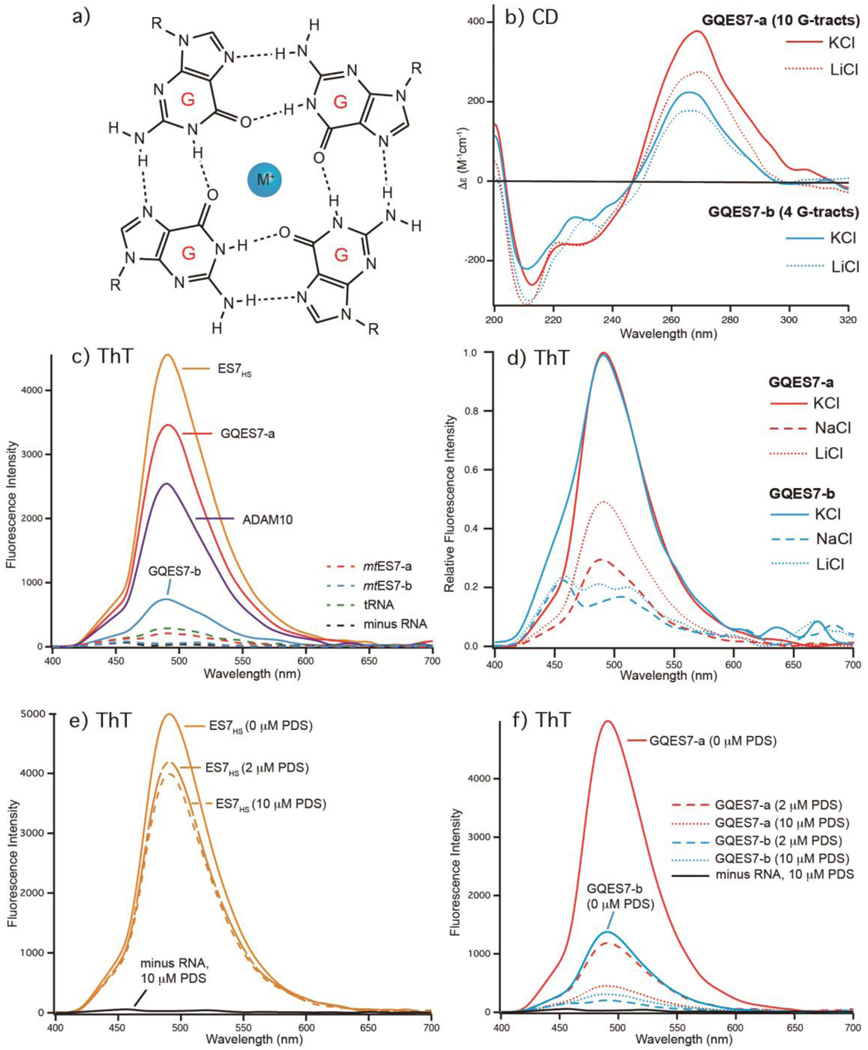
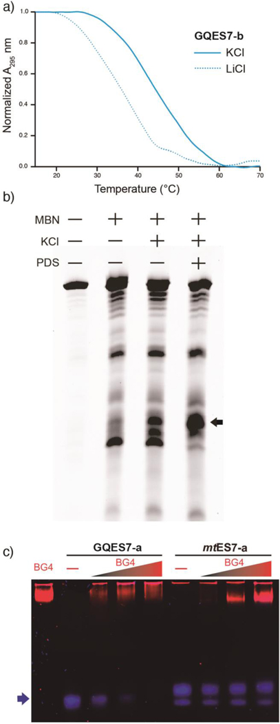
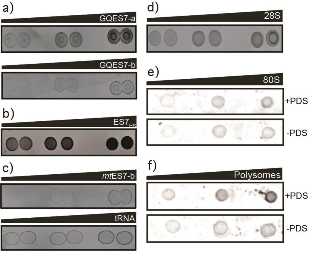
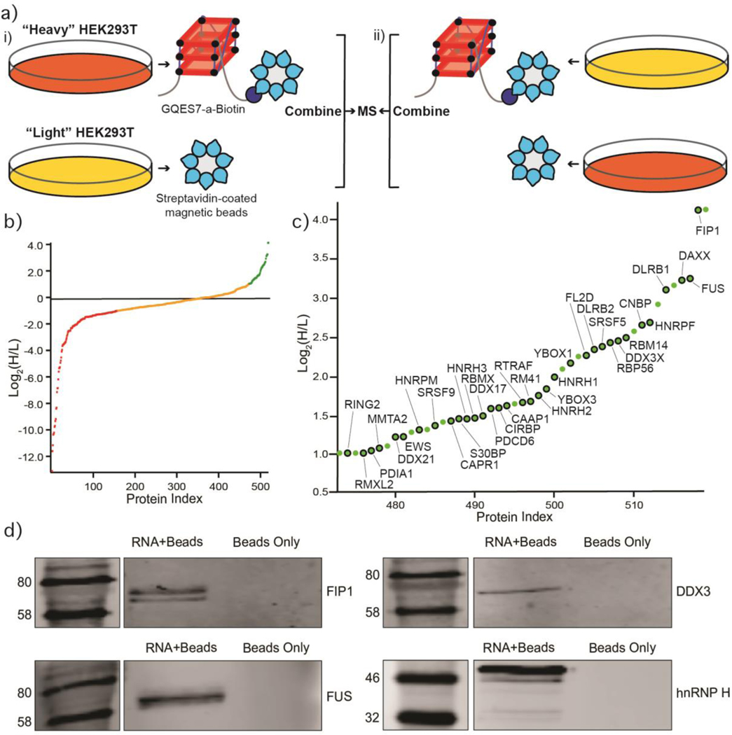
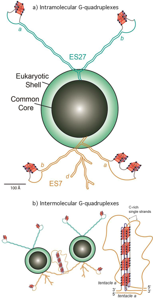
rRNA is the single most abundant polymer in most cells. Mammalian rRNAs are nearly twice as large as those of prokaryotes. Differences in rRNA size are due to expansion segments, which contain extended tentacles in metazoans. Here we show that the terminus of an rRNA tentacle of Homo sapiens contains 10 tandem G-tracts that form highly stable G-quadruplexes in vitro. We characterized rRNA of the H. sapiens large ribosomal subunit by computation, circular dichroism, UV melting, fluorescent probes, nuclease accessibility, electrophoretic mobility shifts, and blotting. We investigated Expansion Segment 7 (ES7), oligomers derived from ES7, intact 28S rRNA, 80S ribosomes, and polysomes. We used mass spectrometry to identify proteins that bind to rRNA G-quadruplexes in cell lysates. These proteins include helicases (DDX3, CNBP, DDX21, DDX17) and heterogeneous nuclear ribonucleoproteins. Finally, by multiple sequence alignments, we observe that G-quadruplex-forming sequences are a general feature of LSU rRNA of Chordata but not, as far as we can tell, of other species. Chordata ribosomes present polymorphic tentacles with the potential to switch between inter- and intramolecular G-quadruplexes. To our knowledge, G-quadruplexes have not been reported previously in ribosomes.

Keywords: Chordates; expansion segments; helicases; polysomes; rRNA.


Conflict of interest statement

CONFLICT OF INTEREST

The authors declare that they have no conflict of interest with the contents of this article.

Figures

Figure 1.
Figure 1.
Model of the secondary structure of the LSU rRNA of Homo sapiens. G-quadruplex-forming regions (defined by G≥3N1–7G≥3N1–7G≥3N1–7G≥3) are highlighted. a) Expansion segment ES7HS is orange. Tentacles a, b and d of ES7HS are indicated. G-quadruplex-forming regions of ES7HS are GQES7-a (red, in tentacle a) and GQES7-b (cyan, in tentacle b) and are boxed by dashed lines. Expansion segment ES27HS is green with purple G-tracts. Helix 63, at the base of ES27HS, contains a G-quadruplex motif (purple). Tentacles a and b of ES27HS are indicated. b) An expanded view of GQES7-b indicates the nucleotide sequence. c) An expanded view of GQES7-a indicates the nucleotide sequence.
Figure 2.
Figure 2.
Formation of G-quadruplexes by rRNA fragments GQES7-a, GQES7-b, and ES7HS. a) G-quadruplexes preferentially coordinate K+. b) CD spectra of GQES7-a and GQES7-b in the presence of either K+ or Li+. c) Fluorescence emission at 487 nm of the G-quadruplex probe ThT in the presence of ES7HS, GQES7-a, GQES7-b, or positive control ADAM10. Negative controls (dashed) are tRNA, mtES7-a, mtES7-b and minus RNA. d) ThT fluorescence emission of GQES7-a and GQES7-b in the presence of various monovalent cations. Intensities of GQES7-a and GQES7-b are normalized in the presence of K+ to highlight cation-induced differences. e) PDS competes with ThT in association with ES7HS. f) PDS competes with ThT in association with GQES7-a and GQES7-b.
Figure 3.
Figure 3.
a) UV thermal melting profile of GQES7-b at 295 nm. Before melting RNA was annealed in the presence of either 100 mM KCl or 100 mM LiCl. b) ES7HS cleavage by mung bean nuclease. ES7 was annealed with or without KCl and with or without PDS. The black arrow indicates cleaved rRNA. c) EMSA of the BG4 antibody with GQES7-a and its non-G-quadruplex-forming mutant mtES7-a, visualized on a native gel. GQES7-a and mtES7-a RNAs were loaded at a constant strand concentration with increasing concentrations of BG4 antibody. The RNA (arrow) is blue and the protein is red.
Figure 4.
Figure 4.
Dot blots performed with the BG4 antibody on a) GQES7-a and GQES7-b, b) intact ES7HS, c) the negative controls mtES7-b and tRNA, d) the 28S rRNA extracted from HEK293T cells and on e) human 80S ribosomes and f) polysomes purified from HEK293 cells. All samples were incubated in the presence of 50mM KCl and ribosomes and polysomes were further analyzed with or without 10 μM PDS, which stabilizes G-quadruplexes. Samples were loaded onto the membrane in increasing amounts from left to right.
Figure 5.
Figure 5.
G-tracts are observed in ES7 tentacles. a) Conventional secondary structural models of ES7 from various eukaryotes. G-tracts within the G≥3N1–7G≥3N1–7G≥3N1–7G≥3 motif are highlighted in red. b) Sequence alignment of ES7 tentacle a showing G-quadruplex-forming sequences are common in chordates. Individual G-tracts in both panels are labeled with Greek symbols. Nucleotides are colored by type. G’s within G-tracts are dark red. Other G’s are pink. All nucleotides are numbered in accordance with H. sapiens 28S rRNA. Sizes of eukaryotic ES7 secondary structures are not to scale. Complete species nomenclature is provided in Table S.3.
Figure 6.
Figure 6.
Identification of GQES7-a-binding proteins. a) Scheme of the SILAC experiment. “RNA+beads” samples were combined in HEK293T grown in heavy media (panel i). The “Beads Only” control sample was combined in HEK293T grown in light media. To verify the proteins identified by this method, the experiment was performed using reverse labeling (panel ii). b) Scatter plot representing fold enrichment of the proteins binding to GQES7-a in “Heavy” HEK293T. Color representation indicates specific proteins that bound more tightly to GQES7-a than to the beads (green), to the beads than to GQES7-a (red) or bound to the beads and GQES7-a to a similar extent (orange). c) A close-up view of the green region of the scatter plot represented in panel b. Dots with a black contour are used to indicate proteins that appeared in the green region of the two replicate experiments described in panel a. d) Western blotting analyses of the eluted proteins from the RNA pull-down of HEK293T. All four blotted proteins (FIP1, FUS, DDX3 and hnRNP H) eluted from the GQES7-a sample (RNA+Beads) but not from the control (Beads Only), confirming the SILAC results.
Figure 7.
Figure 7.
a) Schematic representation of the common core, the eukaryotic shell and the tentacles of the LSU of the Homo sapiens ribosome. G-quadruplexes are indicated on ES7 and ES27. The lengths of ES7HS (orange) and ES27HS (green) tentacles are roughly scaled to the size of the common core. The G-quadruplexes represented in tentacle b of ES27HS do not fall within the G≥3N1–7G≥3N1–7G≥3N1–7G≥3 motif and are speculative. The G-quadruplex region found in Helix 63 of ES27HS is not indicated here. b) Schematic representation of interactions between ribosomes via intermolecular G-quadruplexes. G-tracts on ES7 tentacle a from different ribosomes contribute to the formation of G-quadruplexes (an expanded view is presented on the right).

Similar articles

Cited by

References

    1. Milo R, Phillips R. Cell biology by the numbers. New York, New York: Garland Science; 2016.
    1. Bernier CR, Petrov AS, Kovacs NA, Penev PI, Williams LD. Translation: The universal structural core of life. Mol. Biol. Evol. 2018;34:2065–76. - PMC - PubMed
    1. Ware VC, Tague BW, Clark CG, Gourse RL, Brand RC, Gerbi SA. Sequence analysis of 28S ribosomal DNA from the amphibian Xenopus laevis. Nucleic Acids Res. 1983;11:7795–817. - PMC - PubMed
    1. Clark CG, Tague BW, Ware VC, Gerbi SA. Xenopus laevis 28S ribosomal RNA: A secondary structure model and its evolutionary and functional implications. Nucleic Acids Res. 1984;12:6197–220. - PMC - PubMed
    1. Hassouna N, Michot B, Bachellerie JP. The complete nucleotide sequence of mouse 28S rRNA gene. Implications for the process of size increase of the large subunit rRNA in higher eukaryotes. Nucleic Acids Res. 1984;12:3563–83. - PMC - PubMed

Publication types