. 2019 May 10;294(19):7632-7643.
doi: 10.1074/jbc.RA118.005415. Epub 2019 Mar 18.

XPG-related nucleases are hierarchically recruited for double-stranded rDNA break resection

Affiliations

XPG-related nucleases are hierarchically recruited for double-stranded rDNA break resection

Kevin J Barnum et al. J Biol Chem. .

Abstract

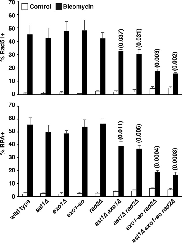
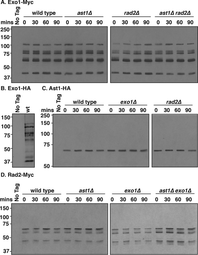
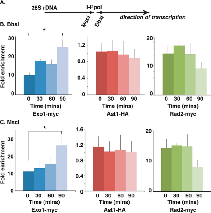
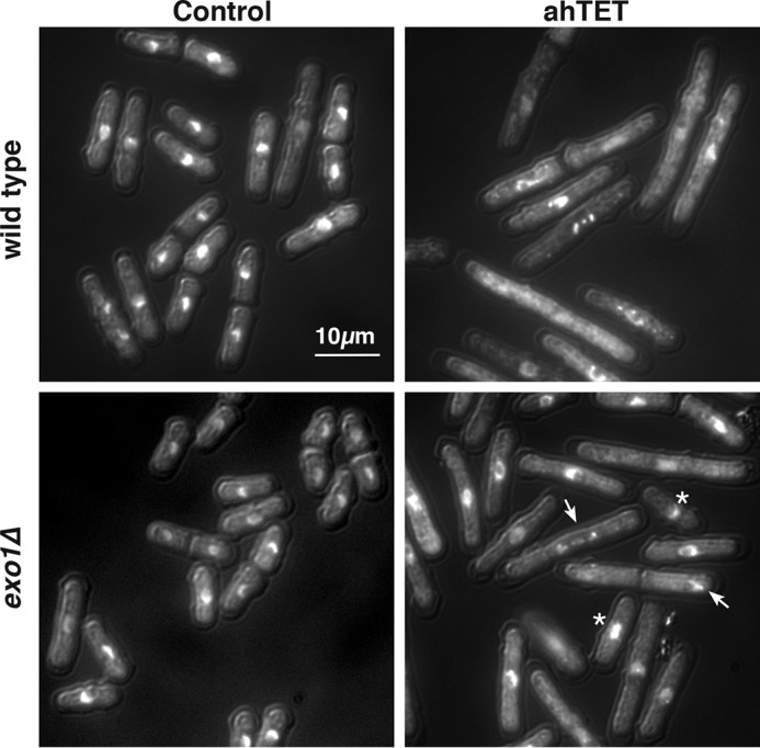
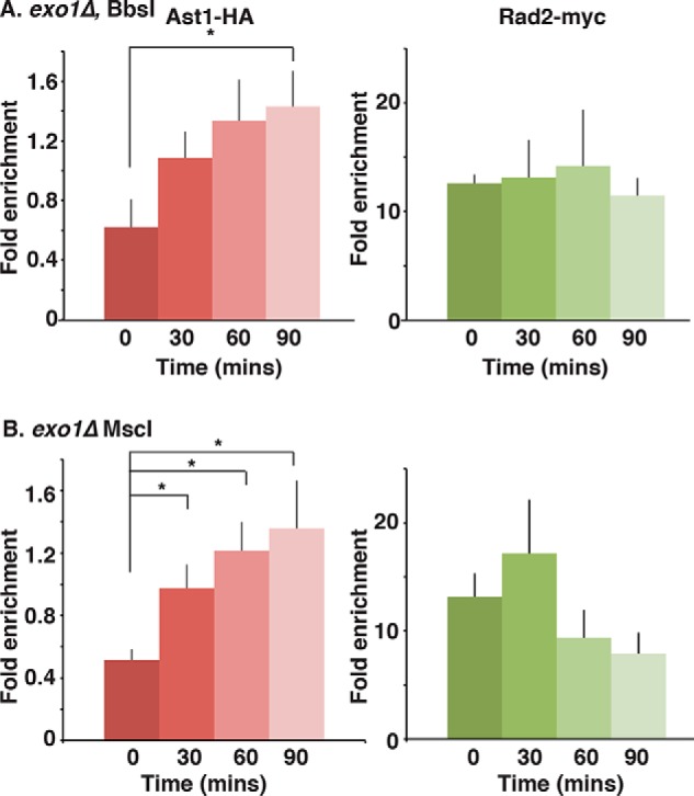
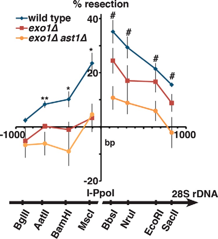
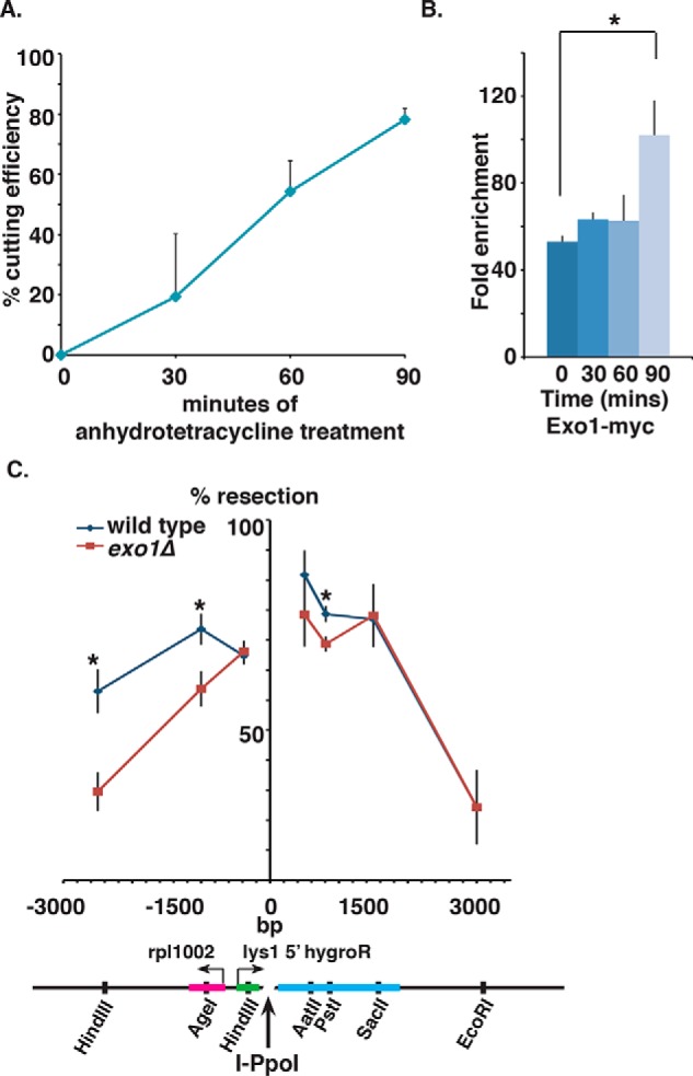
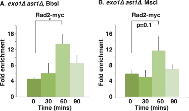
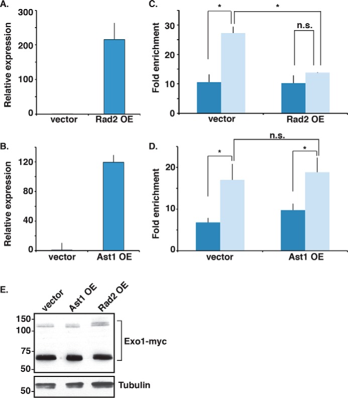
dsDNA breaks (DSBs) are resected in a 5'→3' direction, generating single-stranded DNA (ssDNA). This promotes DNA repair by homologous recombination and also assembly of signaling complexes that activate the DNA damage checkpoint effector kinase Chk1. In fission yeast (Schizosaccharomyces pombe), genetic screens have previously uncovered a family of three xeroderma pigmentosum G (XPG)-related nucleases (XRNs), known as Ast1, Exo1, and Rad2. Collectively, these XRNs are recruited to a euchromatic DSB and are required for ssDNA production and end resection across the genome. Here, we studied why there are three related but distinct XRN enzymes that are all conserved across a range of species, including humans, whereas all other DSB response proteins are present as single species. Using S. pombe as a model, ChIP and DSB resection analysis assays, and highly efficient I-PpoI-induced DSBs in the 28S rDNA gene, we observed a hierarchy of recruitment for each XRN, with a progressive compensatory recruitment of the other XRNs as the responding enzymes are deleted. Importantly, we found that this hierarchy reflects the requirement for different XRNs to effect efficient DSB resection in the rDNA, demonstrating that the presence of three XRN enzymes is not a simple division of labor. Furthermore, we uncovered a specificity of XRN function with regard to the direction of transcription. We conclude that the DSB-resection machinery is complex, is nonuniform across the genome, and has built-in fail-safe mechanisms, features that are in keeping with the highly pathological nature of DSB lesions.

Keywords: Checkpoint; DNA Damage Response; DNA Repair; DNA damage response; DNA recombination; DNA repair; Schizosaccharomyces pombe; XPG-Related Nuclease; cell cycle; chromosomes.


Conflict of interest statement

The authors declare that they have no conflicts of interest with the contents of this article

Figures

Figure 1.
Figure 1.
Formation of Rad51 and RPA foci following bleomycin treatment. G2 cells of the indicated strains were left untreated or treated with 0.5 milliunits of bleomycin as described (34). Rad51 foci were imaged by immunofluorescence, and RPA foci were imaged by GFP–Rad11. Data are mean ± S.D. from three counts of 100 cells. Numbers in parentheses are p values (Student's two-sided t test) of strains compared with WT. Absence of parentheses indicates a nonsignificant difference.
Figure 2.
Figure 2.
XPG-related nuclease protein levels during DSB induction at the 28S rDNA PpoI site. Exponentially growing cells in YES medium with the I–PpoI system and the indicated genotypes were treated with 5 μm anhydrotetracycline for 0, 30, 60, or 90 min, and total protein was extracted and submitted to SDS-PAGE. Expression of Exo1–Myc (predicted 77 kDa) (A), Exo1–HA (predicted 68 kDa) (B), Ast1–HA (predicted 62 kDa) (C), and Rad2–Myc (predicted 56 kDa) (D) is shown. Gels reveal extensive processing and post-translational modification of Exo1–Myc and Rad2–Myc in contrast to Ast1–HA, which runs as a single band. The protein level of each of the XPG-related nucleases does not change significantly over the time course or genetic background. No Tag lanes contain extracts from isogenic untagged controls.
Figure 3.
Figure 3.
XPG-related nuclease recruitment to an I–PpoI-induced DSB in the rDNA. A, scheme of the I–PpoI site in the 28S rDNA gene. The BbsI primer set is 150 bp transcriptionally 3′ from the I–PpoI cut site. The MscI primer set is ∼100 bp 5′ to the cut site. B and C, cells were grown in YES medium, treated with 5 μm anhydrotetracycline for 0, 30, 60, or 90 min, and then fixed with 1% formaldehyde and submitted to ChIP. Primers used were BbsI (3′ to the DSB) and MscI (5′ to the BSB). Data (mean ± S.E.) are displayed as enrichment over isogenic untagged strains. Exo1–Myc occupancy significantly increased over the time course, whereas the occupancies of the other two nucleases did not change. *, p < 0.05 WT versus exo1Δ by Student's two-sided t test with n = 3–5.
Figure 4.
Figure 4.
Low level mitotic abnormality of I–PpoI exo1Δ cells after anhydrotetracycline treatment. The tet-I–PpoI cells with either WT or exo1Δ background were either treated with 5 μm anhydrotetracycline for 4 h or left untreated (control). Cells were stained with DAPI, and images were captured. Cells lacking exo1 inefficiently elongated (asterisks) and displayed abnormal mitoses (arrow).
Figure 5.
Figure 5.
XPG-related nuclease recruitment to an I–PpoI-induced DSB in the rDNA in exo1Δ cells. Cells with the I–PpoI system and exo1Δ background were grown in exponentially growing cultures in YES medium and either left untreated or treated with 5 μm anhydrotetracycline for 30, 60, or 90 min. ChIP was performed and analyzed for occupancy at the BbsI site 3′ to the DSB (A) or occupancy at the MscI site 5′ to the DSB (B). Data (mean ± S.E.) are displayed as enrichment over isogenic untagged strains. Ast1–HA occupancy increased by 90 min of DSB induction in cells lacking exo1. *, p < 0.05 by Student's two-sided t test with n = 5.
Figure 6.
Figure 6.
DSB resection efficiency at an I–PpoI-induced DSB in the rDNA. Cells with the I–PpoI system in either WT, exo1Δ, or exo1Δast1Δ backgrounds were grown to exponential phase in YES medium and were then either left untreated or treated with 5 μm anhydrotetracycline for 2 h. Digests at indicated sites were performed, and qPCR was performed across the respective restriction sites. qPCR data were used to calculate % restriction. Resection efficiency (mean ± S.E., n = 4) was reduced significantly in exo1Δ cells specifically in the direction opposing transcription. *, p < 0.05; **, p < 0.005 WT versus exo1Δ; and #, p < 0.05 for WT versus ast1Δ exo1Δ by Student's two-sided t test.
Figure 7.
Figure 7.
DSB resection efficiency at a PpoI-induced DSB in the euchromatic lys1 locus. WT cell expression in Exo1–Myc (plus untagged controls) with an I–PpoI site at lys1 (but lacking from the 28S rDNA gene) was grown to exponential phase and then either left untreated or treated with 5 μm anhydrotetracycline. A, DSB cutting efficiency at an I–PpoI-induced DSB in the lys1 ORF. Input ChIP DNA was analyzed by qPCR performed with primers across the PpoI cut site (PpoLysCut, see Table 2). qPCR of the ORF of the ade6 gene was used as a control for DNA content. Data are mean ± S.E., n = 3. Cutting efficiency rises to ∼80% by 90 min of anhydrotetracycline treatment. B, ChIP of Exo1–Myc to the lys1 I–PpoI site. Data (mean ± S.E.) are displayed as enrichment over an isogenic untagged strain. Exo1–Myc occupancy increased by 90 min of DSB induction in cells lacking exo1. *, p < 0.05 by Student's two-sided t test with n = 5. C, WT and exo1Δ cells with an I–PpoI site at lys1 (but lacking from the 28S rDNA gene) were treated with 5 μm anhydrotetracycline for 2 h, and resection was assayed at the indicated restriction sites. Data are mean ± S.E., n = 3. Resection efficiency was reduced significantly in exo1Δ cells in the direction opposing transcription of the lys1 5′ region. *, p < 0.05 WT versus exo1Δ by Student's two-sided t test.
Figure 8.
Figure 8.
Rad2–Myc recruitment to an I–PpoI-induced DSB in the rDNA in exo1Δ ast1Δ cells. Cells with the I–PpoI system and exo1Δ ast1Δ background were grown in exponentially growing cultures in YES and either left untreated or treated with 5 μm anhydrotetracycline for 30, 60, or 90 min. ChIP was performed. Occupancy at the BbsI site 3′ to the DSB (A) and the MscI site 5′ to the DSB (B) was measured. Data (mean ± S.E.) are displayed as enrichment over isogenic untagged strains. Rad2–Myc occupancy increased by 60 min of DSB induction in cells lacking exo1 and ast1. *, p < 0.05 at Bbs1, p = 0.1 at MscI, by Student's two-sided t test with n = 5.
Figure 9.
Figure 9.
Competition of the XPG-related nucleases for recruitment to the I–PpoI-induced DSB in the rDNA. Overexpression of Rad2 (A) and Ast1 (B) was from the nmt1 promoter. Thiamine was washed out from cultures, and cells were harvested 16 h later. RNA was isolated from cells; cDNA was made, and mRNA levels were quantified by qPCR normalized to act1. C and D, cells were treated for 0 or 90 min with anhydrotetracycline after 16 h of thiamine washout, and ChIP was performed and analyzed using the BbsI primer set. Data are mean ± S.E., n = 3. Neither Rad2 nor Ast1 overexpression had an effect on the baseline levels of Exo1–Myc in the rDNA. Rad2 overexpression, but not Ast1 overexpression, abrogated the recruitment of Exo1–Myc to the DSB. *, p < 0.05 by Student's two-sided t test. E, Western blot analysis of Exo1–Myc expression (plus tubulin as a loading control) in the strains and time points used for the ChIP analysis in C and D.

Similar articles

Cited by

References

    1. Symington L. S., and Gautier J. (2011) Double-strand break end resection and repair pathway choice. Annu. Rev. Genet. 45, 247–271 10.1146/annurev-genet-110410-132435 - DOI - PubMed
    1. O'Connell M. J., Walworth N. C., and Carr A. M. (2000) The G2-phase DNA-damage checkpoint. Trends Cell Biol. 10, 296–303 10.1016/S0962-8924(00)01773-6 - DOI - PubMed
    1. MacDougall C. A., Byun T. S., Van C., Yee M. C., and Cimprich K. A. (2007) The structural determinants of checkpoint activation. Genes Dev. 21, 898–903 10.1101/gad.1522607 - DOI - PMC - PubMed
    1. O'Connell M. J., and Cimprich K. A. (2005) G2 damage checkpoints: what is the turn-on? J. Cell Sci. 118, 1–6 10.1242/jcs.01626 - DOI - PubMed
    1. Walworth N. C., and Bernards R. (1996) rad-dependent response of the chk1-encoded protein kinase at the DNA damage checkpoint. Science 271, 353–356 10.1126/science.271.5247.353 - DOI - PubMed

Publication types

MeSH terms

LinkOut - more resources