. 2019 May 10;294(19):7692-7710.
doi: 10.1074/jbc.RA119.007640. Epub 2019 Mar 18.

Protein arginine methyltransferase 5 (PRMT5) promotes survival of lymphoma cells via activation of WNT/β-catenin and AKT/GSK3β proliferative signaling

Affiliations

Protein arginine methyltransferase 5 (PRMT5) promotes survival of lymphoma cells via activation of WNT/β-catenin and AKT/GSK3β proliferative signaling

Jihyun Chung et al. J Biol Chem. .

Abstract

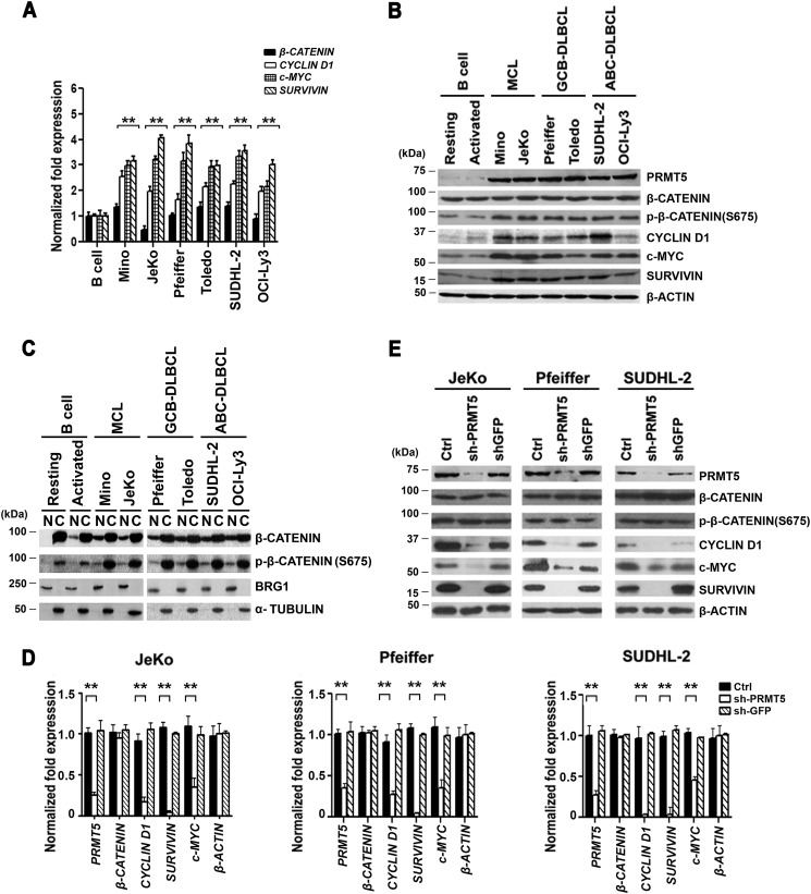
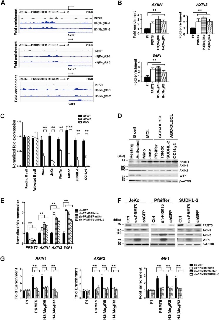
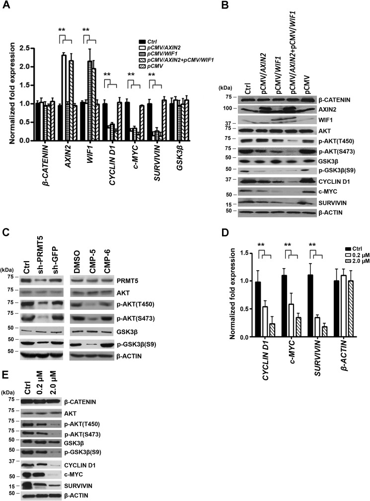
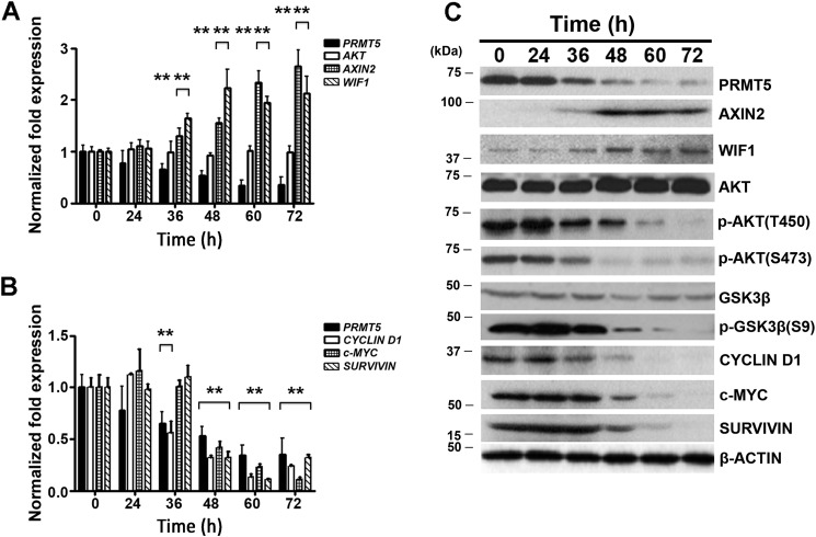
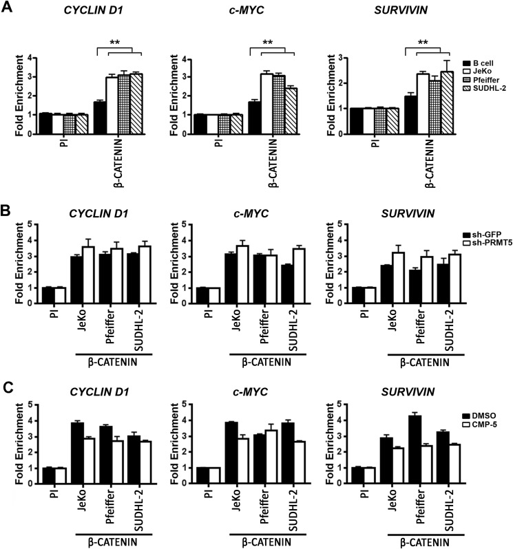
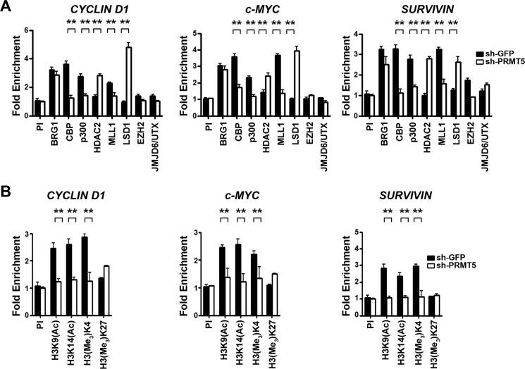
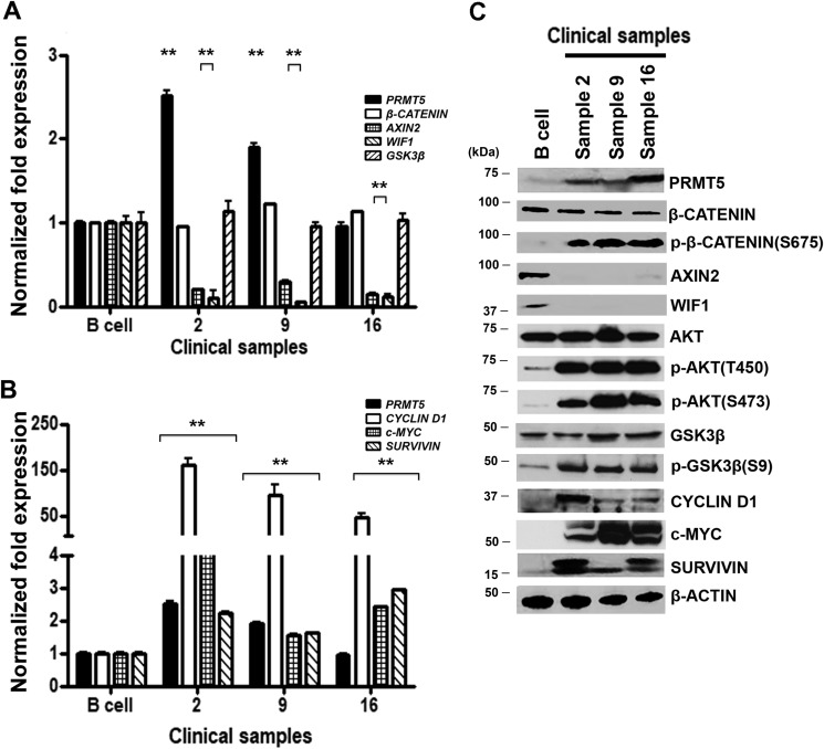
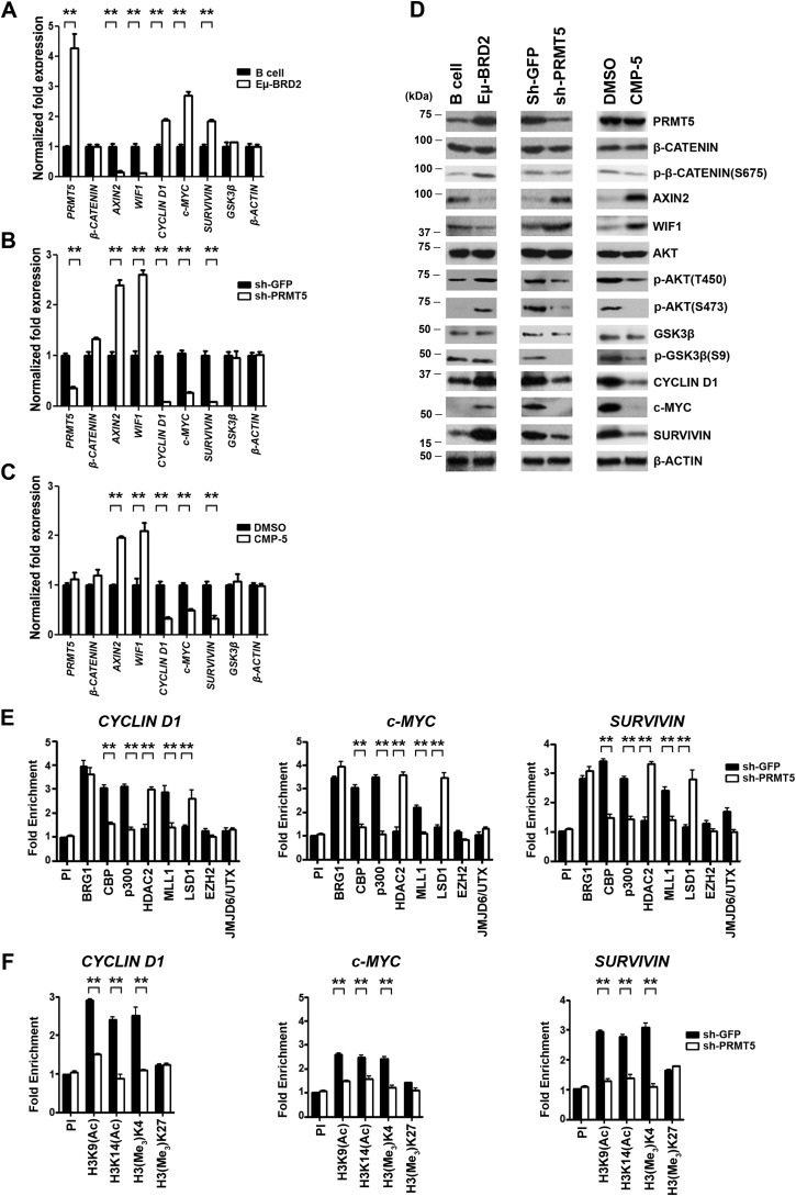
Epigenetic regulation by the type II protein arginine methyltransferase, PRMT5, plays an essential role in the control of cancer cell proliferation and tumorigenesis. In this report, we investigate the relationship between PRMT5 and WNT/β-CATENIN as well as AKT/GSK3β proliferative signaling in three different types of non-Hodgkin's lymphoma cell lines, clinical samples, and mouse primary lymphoma cells. We show that PRMT5 stimulates WNT/β-CATENIN signaling through direct epigenetic silencing of pathway antagonists, AXIN2 and WIF1, and indirect activation of AKT/GSK3β signaling. PRMT5 inhibition with either shRNA-mediated knockdown or a specific small molecule PRMT5 inhibitor, CMP-5, not only leads to derepression of WNT antagonists and decreased levels of active phospho-AKT (Thr-450 and Ser-473) and inactive phospho-GSK3β (Ser-9) but also results in decreased transcription of WNT/β-CATENIN target genes, CYCLIN D1, c-MYC, and SURVIVIN, and enhanced lymphoma cell death. Furthermore, PRMT5 inhibition leads to reduced recruitment of co-activators CBP, p300, and MLL1, as well as enhanced recruitment of co-repressors HDAC2 and LSD1 to the WNT/β-CATENIN target gene promoters. These results indicate that PRMT5 governs expression of prosurvival genes by promoting WNT/β-CATENIN and AKT/GSK3β proliferative signaling and that its inhibition induces lymphoma cell death, which warrants further clinical evaluation.

Keywords: Akt PKB; WNT antagonists; WNT/β-catenin signaling; Wnt signaling; histone; lymphoma; non-Hodgkin's lymphoma; protein arginine N-methyltransferase 5 (PRMT5); protein arginine methyltransferase 5 (PRMT5); protein kinase B (AKT)/glycogen synthase kinase 3β.


Conflict of interest statement

The authors declare that they have no conflicts of interest with the contents of this article

Figures

Figure 1.
Figure 1.
PRMT5 regulates WNT/β-CATENIN signaling in patient-derived lymphoma cell lines. A, levels of PRMT5, β-CATENIN, CYCLIN D1, c-MYC, and SURVIVIN mRNAs were measured in normal B (resting or activated), pre-GCB MCL (Mino and JeKo), GCB-DLBCL (Pfeiffer and Toledo), and post-GCB ABC-DLBCL (SUDHL-2 and OCI-Ly3) cells by real-time RT-PCR using gene-specific primers and probe sets. The values represent the average from three biological replicates with three technical replicates each and are reported as mean ± S.D. 18S rRNA was used as an internal control. B, RIPA extracts (20 μg) from either normal or transformed B cells were analyzed by Western blotting using the indicated antibodies. β-ACTIN serves as a loading control and is used in both B and Fig. 2D because these data resulted from the same experiment. This experiment was repeated two times, and representative blots are shown. C, approximately 25 μg of either nuclear (N) or cytosolic (C) extracts were prepared from either normal resting or activated B cells, as well as the indicated lymphoma cell lines, and proteins were detected using the indicated antibodies. Both BRG1 and α-TUBULIN served as control for nuclear and cytosolic fractionation. D, levels of PRMT5, β-CATENIN, CYCLIN D1, c-MYC, and SURVIVIN mRNAs were measured by real-time RT-PCR using total RNA isolated from either uninfected (Ctrl) JeKo, Pfeiffer, and SUDHL-2 cells, or lymphoma cells infected with shGFP or shPRMT5 lentivirus. Specific primers and probe sets were used to detect each of the indicated genes. This experiment was conducted using three biological replicates with three technical replicates as described in A. ** indicates p values < 10−3. E, RIPA extracts (20 μg) were prepared from control-uninfected and -infected (shGFP or shPRMT5) lymphoma cell lines and analyzed by immunoblotting using the indicated antibodies. β-ACTIN served as a loading control.
Figure 2.
Figure 2.
PRMT5 epigenetically regulates AXIN2 and WIF1, and its inhibition reactivates both target genes. A, cross-linked chromatin from Pfeiffer cells was immunoprecipitated using anti-H3(Me2)R8 antibody and analyzed by ChIP-Seq as described under “Experimental procedures.” Enrichment of H3(Me2)R8 on AXIN1, AXIN2, and WIF1 promoter sequences spanning −2 kb to +1 kb was determined using two biological replicates (H3(Me2)R8-1, and H3(Me2)R8-2). Peak detection for H3(Me2)R8 enrichment is reported in blue color compared with control input, which is reported in gray color. B, ChIP assays were performed on cross-linked chromatin from Pfeiffer cells using pre-immune (PI), anti-PRMT5, anti-H3(Me2)R8, or anti-H4(Me2)R3 antibody. ChIP experiments were carried out using two biological replicates with three technical replicates, and AXIN1, AXIN2, and WIF1 promoter sequences were detected by real-time PCR. Fold enrichment with each antibody was calculated relative to the PI sample. Data in each graph represent the mean ± S.D. C, levels of AXIN1, AXIN2, and WIF1 mRNAs were measured in normal B cells (resting and activated) and the indicated transformed B cells by real-time RT-PCR using gene-specific primers and probe sets. This experiment was conducted using three biological replicates with three technical replicates, and 18S rRNA was used as control. The data shown represent the mean ± S.D. D, RIPA extracts (20 μg) from normal (resting and activated) and transformed B cells were analyzed by Western blotting using the indicated antibodies. Anti-β-ACTIN was used to show equal loading. β-ACTIN loading control in both B and D are the same because these data resulted from the same experiment. E, levels of PRMT5, AXIN1, AXIN2, and WIF1 mRNAs were measured using total RNA from either control (Ctrl) uninfected JeKo, Pfeiffer, and SUDHL-2 cells or lymphoma cells infected with shGFP or shPRMT5 lentivirus as described in C. ** indicates p values < 10−3. F, AXIN1, AXIN2, and WIF1 protein levels were analyzed by Western blotting using RIPA extracts (20 μg) prepared from control-uninfected and -infected (shGFP or shPRMT5) JeKo, Pfeiffer, and SUDHL-2 lymphoma cell lines 72 h post-infection. Anti-β-ACTIN was used to show equal loading. G, ChIP assays were carried out using cross-linked chromatin from either control shGFP- or shPRMT5-infected lymphoma cell lines. Enrichment of PRMT5 and its induced epigenetic marks was determined using the indicated antibodies, and PI antibody was used as a control. This experiment was repeated using three biological replicates with three technical replicates each, and the data were plotted as in B.
Figure 3.
Figure 3.
Re-expression of AXIN2 and/or WIF1 inhibits WNT/β-CATENIN target gene expression through inactivation of AKT. A, Pfeiffer cells were transfected with either control (Ctrl) vector (pCMV-entry) or pCMV-entry/AXIN2 and/or pCMV-entry/WIF1 for 72 h, and mRNA levels of AXIN2, WIF1, CYCLIN D1, c-MYC, and SURVIVIN were analyzed by real-time RT-PCR. This experiment was conducted using three biological replicates with three technical replicates each, and β-ACTIN was used as control. The data shown represent the mean ± S.D., and ** indicates p values < 10−3. B, RIPA extracts (20 μg) prepared from either control-untransfected or -transfected Pfeiffer cells were analyzed by immunoblotting using the indicated antibodies. β-ACTIN was detected to show equal loading. C, levels of both active phospho-AKT (Thr-450 or Ser-473) and inactive AKT as well as active GSK3β or inactive phospho-GSK3β (Ser-9) were determined by immunoblotting using RIPA extracts (20 μg) prepared from Pfeiffer cells infected with lentivirus that expresses either control shGFP or shPRMT5 and Pfeiffer cells treated with either control DMSO or CMP-5. Both uninfected and CMP-6–treated Pfeiffer cells were used as internal controls. RIPA cell extracts were prepared 72 h post-infection or 48 h post-treatment. D, total RNA was prepared from Pfeiffer cells treated for 48 h with either control DMSO or increasing concentrations of AKT inhibitor IV (0.2 and 2 μm), and the mRNA levels of CYCLIN D1, c-MYC, and SURVIVIN were evaluated by real-time RT-PCR using gene-specific primers and probe sets as described in A. E, Pfeiffer cells were treated as in D, and RIPA extracts (20 μg) were analyzed by Western blotting using the indicated antibodies. β-ACTIN was detected to show equal loading.
Figure 4.
Figure 4.
PRMT5 knockdown triggers WNT/β-CATENIN target gene suppression through re-expression of AXIN2 and WIF1 and inactivation of AKT signaling. Pfeiffer cells were infected with lentivirus that expresses shPRMT5, and mRNA levels of PRMT5, AKT, AXIN2, and WIF1 (A), and PRMT5, CYCLIN D1, c-MYC, and SURVIVIN (B) were measured by real-time RT-PCR at different time points using 18S rRNA as internal control. For comparison, mRNA levels were also determined before infection at time 0 h. Values represent the average of three biological replicates used with three technical replicates each and are reported as mean ± S.D. C, RIPA extracts (20 μg) were prepared from control uninfected (0 h) and infected (shPRMT5) Pfeiffer cell lines and analyzed by immunoblotting using the indicated antibodies. β-ACTIN was detected to show equal loading.
Figure 5.
Figure 5.
β-CATENIN recruitment to the promoter region of CYCLIN D1, c-MYC, and SURVIVIN is increased in lymphoma cells and is unaffected by PRMT5 knockdown. A, ChIP assays were conducted using cross-linked chromatin from either normal or the indicated transformed B cells using PI or immune anti-β-CATENIN antibody. This experiment was conducted using three biological replicates with three technical replicates each, and CYCLIN D1, c-MYC, and SURVIVIN promoter sequences were detected by real-time PCR. Fold enrichment was calculated relative to the PI sample, and the data in each graph are represented as mean ± S.D. B, cross-linked chromatin from the indicated lymphoma cells infected with lentivirus that expresses either control shGFP or shPRMT5 was immunoprecipitated using PI or immune anti-β-CATENIN antibody. The values in each graph were generated using two biological replicates with three technical replicates each and are represented as described in A. C, JeKo, Pfeiffer, and SUDHL-2 cells were treated with either DMSO or CMP-5, and cross-linked chromatin was immunoprecipitated with either PI or immune anti-β-CATENIN antibody. CYCLIN D1, c-MYC, and SURVIVIN promoter sequences were detected as described in B.
Figure 6.
Figure 6.
PRMT5 knockdown alters recruitment of co-activators and co-repressors to the WNT/β-CATENIN target genes. A and B, cross-linked chromatin from Pfeiffer cells infected with lentivirus that expresses either control shGFP or shPRMT5 was immunoprecipitated using the indicated antibodies, and PI antibody was used as a control. ChIP assays were carried out using two biological replicates with three technical replicates each. Fold enrichment with each antibody was calculated relative to the PI sample, and the data in each graph are represented as mean ± S.D.
Figure 7.
Figure 7.
PRMT5 overexpression correlates with enhanced WNT/β-CATENIN and AKT/GSK3β signaling in NHL clinical samples. Levels of PRMT5, β-CATENIN, AXIN2, WIF1, GSK3β (A) and CYCLIN D1, c-MYC, and SURVIVIN (B) mRNAs were measured in normal B cells and NHL patient samples (2, 9, and 16) by real-time RT-PCR using gene-specific primers and probe sets. This experiment was conducted using three biological replicates with three technical replicates each, and the values represent the mean ± S.D. 18S rRNA was used as internal control. C, RIPA extracts (40 μg) prepared from normal B cells and NHL patient samples were analyzed by immunoblotting using the indicated antibodies, and β-ACTIN was detected to show equal loading.
Figure 8.
Figure 8.
PRMT5 knockdown or inhibition results in Axin2 and Wif1 derepression and inactivation of WNT/β-CATENIN and AKT/GSK3β signaling in mouse primary lymphoma cells. A, real-time RT-PCR was performed on total RNA from normal mouse B cells and Eμ-BRD2 cells, and steady-state mRNA levels of the indicated target genes were measured using gene-specific primer sets and probes. B, Eμ-BRD2 cells were activated by adding recombinant human interleukin-4 (15 ng/ml) and goat anti-human IgG + IgM (15 μg/ml) before infection with lentivirus that expresses either control shGFP–GFP or shPRMT5–GFP. Total RNA was isolated 72 h post-infection, and the mRNA levels of the indicated genes were measured by real-time RT-PCR using gene-specific primers and probe sets. C, total RNA was isolated from Eμ-BRD2 cells treated with either control DMSO or CMP-5, and mRNA levels of the indicated target genes were measured 48 h post-treatment as described in B. D, RIPA extracts (40 μg) were prepared from the indicated cells and analyzed by immunoblotting using the specified antibodies. β-ACTIN serves as a loading control. E and F, ChIP assays were carried out using cross-linked chromatin from activated Eμ-BRD2 cells infected with lentivirus that expresses either shGFP or shPRMT5. The values in each graph were generated from two biological replicates with three technical replicates and are plotted as mean ± S.D.

Similar articles

Cited by

References

    1. Kuppers R., Klein U., Hansmann M. L., and Rajewsky K. (1999) Cellular origin of human B-cell lymphoma. N. Engl. J. Med. 341, 1520–1529 10.1056/NEJM199911113412007 - DOI - PubMed
    1. Stevenson F. K., Sahota S. S., Ottensmeier C. H., Zhu D., Forconi F., and Hamblin T. J. (2001) The occurrence and significance of V gene mutations in B cell-derived human malignancy. Adv. Cancer Res. 83, 81–116 10.1016/S0065-230X(01)83004-9 - DOI - PubMed
    1. Nogai H., Dörken B., and Lenz G. (2011) Pathogenesis of non-Hodgkin's lymphoma. J. Clin. Oncol. 29, 1803–1811 10.1200/JCO.2010.33.3252 - DOI - PubMed
    1. Swerdlow S. H., Campo E., Seto M., and Müller-Hemrelink H. K. (2008) in WHO Classification of Tumours of Haematopoietic and Lymphoid Tissues (Swerdlow S. H., Campo E., Harris N. L., Jaffe E. S., Pileri S. A., Stein H., Thiele J., and Vardiman J. W., eds) pp. 229–232. IARC Publishing Group, Lyon, France
    1. Alizadeh A. A., Eisen M. B., Davis R. E., Ma C., Lossos I. S., Rosenwald A., Boldrick J. C., Sabet H., Tran T., Yu X., Powell J. I., Yang L., Marti G. E., Moore T., Hudson J. Jr., et al. (2000) Distinct types of diffuse large B-cell lymphoma identified by gene expression profiling. Nature 403, 503–511 10.1038/35000501 - DOI - PubMed

Publication types

MeSH terms

Substances