. 2019 May 1;202(9):2737-2746.
doi: 10.4049/jimmunol.1801627. Epub 2019 Mar 18.

Dimethyl Fumarate Disrupts Human Innate Immune Signaling by Targeting the IRAK4-MyD88 Complex

Affiliations

Dimethyl Fumarate Disrupts Human Innate Immune Signaling by Targeting the IRAK4-MyD88 Complex

Balyn W Zaro et al. J Immunol. .

Abstract

Dimethyl fumarate (DMF) is a prescribed treatment for multiple sclerosis and has also been used to treat psoriasis. The electrophilicity of DMF suggests that its immunosuppressive activity is related to the covalent modification of cysteine residues in the human proteome. Nonetheless, our understanding of the proteins modified by DMF in human immune cells and the functional consequences of these reactions remains incomplete. In this study, we report that DMF inhibits human plasmacytoid dendritic cell function through a mechanism of action that is independent of the major electrophile sensor NRF2. Using chemical proteomics, we instead identify cysteine 13 of the innate immune kinase IRAK4 as a principal cellular target of DMF. We show that DMF blocks IRAK4-MyD88 interactions and IRAK4-mediated cytokine production in a cysteine 13-dependent manner. Our studies thus identify a proteomic hotspot for DMF action that constitutes a druggable protein-protein interface crucial for initiating innate immune responses.


Figures

Figure 1.
Figure 1.. DMF inhibits IFN-α release from human pDCs.
(A) Structures of dimethyl fumarate (DMF), monomethyl fumarate (MMF), and dimethyl succinate (DMS). (B) DMF, but not MMF or DMS, blocks IFN-α release from primary human pDCs. pDCs were isolated from human blood and treated with the indicated compounds (50 μM each) and concomitantly activated with CpGA. After 18 h, IFN-α was measured in the supernatant by ELISA. (C) DMF inhibits IFN-α release in a concentration-dependent manner. pDCs were isolated from human blood and treated with indicated concentrations of DMF for 18 h with concomitant CpGA stimulation. (D) FACS dot plot showing reduced intracellular IFN-α production in DMF-treated human pDCs. pDCs harvested from human blood were stimulated with CpGA and treated concomitantly with DMF (50 μM), MMF (50 μM), or DMS (50 μM) for 6 h. Frequency of IFN-α producing pDC (E) and mean fluorescent intensity of IFN-α levels gated on IFN-α+ pDCs (F). Results are representative of at least two independent experiments and ** P < 0.01, *** P < 0.005 by two-tailed unpaired t test.
Figure 2.
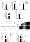
Figure 2.. Inhibition of IFN-α production by DMF occurs through multiple TLR signaling pathways and is mostly independent of NRF2 or GSH abundance.
(A) DMF inhibits TLR7 and TLR9-induced pDC IFN-α production. pDCs were isolated from human blood and treated with 50μM DMF for 18 h along with 1 μM CpG-A, 1 μM CpG-B, one multiplicity of infection with Influenza virus, 100nM R-848 or 500uM Loxoribine and the levels of IFN-α were quantified by ELISA. Results are representative of at least two independent experiments and *** P < 0.005, **** P < 0.0001 by two-tailed unpaired t test. (B) Structures of dimethyl fumarate (DMF) analogs. DEF: diethyl fumarate; DMM: dimethyl maleate; DEM: diethyl maleate; DiPrF: diisopropyl fumarate; DBnF: dibenzyl fumarate; DMenF: dimenthyl fumarate; TMA: tetramethyl fumaramide; DMS: dimethyl succinate; MMF: monomethyl fumarate. (C) IFN-α release from purified human pDCs stimulated with CpG-A and treated concomitantly with 50 μM of indicated compounds in (B) for 18 h. (D) IFN-α release measured by ELISA from purified human pDCs stimulated with CpG-A and treated with either the NRF2 inhibitor ML385 (5 μM), DMF (50 μM), or ML385 and DMF. (E) IFN-α release measured by ELISA from purified human pDCs stimulated with CpG-A and treated with either the glutathione inhibitor BSO (2 mM), DMF (50 μM), or BSO and DMF. Results are representative of at least two independent experiments and *** P < 0.005 by two-tailed unpaired t test.
Figure 3.
Figure 3.. Cysteine-13 (C13) of IRAK4 is a proteomic hot spot for DMF in Cal-1 cells.
(A) IFN-α release from Cal-1 cells stimulated with Sendai virus and treated concomitantly with 50 μM of indicated compounds for 18 h. (B) Schematic diagram depicting competitive IsoTOP for assessing DMF-reactive cysteines. (C) Scatter plot of ratio (R) values (DMSO/DMF) for quantified cysteine residues in isoTOP-ABPP experiments from Cal-1 cells treated for 1 or 4 h with 50 μM DMF. (D) Representative parent ion (MS1) profiles for DMF-hypersensitive (C13 of IRAK4), -moderately sensitive (C75 of ADA), and -insensitive (C152 of GAPDH) cysteines from isoTOP-ABPP experiments in Cal-1 cells. (E) Crystal structure of the Myddosome comprising six MyD88 (green), four IRAK4 (blue), and four IRAK2 (gray) molecules (PDB accession number 3MOP). In magenta is C13 of IRAK4. (F, G) HEK293T cells were transfected with either IRAK4 and GFP or IRAK4 and MyD88. 20 hours later, cells were treated with DMF (50 μM) for 4 hours and lysed. The isoTOP-ABPP protocol was then performed as previously described. The R values (DMSO/DMF) from the isoTOP-ABPP experiment for each quantified cysteine are shown in the bar graph.
Figure 4.
Figure 4.. DMF disrupts IRAK4-MyD88 complexes and signaling in a C13-dependent manner.
(A) DMF, but not MMF or DMS, impairs interactions between IRAK4 and MyD88. MyD88-FLAG (immunoprecipitated from HEK293T cells expressing MyD88-FLAG) bound to anti-FLAG beads was exposed to lysate from IRAK4-K2D-expressing HEK293T cells pre-treated with DMF, MMF, or DMS (100 μM compound, 4 h), or DMSO control. Shown Western blot is representative of three replicates used for quantitation. * P = 0.02 DMF vs. MMF as determined by paired t-test. (B) DMF-mediated disruption of the IRAK4-MyD88 interaction depends on C13 of IRAK4. MyD88 was immunoprecipitated from HEK293T cells expressing FLAG-tagged MyD88 and exposed to lysate from WT- or C13A-IRAK4-K2D-expressing HEK293T cells pre-treated with DMF (100 μM, 4 h) or DMSO control. Shown Western blot is representative of three replicates used for quantitation. (C) Impact of mutagenesis of C13 on IRAK4 interactions with Myd88. MyD88 was immunoprecipitated from HEK293T cells expressing FLAG-tagged MyD88 and exposed to lysate from HEK293T cells expressing the indicated C13 mutants of IRAK4. Western blot is representative of three replicates used for quantitation. * C13E vs C13A P = 0.049 * C13D vs C13A P = 0.039 as determined by paired t-test. (D) Impact of DMF on IRAK4 kinase activity. A reaction of purified IRAK4 (50ng/reaction), MBP (100ng/reaction) and ATP (50μM) was was treated with various concentrations of either Staurosporine (40μM to 0.02nM) or DMF (4mM to 2nM) to generate a dose response curve to assess the efficacy of these compounds as inhibitors of IRAK4 kinase activity. ADP-Glo™ Kinase Assay measures the conversion of ATP to ADP, which correlates with overall kinase activity within the reaction. (E) IRAK4-deficient B-EBV cells were reconstituted with human WT IRAK4 by lentiviral transduction, pre-treated with DMF, MMF, or DMS (50 μM, 4 h) or DMSO control, stimulated with LPS, CpG-A or Poly I:C for 24 h, and TNF-α measured by ELISA. *** LPS stimulated cells, DMSO versus DMF Treatment, P = 0.0002. ***CpG-A Treated cells, DMSO versus DMF treatment, P = 0.001. (F) IRAK4-deficient B-EBV cells were reconstituted with WT-IRAK4 or C13A-IRAK4 by lentiviral transduction, pre-treated with DMF (50 μM, 4 h) or DMSO control, stimulated with LPS for 24 hours, and TNF-α levels measured by ELISA. **** DMF treated cells versus DMSO treated cells, P < 0.0001.

Similar articles

Cited by

References

    1. Reizis B, Bunin A, Ghosh HS, Lewis KL, and Sisirak V 2011. Plasmacytoid dendritic cells: recent progress and open questions. Annu Rev Immunol 29: 163–183. - PMC - PubMed
    1. Gilliet M, Cao W, and Liu YJ 2008. Plasmacytoid dendritic cells: sensing nucleic acids in viral infection and autoimmune diseases. Nat Rev Immunol 8: 594–606. - PubMed
    1. Reizis B, Colonna M, Trinchieri G, Barrat F, and Gilliet M 2011. Plasmacytoid dendritic cells: one-trick ponies or workhorses of the immune system? Nat Rev Immunol 11: 558–565. - PMC - PubMed
    1. Colonna M, Trinchieri G, and Liu YJ 2004. Plasmacytoid dendritic cells in immunity. Nat Immunol 5: 1219–1226. - PubMed
    1. Swiecki M, and Colonna M 2015. The multifaceted biology of plasmacytoid dendritic cells. Nat Rev Immunol 15: 471–485. - PMC - PubMed

Publication types

MeSH terms