. 2019 May 1;79(9):2415-2425.
doi: 10.1158/0008-5472.CAN-18-3177. Epub 2019 Mar 18.

Inhibition of NF-κB-Dependent Signaling Enhances Sensitivity and Overcomes Resistance to BET Inhibition in Uveal Melanoma

Affiliations

Inhibition of NF-κB-Dependent Signaling Enhances Sensitivity and Overcomes Resistance to BET Inhibition in Uveal Melanoma

Grazia Ambrosini et al. Cancer Res. .

Erratum in

Abstract

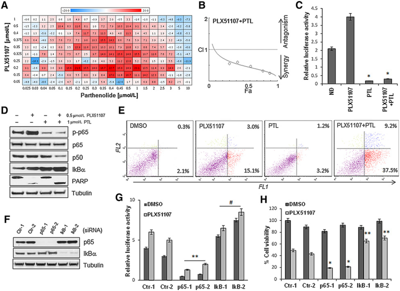
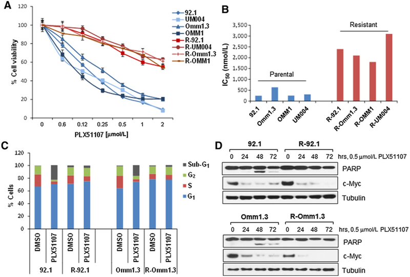
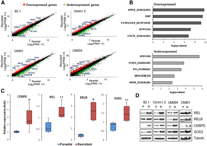
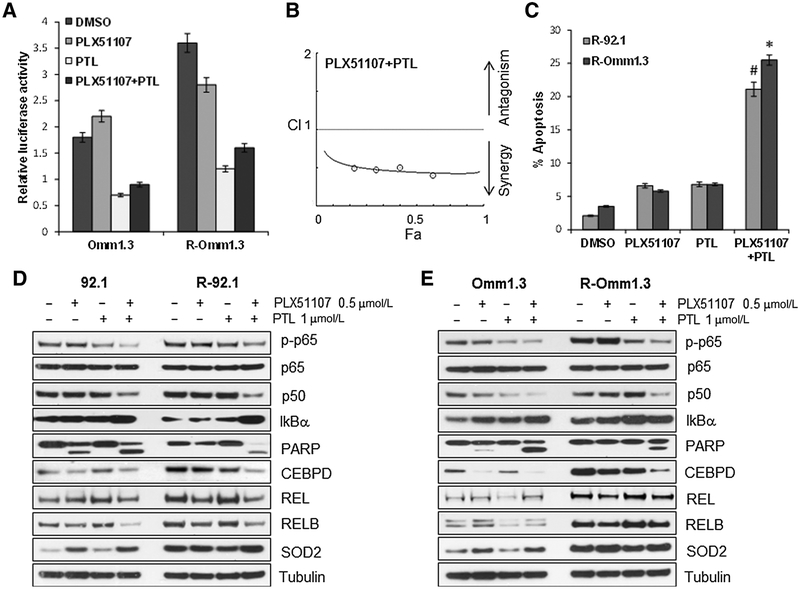
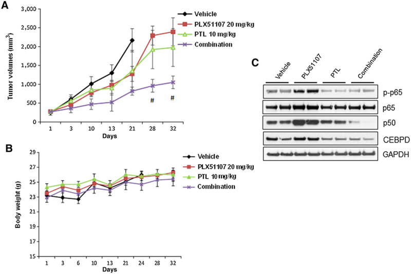
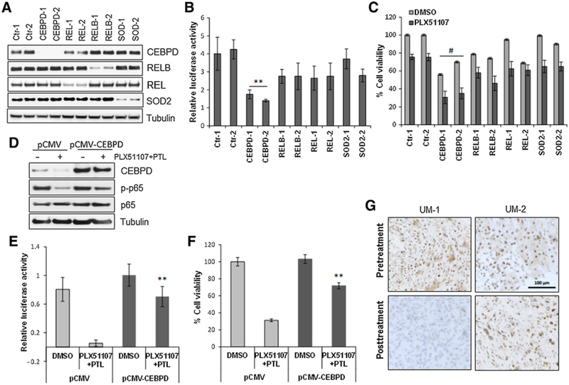
Bromodomain and extraterminal protein inhibitors (BETi) are epigenetic therapies aimed to target dysregulated gene expression in cancer cells. Despite early successes of BETi in a range of malignancies, the development of drug resistance may limit their clinical application. Here, we evaluated the mechanisms of BETi resistance in uveal melanoma, a disease with little treatment options, using two approaches: a high-throughput combinatorial drug screen with the clinical BET inhibitor PLX51107 and RNA sequencing of BETi-resistant cells. NF-κB inhibitors synergistically sensitized uveal melanoma cells to PLX51107 treatment. Furthermore, genes involved in NF-κB signaling were upregulated in BETi-resistant cells, and the transcription factor CEBPD contributed to the mechanism of resistance. These findings suggest that inhibitors of NF-κB signaling may improve the efficacy of BET inhibition in patients with advanced uveal melanoma. SIGNIFICANCE: These findings provide evidence that inhibitors of NF-κB signaling synergize with BET inhibition in in vitro and in vivo models, suggesting a clinical utility of these targeted therapies in patients with uveal melanoma.


Figures

Figure 1.
Figure 1.
Parthenolide has synergistic activity in combination with PLX51107 in a high-throughput drug screening. A, Uveal melanoma cells (92.1) were treated in secondary screenings with increasing concentrations of PLX51107 (0.05–1 μmmol/L) and parthenolide (0.025–10 μmmol/L) for 72 hours. The Bliss values for positive interactions (additive/synergy) are indicated in red and negative interactions in blue. B, Chou–Talalay plot (x-axis, Fa, or fractional activity, reflects the fraction of cells affected by the drug treatment relative to vehicle controls; y-axis, combination index, with <1, >1, and = 1 indicating synergistic, antagonistic, and additive effects, respectively). Each point represents a different combination of drug concentrations. C, The cells were transfected with a vector containing an NF-κB reporter gene along with a Renilla luciferase vector (1:10 ratio), then treated with 0.5 μmmol/L PLX51107, 1 μmmol/L PTL, and the combination for 24 hours. Luciferase activity was determined by chemiluminescence. Results are normalized to Renilla luciferase activity and represent the mean ± SD.*, P < 0.005.D, Western blot analysis of 92.1 cells treated with 0.5 μmmol/L PLX51107 and 1 μmmol/L PTL alone and in combination for 48 hours, showing inhibition of p65 phosphorylation, decreased expression of p50, induction of IkBα, and PARP cleavage. E, Apoptosis assay of 92.1 cells treated with 0.5 μmmol/L PLX51107 and 1 μmmol/L PTL alone and in combination for 48 hours, measuring cell permeability to fluorescent stains YO-PRO (FL1, for early apoptosis) and propidium iodide (FL2, for late apoptosis). F, Immunoblotting of cells transfected with two control (siCtr-1, −2), two p65 (sip65–1, −2), and two IkBα (IkB-1, −2) siRNA. G, The siRNA-transfected cells were then transfected with the NF-κB reporter gene vector/Renilla and tested for luciferase activity.**, P < 0.01; #, P < 0.05, comparing sip65–1,−2 with control siRNA. H, Cell viability assays of cells depleted of the indicated proteins after treatment with PLX51107 for 72 hours. Bars, mean ± SD.*, P < 0.005; **, P < 0.01.
Figure 2.
Figure 2.
Uveal melanoma cells develop resistance to BET inhibitors. A, Proliferation assays of uveal melanoma cell lines 92.1, Omm1.3, UM004, OMM1 (blue), and their resistant counterpart R-92.1, R-Omm1.3, R-UM004, R-OMM1 (red). Cell viability after 72 hours was calculated as percentage of untreated controls. Each point is a mean ± SD. B, The IC50s were calculated using the CompuSync software. The parental cell lines showed IC50 = 200–600 nmol/L, while for the resistant cells IC50 = 1,600–2,500 nmol/L. C, Parental and BETi-resistant cells were treated with DMSO or 0.5 μmmol/L PLX51107 for 48 hours, then stained with propidium iodide and analyzed for cell-cycle distribution by flow cytometry. The sub-G1 population was 23% for 92.1 and 17% for Omm1.3, while R-92.1 and R-Omm1.3 cells showed 4% and 3%, respectively. D, 92.1 and Omm1.3 cells were treated with 0.5 μmmol/L PLX51107 for up to 72 hours, and cell lysates were analyzed by immunoblotting for expression of c-Myc, PARP, and tubulin.
Figure 3.
Figure 3.
RNA sequencing and GSEA. A, Scatter plots of candidate genes in the given cell line showing overexpression (red) and underexpression (green) in at least two cell lines. Selected genes with high differential expression are indicated. B, GSEA for differentially over- and underexpressed genes in the resistant cells at baseline. The bars show the log(P value) from a Fisher exact test for enrichment for each pathway. C, Validation of gene expression by qPCR analysis using gene-specific primers for CEBPD, REL, RELB, and SOD2 in all four parental and four BETi-resistant cell lines. Triplicate values were normalized with GAPDH using the ΔΔCt method and reported as distributions of fold change relative to untreated 92.1 cells (set at 1) between the two groups (parental and resistant cells).#, P < 0.05; **, P < 0.01. D, Immunoblotting of parental and BETi-resistant cells using antibodies for the indicated proteins. Each blot is representative of at least two experiments showing same results.
Figure 4.
Figure 4.
Inhibition of NF-κB restores sensitivity to PLX51107 in BETi-resistant cells. A, NF-κB luciferase reporter gene assay of parental and resistant cells after treatment with PLX51107 (0.5 μmmol/L), PTL (1 μmmol/L), and the combination for 24 hours. B, The combination PLX55107 + PTL is synergistic (CI < 1). Each point represents a different combination of drug concentrations. C, Quantification of apoptotic cells for the experiments shown inþSupplementary Fig. S7. #, P < 0.05;*, P < 0.001.D and E, Parental and resistant 92.1 and Omm1.3 cells were analyzed by immunoblotting for the indicated proteins after 48-hour treatments with 0.5 μmmol/L PLX51107, 1 μmmol/L PTL, and the combination. The concurrent inhibition of BET and NF-κB reduces the expression of p-p65, p50, and CEBPD, while inducing PARP cleavage.
Figure 5.
Figure 5.
Inhibition of NF-κB increases sensitivity to BET inhibition in vivo. A, Tumors derived from R-Omm1.3 cells were implanted in athymic nu/nu mice. When these tumors reached an average of 270 mm3 diameter, the mice were administered (7/group) with vehicle or PLX51107 20 mg/kg orally, PTL 10 mg/kg i.p., and the combination of the two drugs. The treatment duration was 3 weeks for vehicle and 5 weeks for the drug treatments. The tumor size was measured twice a week. Each value represents the mean measurement of at least 5 animals. ± SEM, #, P < 0.05. B, Body weight of the host mice was measured twice a week. C, Two xenograft tumors per group were lysed at the end of the treatments and analyzed by Western blotting with the indicated antibodies.
Figure 6.
Figure 6.
Effects of gene silencing in the resistant phenotype. A, The indicated genes were silenced with two independent siRNA in the cell line R-92.1. Two nonspecific siRNAs were used as controls. B, siRNA-depleted cells were than transfected with an NF-κB-Luc vector and tested for NF-κB activity.**, P < 0.01. C, Cell viability assay of siRNA-transfected cells in the presence of 0.5 μmmol/L PLX51107 after 72 hours. Bars, mean ± SD. #, P < 0.05. D, R-92.1 cells were transfected with an empty vector (pCMV) or a CEBPD construct (pCMV-CEBPD). Cell lysates were subjected to immunoblotting using antibodies for CEBPD, p-p65, p65, and tubulin. E, NF-κB-Luc activity in vector and CEBPD-expressing cells with or without the combination treatment PLX51107 + PTL. **, P < 0.01. F, Viability assay of CEBPD-transfected cells after the combination treatment for 72 hours. Columns, mean ± SD of three independent experiments. **, P < 0.01. G, IHC analysis of CEBPD expression at baseline and after 2 weeks of treatment with PLX51107 in specimens from two representative patients with uveal melanoma. Suppression of CEBPD staining was observed after treatment in patient UM-1 who achieved prolonged stable disease lasting more than 1 year. Patient UM-2 had no significant changes in CEBPD expression and experienced disease progression.

Similar articles

Cited by

References

    1. Diener-West M, Reynolds SM, Agugliaro DJ, Caldwell R, Cumming K, Earle JD, et al. Development of metastatic disease after enrollment in the COMS trials for treatment of choroidal melanoma: Collaborative Ocular Melanoma Study Group Report No. 26. Arch Ophthalmol 2005;123: 1639–43. - PubMed
    1. Kuk D, Shoushtari AN, Barker CA, Panageas KS, Munhoz RR, Momtaz P, et al. Prognosis of mucosal, uveal, acral, nonacral cutaneous, and unknown primary melanoma from the time of first metastasis. Oncologist 2016;21: 848–54. - PMC - PubMed
    1. Kujala E, Makitie T, Kivela T. Very long-term prognosis of patients with malignant uveal melanoma. Invest Ophthalmol Vis Sci 2003;44: 4651–9. - PubMed
    1. Van Raamsdonk CD, Bezrookove V, Green G, Bauer J, Gaugler L, O’Brien JM, et al. Frequent somatic mutations of GNAQ in uveal melanoma and blue naevi. Nature 2009;457:599–602. - PMC - PubMed
    1. Van Raamsdonk CD, Griewank KG, Crosby MB, Garrido MC, Vemula S, Wiesner T, et al. Mutations in GNA11 in uveal melanoma. N Engl J Med 2010;363:2191–9. - PMC - PubMed

Publication types

MeSH terms