. 2019 May 1;79(9):2208-2219.
doi: 10.1158/0008-5472.CAN-18-2565. Epub 2019 Mar 18.

Myc and Loss of p53 Cooperate to Drive Formation of Choroid Plexus Carcinoma

Affiliations

Myc and Loss of p53 Cooperate to Drive Formation of Choroid Plexus Carcinoma

Jun Wang et al. Cancer Res. .

Abstract

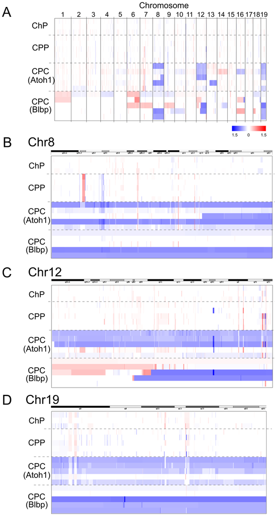
Choroid plexus carcinoma (CPC) is a rare brain tumor that occurs most commonly in very young children and has a dismal prognosis despite intensive therapy. Improved outcomes for patients with CPC depend on a deeper understanding of the mechanisms underlying the disease. Here we developed transgenic models of CPCs by activating the Myc oncogene and deleting the Trp53 tumor suppressor gene in murine neural stem cells or progenitors. Murine CPC resembled their human counterparts at a histologic level, and like the hypodiploid subset of human CPC, exhibited multiple whole-chromosome losses, particularly of chromosomes 8, 12, and 19. Analysis of murine and human CPC gene expression profiles and copy number changes revealed altered expression of genes involved in cell cycle, DNA damage response, and cilium function. High-throughput drug screening identified small molecule inhibitors that decreased the viability of CPC. These models will be valuable tools for understanding the biology of choroid plexus tumors and for testing novel approaches to therapy. SIGNIFICANCE: This study describes new mouse models of choroid plexus carcinoma and uses them to investigate the biology and therapeutic responsiveness of this highly malignant pediatric brain tumor.


Conflict of interest statement

The authors declare no potential conflicts of interest.

Figures

Figure 1.
Figure 1.
Expression of Myc and loss of p53 induce choroid plexus carcinoma. A. Mice used in this study. Crossing Atoh1-Cre mice to lox-STOP-lox (LSL) MycT58A mice results in progeny that develop choroid plexus papilloma (CPP); further crossing of these animals to p53-flox mice results in progeny that develop choroid plexus carcinoma (CPC). B. Kaplan-Meier survival curve shows median survival times for murine CPC (Atoh1-Cre;Myc/Myc;p53fl/fl) and CPP are 131.5 and 387 (Atoh1-Cre;Myc/Myc;p53fl/+) or 400 (Atoh1-Cre;Myc/Myc) days respectively, whereas mice that have p53 loss without MycT58A expression or mice that do not express Atoh1-Cre do not develop tumors. C and D. Hematoxylin and eosin staining shows histology of murine CPP and CPC. E and F. Ki67 staining shows proliferation in murine CPP and CPC. G-J. Aqp1 and Otx2 immunostaining on brain sections of murine CPP and CPC. * indicates normal choroid plexus, dotted line separates normal choroid plexus from the tumor. K-P. p-H2A.X and Ki67 immunostaining shows DNA damage and proliferation status in murine CPC, CPP or normal choroid plexus.
Figure 2.
Figure 2.
Murine CPCs originate from the Atoh1+ lineage. A. Schematic shows breeding strategy used to generate mice for lineage tracing of Atoh1+ progenitors. In Atoh1-Cre mice x Rosa26-LoxP-Stop-LoxP-tdTomato mice, Atoh1+ progenitors and their progeny express tdTomato. B. Immunohistochemistry shows that at embryonic stage E15.5 and postnatal day 7, cells originating from Atoh1+ progenitors express choroid plexus epithelial cell markers Aquaporin1 (Aqp1) and Orthodenticle homolog 2 (Otx2). Insets show magnified regions of choroid plexus tissue. C. Murine CPCs (Atoh1-Cre; Myc/Myc; p53flox/flox; Rosa26-Loxp-Stop-LoxP-tdTomato) express tdTomato reporter ubiquitously. Dotted line highlights the boundary between tumor (left) and normal choroid plexus tissue (right).
Figure 3.
Figure 3.
Choroid plexus tumors are dependent on Myc. A, B. Quantitative RT-PCR showing Myc and p53 expression in choroid plexus carcinoma (CPC) and choroid plexus papilloma (CPP) compared to normal mouse cerebellum (CB) and normal choroid plexus (ChP). Error bars represent standard error of the mean (SEM) C. Western blot showing MYC expression in a collection of human CPC and CPP specimens. D. Effects of Myc inhibitor 10058-F4 on viability of murine CPC cells. E. Effects of Myc inhibitor KJ-Pyr-9 on viability of murine CPC cells. F. Effects of CD532 on viability of murine CPC cells. G and H. Effects of various compounds that inhibit MYC on viability of human CPC cells (CHLA-CPC02 in G and CHLA-CPC03 in H). Error bars represent standard error of the mean (SEM).
Figure 4.
Figure 4.
Mouse choroid plexus carcinomas exhibit large chromosomal losses. A. Array CGH analysis of normal mouse choroid plexus (ChP) (n=3), choroid plexus papilloma (CPP) (n=5) and choroid plexus carcinoma (CPC) (n=10). B-D. High resolution image showing DNA copy number status on mouse chromosomes 8, 12 and 19. DNA copy number status is shown in integrative genomics viewer.
Figure 5.
Figure 5.
CPC and CPP exhibit distinct gene expression profiles. A. Principal component analysis shows normal choroid plexus (ChP), choroid plexus papilloma (CPP) and choroid plexus carcinoma (CPC) cluster separately. B. Heat map of gene expression data shows CPP and CPC share common gene expression signatures but are distinct from each other. C, D. Pathway analysis reveals enrichment (C) or downregulation (D) of pathways in CPC compared to ChP.
Figure 6.
Figure 6.
High throughput drug screening identifies compounds active against choroid plexus tumors. A. Compounds identified in high throughput drug screening using mouse choroid plexus carcinoma cells. Percent viability (normalized to vehicle treat control) of mouse CPC, mouse CPP and patient-derived human CPC cells treated with each compound is shown. B. Effects of CDK inhibitors on viability of mouse CPC cells; IC50’s are shown in the legend. C. Effects of CDK inhibitors Flavopiridol and Dinaciclib on phosphorylation of Rb at Ser 807/811. D. Effects of Triptolide on viability of mouse CPC cells. Error bars represent standard error of the mean (SEM). E. qRT-PCR showing effects of triptolide on Hspa1b, 3 hours after treatment of mouse CPC cells. Error bars represent standard error of the mean (SEM) F. Western Blot showing effects of triptolide on RNA polymerase II expression in mouse CPC cells.

Similar articles

Cited by

References

    1. Lam S, Lin Y, Cherian J, Qadri U, Harris DA, Melkonian S, et al. Choroid plexus tumors in children: a population-based study. Pediatr Neurosurg 2013;49:331–8 - PubMed
    1. Mazloom A, Wolff JE, Paulino AC. The impact of radiotherapy fields in the treatment of patients with choroid plexus carcinoma. Int J Radiat Oncol Biol Phys 2010;78:79–84 - PubMed
    1. Mallick S, Benson R, Melgandi W, Rath GK. Effect of Surgery, Adjuvant Therapy, and Other Prognostic Factors on Choroid Plexus Carcinoma: A Systematic Review and Individual Patient Data Analysis. Int J Radiat Oncol Biol Phys 2017;99:1199–206 - PubMed
    1. Tabori U, Shlien A, Baskin B, Levitt S, Ray P, Alon N, et al. TP53 alterations determine clinical subgroups and survival of patients with choroid plexus tumors. Journal of clinical oncology : official journal of the American Society of Clinical Oncology 2010;28:1995–2001 - PubMed
    1. Tong Y, Merino D, Nimmervoll B, Gupta K, Wang YD, Finkelstein D, et al. Cross-Species Genomics Identifies TAF12, NFYC, and RAD54L as Choroid Plexus Carcinoma Oncogenes. Cancer Cell 2015;27:712–27 - PMC - PubMed

Publication types

MeSH terms

Supplementary concepts