Review
. 2019 Mar 18;132(6):jcs224212.
doi: 10.1242/jcs.224212.

Message in a vesicle - trans-kingdom intercommunication at the vector-host interface

Affiliations
Review

Message in a vesicle - trans-kingdom intercommunication at the vector-host interface

Adela S Oliva Chávez et al. J Cell Sci. .

Abstract

Vector-borne diseases cause over 700,000 deaths annually and represent 17% of all infectious illnesses worldwide. This public health menace highlights the importance of understanding how arthropod vectors, microbes and their mammalian hosts interact. Currently, an emphasis of the scientific enterprise is at the vector-host interface where human pathogens are acquired and transmitted. At this spatial junction, arthropod effector molecules are secreted, enabling microbial pathogenesis and disease. Extracellular vesicles manipulate signaling networks by carrying proteins, lipids, carbohydrates and regulatory nucleic acids. Therefore, they are well positioned to aid in cell-to-cell communication and mediate molecular interactions. This Review briefly discusses exosome and microvesicle biogenesis, their cargo, and the role that nanovesicles play during pathogen spread, host colonization and disease pathogenesis. We then focus on the role of extracellular vesicles in dictating microbial pathogenesis and host immunity during transmission of vector-borne pathogens.

Keywords: Arthropod-borne disease; Cell communication; Extracellular vesicle; Immunomodulation; Microbial transmission.


Conflict of interest statement

Competing interestsThe authors declare no competing or financial interests.

Figures
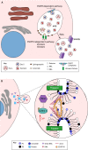
Fig. 1.
Fig. 1.
Exosome and microvesicle biogenesis. (A) Exosomes are formed in the lumen of multivesicular bodies (MVBs) through two biological signaling cascades: (1) the endosomal sorting complex required for transport (ESCRT)-dependent, and (2) the ESCRT-independent pathways. In the ESCRT-dependent pathway, exosomes are produced by the action of four different ESCRT complexes (ESCRT-0, -I, -II, and III). In the ESCRT-independent pathway, exosomes are created by the accumulation of ceramide when acid sphingomyelinases (aSmases) and neutral sphingomyelinases (nSmases) hydrolyze sphingomyelin. Rab proteins mediate the transport of the MVB, and SNARE molecules drive the fusion of vesicles with the plasma membrane. Fusion of membranes leads to the secretion of different subpopulations of vesicles (e.g. Exo-L, Exo-S and exomeres), which exhibit distinct DNA, RNA and protein signatures. (B) Microvesicles are secreted through the invagination of the plasma membrane. Flippases and floppases rearrange the lipid content of the outer layer of the plasma membrane, resulting in the enrichment of phosphatidylserine (PS), phosphatidylethanolamine (PE) and ceramide. Lipid remodeling changes the curvature of the plasma membrane, thereby forming the budding of the microvesicle, which is triggered by increases in Ca2+ concentration. The exact mechanism of microvesicle excision remains mostly unknown. However, the ESCRT-III complex and changes in actin dynamics driven by the small GTPase RhoA and cofilin 1 may be involved.
Fig. 2.
Fig. 2.
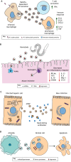
Extracellular vesicles and infection. (A) Immune activation in the mouse model. M. tuberculosis modifies the protein content of host extracellular vesicles (EVs) during intracellular infection. Secretion of modified EVs is dependent on Rab27a. Interaction of modified EVs with uninfected macrophages and activated T cells leads to the secretion of chemokines and cytokines, thus, eliciting an immune response. (B) Immune evasion in the mouse model. Parasitic nematodes manipulate host immune responses in the intestine by secreting exosomes. These exosomes contain regulatory small RNAs, which decrease pro-inflammatory responses, activation of alternatively activated macrophages (AAMФ) and eosinophil migration. Binding of these nematode vesicles to mammalian cells suppresses the expression of Il1rl1, Mpk1 and the mannose receptor CD206, as well as the secretion of resistin-like molecule α (RELMα), Ym1 and several cytokines. (C) Invasion of hepatic cells. Hepatitis virus A (HAV) hijacks the exosome biogenesis accessory proteins ALIX and VSP4 to form an envelope (eHAV) that protects the virus from neutralizing antibodies. (D) Pathogenesis of amoeba to mammalian cells. The pathogenic free-living amoeba Acanthamoeba castellanii secretes EVs that contain metalloproteases and serine proteases that are taken up in the model CHO and T98G cell lines. Once EVs are endocytosed, the cargo leads to apoptosis of host cells, possibly, causing tissue damage that is associated with Acanthamoeba keratitis.
Fig. 3.
Fig. 3.
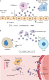
Extracellular vesicles affect host responses during vector-borne pathogen infection. (A) Cerebral malaria in humans is linked to damage of the blood–brain barrier. These symptoms may be associated with extracellular vesicle (EV) secretion by P. falciparum-infected red blood cells (iRBCs). These vesicles contain the P. falciparum erythrocyte membrane protein 1 (PfEMP1), host miR-451a and DNA. PfEMP1 and miR-451a increase the permeability of the cell barrier through the downregulation of host gene expression. In addition, P. falciparum DNA in these vesicles activate immune responses in circulating monocytes through the stimulator of interferon genes (STING) pathway. (B) Leishmania spp., the causative agent of Leishmaniasis, secrete EVs during host infection. As shown in mice, these vesicles are taken up by monocytes and T cells, where they suppress the secretion of pro-inflammatory cytokines and steer T cell differentiation towards Th2 responses. (C) The Chagas disease parasite Trypanosoma cruzi secretes EVs that exert an immunosuppressive activity in the human host by blocking complement formation, suppressing macrophage antigen presentation (MHC-II), inhibiting T cell stimulation through CD86, dampening the secretion of TNF and the expression of iNOS, and increasing invasion of cardiac tissue through the elevated levels of IL-4 and IL-10, leading to tissue damage in the heart.
Fig. 4.
Fig. 4.
Parasite-derived extracellular vesicles influence vector–host interactions. (A) During replication within the sandfly midgut, Leishmania spp. secrete extracellular vesicles (EVs) that contain virulence factors and heat-shock proteins, such as GP63 and Hsp7. Blood-feeding sandflies inject these EVs into the bite site, along with the parasites, thereby stimulating the secretion of pro-inflammatory cytokines, increasing tissue damage and enhancing pathogen replication, which aggravates the disease. (B) EVs are involved in environmental sensing and signaling during replication within arthropod vectors. T. brucei uses EVs to signal stress conditions to other replicating parasites inside the midgut of tsetse flies. Unfit parasites (dark purple) secrete EVs containing SL RNA, which accumulates when mRNA trans-splicing is disrupted. These EVs are taken up by healthy parasites (light purple), leading to an arrest in their movement and/or change in direction. Intercommunication through EVs may enable successful vector colonization by these parasites to avoid an environment that is unfit for replication.

Similar articles

Cited by

References

    1. Altan-Bonnet N. (2016). Extracellular vesicles are the Trojan horses of viral infection. Curr. Opin. Microbiol. 32, 77-81. 10.1016/j.mib.2016.05.004 - DOI - PMC - PubMed
    1. Arakelyan A., Fitzgerald W., Zicari S., Vanpouille C. and Margolis L. (2017). Extracellular vesicles carry HIV Env and facilitate HIV infection of human lymphoid tissue. Sci. Rep. 7, 1695 10.1038/s41598-017-01739-8 - DOI - PMC - PubMed
    1. Atayde V. D., Aslan H., Townsend S., Hassani K., Kamhawi S. and Olivier M. (2015). Exosome secretion by the parasitic protozoan Leishmania within the sand fly midgut. Cell Rep. 13, 957-967. 10.1016/j.celrep.2015.09.058 - DOI - PMC - PubMed
    1. Babatunde K. A., Mbagwu S., Hernández-Castañeda M. A., Adapa S. R., Walch M., Filgueira L., Falquet L., Jiang R. H. Y., Ghiran I. and Mantel P.-Y. (2018). Malaria infected red blood cells release small regulatory RNAs through extracellular vesicles. Sci. Rep. 8, 884 10.1038/s41598-018-19149-9 - DOI - PMC - PubMed
    1. Baietti M. F., Zhang Z., Mortier E., Melchior A., Degeest G., Geeraerts A., Ivarsson Y., Depoortere F., Coomans C., Vermeiren E. et al. (2012). Syndecan-syntenin-ALIX regulates the biogenesis of exosomes. Nat. Cell Biol. 14, 677-685. 10.1038/ncb2502 - DOI - PubMed

Publication types

MeSH terms