. 2019 May 8;39(19):3770-3781.
doi: 10.1523/JNEUROSCI.3173-18.2019. Epub 2019 Mar 18.

The Divergent Roles of Dietary Saturated and Monounsaturated Fatty Acids on Nerve Function in Murine Models of Obesity

Affiliations

The Divergent Roles of Dietary Saturated and Monounsaturated Fatty Acids on Nerve Function in Murine Models of Obesity

Amy E Rumora et al. J Neurosci. .

Abstract

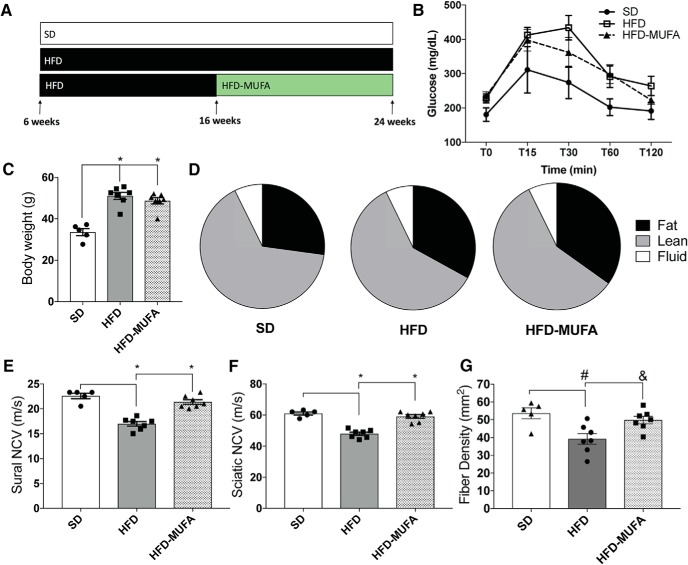
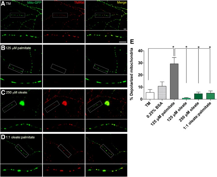
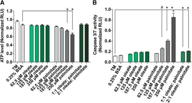
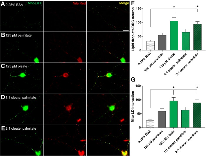
Neuropathy is the most common complication of prediabetes and diabetes and presents as distal-to-proximal loss of peripheral nerve function in the lower extremities. Neuropathy progression and disease severity in prediabetes and diabetes correlates with dyslipidemia in man and murine models of disease. Dyslipidemia is characterized by elevated levels of circulating saturated fatty acids (SFAs) that associate with the progression of neuropathy. Increased intake of monounsaturated fatty acid (MUFA)-rich diets confers metabolic health benefits; however, the impact of fatty acid saturation in neuropathy is unknown. This study examines the differential effect of SFAs and MUFAs on the development of neuropathy and the molecular mechanisms underlying the progression of the complication. Male mice Mus musculus fed a high-fat diet rich in SFAs developed robust peripheral neuropathy. This neuropathy was completely reversed by switching the mice from the SFA-rich high-fat diet to a MUFA-rich high-fat diet; nerve conduction velocities and intraepidermal nerve fiber density were restored. A MUFA oleate also prevented the impairment of mitochondrial transport and protected mitochondrial membrane potential in cultured sensory neurons treated with mixtures of oleate and the SFA palmitate. Moreover, oleate also preserved intracellular ATP levels, prevented apoptosis induced by palmitate treatment, and promoted lipid droplet formation in sensory neurons, suggesting that lipid droplets protect sensory neurons from lipotoxicity. Together, these results suggest that MUFAs reverse the progression of neuropathy by protecting mitochondrial function and transport through the formation of intracellular lipid droplets in sensory neurons.SIGNIFICANCE STATEMENT There is a global epidemic of prediabetes and diabetes, disorders that represent a continuum of metabolic disturbances in lipid and glucose metabolism. In the United States, 80 million individuals have prediabetes and 30 million have diabetes. Neuropathy is the most common complication of both disorders, carries a high morbidity, and, despite its prevalence, has no treatments. We report that dietary intervention with monounsaturated fatty acids reverses the progression of neuropathy and restores nerve function in high-fat diet-fed murine models of peripheral neuropathy. Furthermore, the addition of the monounsaturated fatty acid oleate to sensory neurons cultured under diabetic conditions shows that oleate prevents impairment of mitochondrial transport and mitochondrial dysfunction through a mechanism involving formation of axonal lipid droplets.

Keywords: diabetes; monounsaturated fatty acid; neuropathy; prediabetes; saturated fatty acid; sensory neuron.


Figures

Figure 1.
Figure 1.
A MUFA-rich diet reverses neuropathy in prediabetic mice. A, Dietary intervention murine model whereby C57BL/6J mice were fed a SD, 60% HFD, or 60% HFD followed by 60% HFD-MUFA (Figure 1-1). BD, Metabolic phenotyping of mice at 24 weeks of age, including (B) glucose tolerance test (mg/dl), (C) terminal body weight (g), and (D) terminal body composition. E, F, Neuropathy phenotyping using NCV (m/s) for sural and sciatic nerves, respectively (Figure 1-2). G, IENFD of mouse footpad. All data represent mean ± SEM from 5 to 8 animals per group. One-way ANOVA with Tukey's multiple-comparisons test. *p < 0.0001, #p = 0.0071, &p = 0.0297.
Figure 2.
Figure 2.
MUFA treatment preserves axonal mitochondrial motility in cultured DRG neurons. A, Kymograph analysis of DRG axons treated for 24 h with TM, vehicle only (0.25% BSA), and varying concentrations (31.25–250 μm) of oleate. B, Percentage of motile mitochondria as observed in A. C, Kymograph analysis of DRG axons treated for 24 h with TM, vehicle only (0.25% BSA), 125 μm palmitate alone, and 125 μm palmitate with 125 or 250 μm oleate. D, Percentage of motile mitochondria as observed in C. E, Kymograph analysis of DRG neurons treated for 24 h with TM, vehicle alone (0.25% BSA), pre-treatment (12 h) with 125 μm palmitate followed by post-treatment (12 h) with 125 or 250 μm oleate, or pre-treatment (12 h) with 125 or 250 μm oleate followed by post-treatment (12 h) with 125 μm palmitate. F, Percentage of motile mitochondria as observed in E. All data represent n = 16–23 neurons per condition (A, B), n = 18–23 neurons per condition (C, D), and 12–16 neurons per condition (E, F): one-way ANOVA with Tukey's multiple-comparisons test. *p < 0.0001.
Figure 3.
Figure 3.
MUFA treatment preserves and protects DRG neuronal MMP. Fluorescence microscopy imaging of DRG neurons expressing mito-GFP that were stained with TMRM dye specific for polarized mitochondrial membranes, following treatment with TM (A), 125 μm palmitate (B), 250 μm oleate (C), or a mix of 125 μm palmitate and oleate (D). Green signal (mito-GFP) indicates mitochondria, red signal (TMRM) indicates mitochondria with membrane potential, and yellow signal (Merge) shows overlap of mito-GFP and TMRM signals indicative of polarized mitochondria. Scale bars represent 10 μm. E, Percentage of depolarized mitochondria as shown in AD; i.e., mitochondria fluorescing green (mito-GFP) but lacking red TMRM signal, as a percentage of total mitochondria. All data represent n = 31–35 neurons per condition: one-way ANOVA with Tukey's multiple-comparisons test. *p < 0.0001.
Figure 4.
Figure 4.
MUFA treatment prevents ATP production decreases and apoptotic signaling induced by palmitate. A, Relative ATP production (RLU) as measured by CellTiter-Glo assay in 50B11 DRG neuronal cells treated for 24 h with TM, vehicle alone (0.25% BSA), varying concentrations (62.5–250 μm) of oleate and palmitate alone, or either 125 or 250 μm oleate mixed with 125 μm palmitate. Data represent mean ± SEM from n = 6 wells of 50B11 neurons per condition: one-way ANOVA with Tukey's multiple-comparisons test. p < 0.0001 (B) Relative caspase 3/7 activity (RLU) as measured by Caspase 3/7-Glo assay in 50B11 DRG neuronal cells treated with conditions identical to those in A. Data represent mean ± SEM from n = 3 wells of 50B11 neurons per condition: one-way ANOVA with Tukey's multiple-comparisons test. *p < 0.0001 and #p < 0.0056.
Figure 5.
Figure 5.
MUFA treatment induces axonal lipid droplet formation in DRG neurons. Fluorescence microscopy of cultured DRG neurons expressing mito-GFP (green puncta) and stained with lipid droplet-specific Nile red (red puncta), following treatment with vehicle alone (0.25% BSA; A), 125 μm palmitate (B), or oleate alone (C), or 125 μm palmitate mixed with 125 μm (D) or 250 μm oleate (E). Yellow puncta (Merged signal) indicates colocalization between mitochondria and lipid droplets in DRG axons. Scale bars represent 20 μm. F, Number of lipid droplets (red puncta as stained by Nile red) per DRG neuron as shown in AE. n = 20–47 neurons per condition: one-way ANOVA with Tukey's multiple-comparisons test. *p < 0.0001. G, Number of mitochondria colocalizing with lipid droplets (yellow puncta in Mito-GFP/Nile red Merge) per DRG neuron as shown in AE. n = 20–37 neurons per condition: one-way ANOVA with Tukey's multiple-comparisons test. *p < 0.0001.

References

    1. Andersen ST, Witte DR, Dalsgaard EM, Andersen H, Nawroth P, Fleming T, Jensen TM, Finnerup NB, Jensen TS, Lauritzen T, Feldman EL, Callaghan BC, Charles M (2018) Risk factors for incident diabetic polyneuropathy in a cohort with screen-detected type 2 diabetes followed for 13 years: ADDITION-denmark. Diabetes Care 41:1068–1075. 10.2337/dc17-2062 - DOI - PubMed
    1. Bagkos G, Koufopoulos K, Piperi C (2014) A new model for mitochondrial membrane potential production and storage. Med Hypotheses 83:175–181. 10.1016/j.mehy.2014.05.001 - DOI - PubMed
    1. Benador IY, Veliova M, Mahdaviani K, Petcherski A, Wikstrom JD, Assali EA, Acín-Pérez R, Shum M, Oliveira MF, Cinti S, Sztalryd C, Barshop WD, Wohlschlegel JA, Corkey BE, Liesa M, Shirihai OS (2018) Mitochondria bound to lipid droplets have unique bioenergetics, composition, and dynamics that support lipid droplet expansion. Cell Metab 27:869–885.e6. 10.1016/j.cmet.2018.03.003 - DOI - PMC - PubMed
    1. Burhans MS, Flowers MT, Harrington KR, Bond LM, Guo CA, Anderson RM, Ntambi JM (2015) Hepatic oleate regulates adipose tissue lipogenesis and fatty acid oxidation. J Lipid Res 56:304–318. 10.1194/jlr.M054429 - DOI - PMC - PubMed
    1. Callaghan BC, Hur J, Feldman EL (2012a) Diabetic neuropathy: one disease or two? Curr Opin Neurol 25:536–541. 10.1097/WCO.0b013e328357a797 - DOI - PMC - PubMed

Publication types