. 2019 Apr 30;39(10):e00559-18.
doi: 10.1128/MCB.00559-18. Print 2019 May 15.

Targeted Degradation of Glucose Transporters Protects against Arsenic Toxicity

Affiliations

Targeted Degradation of Glucose Transporters Protects against Arsenic Toxicity

Marco Jochem et al. Mol Cell Biol. .

Abstract

The abundance of cell surface glucose transporters must be precisely regulated to ensure optimal growth under constantly changing environmental conditions. We recently conducted a proteomic analysis of the cellular response to trivalent arsenic, a ubiquitous environmental toxin and carcinogen. A surprising finding was that a subset of glucose transporters was among the most downregulated proteins in the cell upon arsenic exposure. Here we show that this downregulation reflects targeted arsenic-dependent degradation of glucose transporters. Degradation occurs in the vacuole and requires the E2 ubiquitin ligase Ubc4, the E3 ubiquitin ligase Rsp5, and K63-linked ubiquitin chains. We used quantitative proteomic approaches to determine the ubiquitinated proteome after arsenic exposure, which helped us to identify the ubiquitination sites within these glucose transporters. A mutant lacking all seven major glucose transporters was highly resistant to arsenic, and expression of a degradation-resistant transporter restored arsenic sensitivity to this strain, suggesting that this pathway represents a protective cellular response. Previous work suggests that glucose transporters are major mediators of arsenic import, providing a potential rationale for this pathway. These results may have implications for the epidemiologic association between arsenic exposure and diabetes.

Keywords: Rsp5; arsenic; glucose transporter; protein degradation; ubiquitin.


Figures

FIG 1
FIG 1
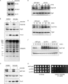
Arsenic-induced downregulation of glucose transporters. (A) Relative protein abundance of glucose transporters Hxt1 to Hxt7 determined by proteomic analysis at 0, 1, and 4 h after treatment with sodium arsenite (1 mM). Error bars represent standard deviations for triplicate cultures. Note that because of the high sequence similarity between Hxt6 and Hxt7 (only 1 amino acid difference), only one unique peptide could be assigned to Hxt6, while all other peptides were assigned to Hxt7. Thus, the result for Hxt7 likely reflects results for a combination of peptides from Hxt6 and Hxt7. (B) Downregulation of Hxt2-HA and Hxt7-HA in response to sodium arsenite, as determined by SDS-PAGE followed by immunoblot analysis. Proteins were extracted from log-phase yeast cells before (lanes −) and 3 h after (lanes +) treatment with 1 mM sodium arsenite. (Top) Anti-HA antibody; (bottom) Pgk1 (loading control). Numbers on the left are molecular masses (in kilodaltons). (C) Strong resistance of the hxt1-7Δ mutant to arsenite toxicity. Cells were spotted in a 3-fold serial dilution series on plates containing or lacking sodium arsenite (5 mM) and incubated at 30°C for 2 (no drug) or 5 (arsenite) days. Because the hxt1-7Δ mutant cannot grow with glucose as the sole carbon source, the medium contained both glucose and maltose (2% each).
FIG 2
FIG 2
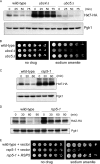
Vacuolar degradation mediates arsenic-induced glucose transporter downregulation. (A) The mRNA levels of the indicated hexose transporters before (lanes −) and 1 h after (lanes +) treatment with sodium arsenite (1 mM) were determined by RT-PCR. Actin (ACT1) served as a loading control. HXT6 and HXT7 differ at only 3 codons (1 amino acid) and so are amplified together by the same primer set. (B) Arsenic-induced downregulation of Hxt2-HA persists in the presence of proteasome inhibitors. Cells were pretreated with dimethyl sulfoxide (DMSO) or bortezomib (Velcade; 30 μM) to inhibit the proteasome and then treated with sodium arsenite (1 mM) for 1 h. Whole-cell extracts were prepared and analyzed by SDS-PAGE, followed by immunoblotting with the indicated antibodies. Pgk1 served as a loading control. Ubiquitin and the known proteasome substrate Tmc1 served to verify the efficiency of proteasome inhibition. This experiment was carried out in the pdr5Δ mutant background to increase the intracellular levels of bortezomib. Note that arsenic alone induces strong proteotoxic effects, as indicated by the increase in both high-molecular-weight ubiquitinated material and Tmc1 (compare the first and second lanes), consistent with prior reports (24, 49). Numbers on the left are molecular masses (in kilodaltons). (C) Arsenic-induced downregulation of Hxt7-HA persists in the presence of proteasome inhibitors. Cells were pretreated with DMSO or bortezomib (100 μM) to inhibit the proteasome and then treated with sodium arsenite (1 mM) for 2 h. Whole-cell extracts were prepared and analyzed by SDS-PAGE, followed by immunoblotting with the indicated antibodies. As in panel B, Pgk1 served as a loading control and Tmc1 served to verify the efficiency of proteasome inhibition. This experiment was carried out in the pdr5Δ mutant background to increase the intracellular levels of bortezomib. (D and E) Stabilization of Hxt2-HA (D) and Hxt7-HA (E) in the doa4Δ mutant. Wild-type and doa4Δ mutant cells were treated with sodium arsenite (1 mM), and whole-cell extracts were prepared at the indicated time points and analyzed by SDS-PAGE and immunoblotting. (Top) Anti-HA antibody; (bottom) Pgk1 (loading control). (F) Stabilization of Hxt7-HA in response to sodium arsenite in the vps36Δ and vps25Δ vacuolar degradation mutants. The experiment was conducted analogously to the experiments whose results are shown in panels D and E. (G) Growth of the wild-type, vps36Δ, and vps25Δ strains in the presence or absence of 1.5 mM sodium arsenite. Cells were spotted in a 3-fold dilution series and incubated at 30°C for 2 days.
FIG 3
FIG 3
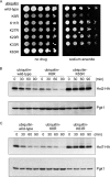
Arsenic-induced glucose transporter degradation requires Ubc4 and Rsp5. (A) Degradation of Hxt7-HA in the wild-type, ubc4Δ, and ubc5Δ strains in response to sodium arsenite (1 mM). Whole-cell extracts were prepared at the indicated time points and analyzed by SDS-PAGE and immunoblotting. (Top) Anti-HA antibody; (bottom) Pgk1 (loading control). (B) Growth of the wild-type, ubc4Δ, and ubc5Δ strains in the presence or absence of sodium arsenite (1.5 mM). Cells were spotted in a 3-fold serial dilution series and incubated at 30°C for 2 to 3 days. (C) Degradation of Hxt7-HA in the wild type and the temperature-sensitive rsp5-1 mutant in response to sodium arsenite (1 mM). Whole-cell extracts were prepared at the indicated time points and analyzed by SDS-PAGE and immunoblotting. (Top) Anti-HA antibody; (bottom) Pgk1 (loading control). The experiment was performed at 30°C. (D) Degradation of Hxt2-HA in the wild type and the temperature-sensitive rsp5-1 mutant in response to sodium arsenite (1 mM). Whole-cell extracts were prepared at the indicated time points and analyzed by SDS-PAGE and immunoblotting. (Top) Anti-HA antibody; (bottom) Pgk1 (loading control). Note that the restrictive temperature used here (37°C) was higher than that used in the experiment whose results are presented in panel C. (E) Growth of the wild type and the rsp5-1 mutant expressing either an empty vector or the RSP5 complementation plasmid, as indicated, in the presence or absence of sodium arsenite (1 mM). Cells were spotted in a 3-fold serial dilution series and incubated at 30°C for 2 (no drug) or 4 (arsenite) days.
FIG 4
FIG 4
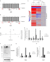
Arsenic-induced degradation of Hxt2 and Hxt7 requires K63 ubiquitin chain linkages. (A) Growth of the wild type and the six indicated ubiquitin mutants in the presence or absence of sodium arsenite (1.5 mM). Cells were spotted in a 3-fold serial dilution series and incubated at 30°C for 2 to 3 days. (B and C) Degradation of Hxt2-HA or Hxt7-HA in strains unable to generate K6 or K63 ubiquitin chain linkages in response to trivalent arsenic (1 mM). Whole-cell extracts were prepared at the indicated time points and analyzed by SDS-PAGE and immunoblotting. (Top) Anti-HA antibody; (bottom) Pgk1 (loading control).
FIG 5
FIG 5
Determination of the ubiquitinated proteome after arsenite treatment. (A and B) Schematic illustration of Hxt2 (A) and Hxt7 (B). The approximate positions of the 17 (Hxt2) and 19 (Hxt7) cytoplasmic lysines, which are potential ubiquitination sites, are indicated. Residues mutated in the Hxt2-9K-to-R and Hxt7-12K-to-R mutants (see Fig. 6) are shown in red. (C) Heat map representation of the relative change in abundance of ubiquitinated peptides at 0, 1, and 4 h after arsenic treatment (1 mM). The three columns per time point represent biological triplicates. A total of 4,125 peptides representing 1,794 unique proteins were quantitated. The results shown here have been normalized to total protein abundance, which was determined in parallel, to provide a more specific readout of arsenic-induced ubiquitination. (D) Principal-component analysis (PCA) of the proteomic data set shown in panel C. Individual points represent the three biological triplicates, confirming a high degree of concordance between triplicates as well as very different ubiquitinated proteomes at 0, 1, and 4 h after arsenic treatment. (E) Relative abundance of the seven ubiquitin chain linkage types after arsenite treatment, as determined by proteomic analysis. Data have been normalized to total ubiquitin abundance. Sodium arsenite treatment was at 1 mM for the indicated times. Error bars reflect standard deviations for biological triplicates. (F) Dynamic changes in the accumulation of high-molecular-weight ubiquitin-immunoreactive material after sodium arsenite treatment (1 mM). Whole-cell extracts were prepared at the indicated time points and analyzed by SDS-PAGE and immunoblotting. (Top) Antiubiquitin antibody; (bottom) Pgk1 (loading control). Numbers on the left are molecular masses (in kilodaltons). (G and H) Relative abundance of all detected cytoplasmic ubiquitinated peptides from Hxt2 and Hxt7, as determined by the proteomic analysis whose results are presented in panel C. Error bars represent standard deviations for triplicate cultures.
FIG 6
FIG 6
Identification of ubiquitination sites in Hxt2 and Hxt7 for arsenic-induced protein degradation. (A) Degradation of the wild type and lysine-to-arginine mutants of Hxt2 in response to sodium arsenite (1 mM). The 6K-to-R mutant affects lysines 37, 257, 524, 527, 534, and 536. The 9K-to-R mutant additionally affects lysines 26, 48, and 309. Whole-cell extracts were prepared at the indicated time points and analyzed by SDS-PAGE and immunoblotting. (Top) Anti-HA antibody; (bottom) Pgk1 (loading control). (B) Degradation of the wild type and the lysine-to-arginine mutants of Hxt7 in response to sodium arsenite (1 mM). The 12K-to-R mutant affects lysines 33, 40, 56, 198, 268, 273, 304, 318, 320, 560, 564, and 570. Whole-cell extracts were prepared at the indicated time points and analyzed by SDS-PAGE and immunoblotting. (Top) Anti-HA antibody; (bottom) Pgk1 (loading control). (C) Growth of the wild type and the hxt1-7Δ mutant expressing plasmids harboring an empty vector, wild-type Hxt2, and the Hxt2-9K-to-R mutant on medium containing glucose as the sole carbon source. Cells were spotted in a 3-fold serial dilution series and incubated at 30°C for 6 days. (D) Growth of the wild type and the hxt1-7Δ mutant expressing plasmids harboring an empty vector, wild-type Hxt7, and the Hxt7-12K-to-R mutant on medium containing glucose as the sole carbon source. Cells were spotted in a 3-fold serial dilution series and incubated at 30°C for 6 days. (E) Growth of the wild type and the hxt1-7Δ mutant expressing plasmids harboring an empty vector, wild-type Hxt2, and the Hxt2-9K-to-R mutant on medium containing or lacking sodium arsenite, as indicated. Cells were spotted in a 3-fold serial dilution series and incubated at 30°C for 3 days (no drug), 9 days (3.5 mM arsenite), or 14 days (5 mM arsenite). Note that this experiment was performed in rich medium with maltose as the sole carbon source to minimize any beneficial effects related to glucose uptake by Hxt2 proteins. (F) Dynamic changes in the accumulation of high-molecular-weight ubiquitin-immunoreactive material in hxt1-7Δ cells expressing either wild-type Hxt2 or the Hxt2-9K-to-R mutant. Sodium arsenite treatment was at 1 mM for the indicated times. Whole-cell extracts were prepared at the indicated time points and analyzed by SDS-PAGE and immunoblotting. (Top) Antiubiquitin antibody; (bottom) Pgk1 (loading control). The effect of the Hxt2-9K-to-R mutant was the most pronounced in the high-molecular-weight range, indicated by a line to the right of the panel. Numbers on the left are molecular masses (in kilodaltons).

References

    1. Rosen BP, Liu Z. 2009. Transport pathways for arsenic and selenium: a minireview. Environ Int 35:512–515. doi:10.1016/j.envint.2008.07.023. - DOI - PMC - PubMed
    1. Maull EA, Ahsan H, Edwards J, Longnecker MP, Navas-Acien A, Pi J, Silbergeld EK, Styblo M, Tseng CH, Thayer KA, Loomis D. 2012. Evaluation of the association between arsenic and diabetes: a National Toxicology Program workshop review. Environ Health Perspect 120:1658–1670. doi:10.1289/ehp.1104579. - DOI - PMC - PubMed
    1. Bonini MG, Sargis RM. 2018. Environmental toxicant exposures and type 2 diabetes mellitus: two interrelated public health problems on the rise. Curr Opin Toxicol 7:52–59. doi:10.1016/j.cotox.2017.09.003. - DOI - PMC - PubMed
    1. Spratlen MJ, Grau-Perez M, Best LG, Yracheta J, Lazo M, Vaidya D, Balakrishnan P, Gamble MV, Francesconi KA, Goessler W, Cole SA, Umans JG, Howard BV, Navas-Acien A. 2018. The association of arsenic exposure and arsenic metabolism with the metabolic syndrome and its individual components: prospective evidence from the Strong Heart Family Study. Am J Epidemiol 187:1598–1612. doi:10.1093/aje/kwy048. - DOI - PMC - PubMed
    1. Yang HC, Fu HL, Lin YF, Rosen BP. 2012. Pathways of arsenic uptake and efflux. Curr Top Membr 69:325–358. doi:10.1016/B978-0-12-394390-3.00012-4. - DOI - PMC - PubMed

Publication types

MeSH terms

Substances