Observational Study
. 2019 Aug;33(8):1232-1239.
doi: 10.1038/s41433-019-0369-3. Epub 2019 Mar 18.

Assessment of the anterior segment of patients with primary congenital glaucoma using handheld optical coherence tomography

Affiliations
Observational Study

Assessment of the anterior segment of patients with primary congenital glaucoma using handheld optical coherence tomography

Anastasia V Pilat et al. Eye (Lond). 2019 Aug.

Abstract

Purpose: To investigate the potential of handheld optical coherence tomography (HH-OCT) in assessing the anterior segment of the eye in patients with primary congenital glaucoma.

Design: A prospective, case-controlled observational study.

Participants: Twenty-two patients with primary congenital glaucoma (PCG, 9 females and 13 males; mean age 4.36 ± 3.4 years) and age-, gender- and ethnicity-matched healthy participants.

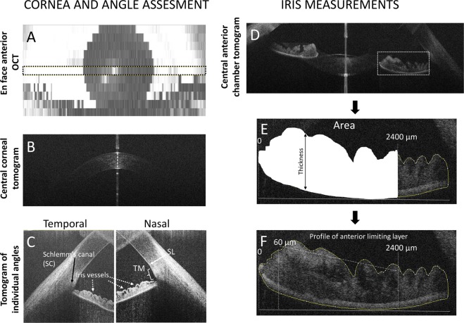
Methods: Anterior OCT was performed in all participants using a high-resolution HH SD-OCT device (Envisu 2300, Leica Microsystems, Germany) without anaesthesia or sedation.

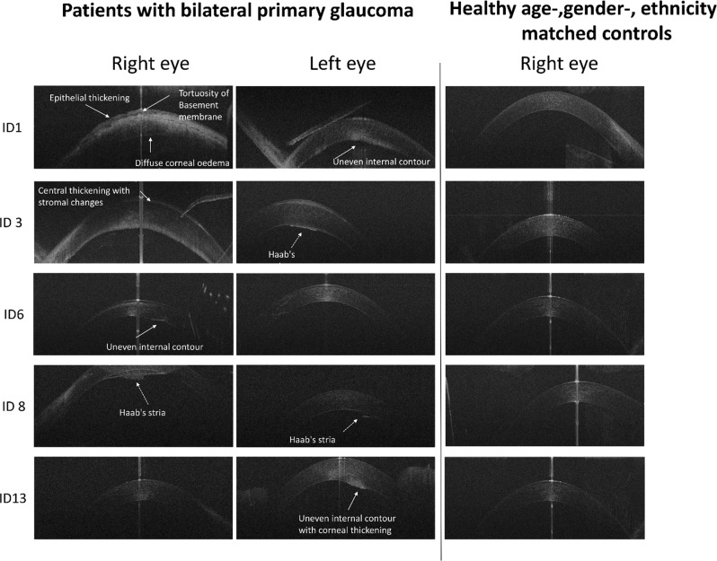
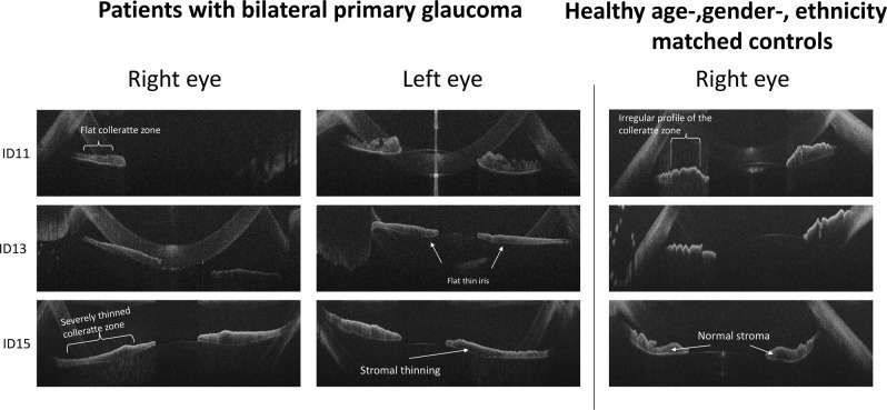
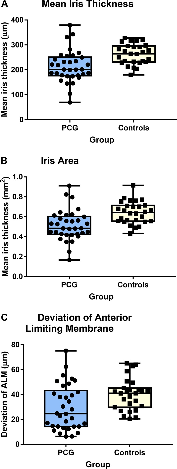
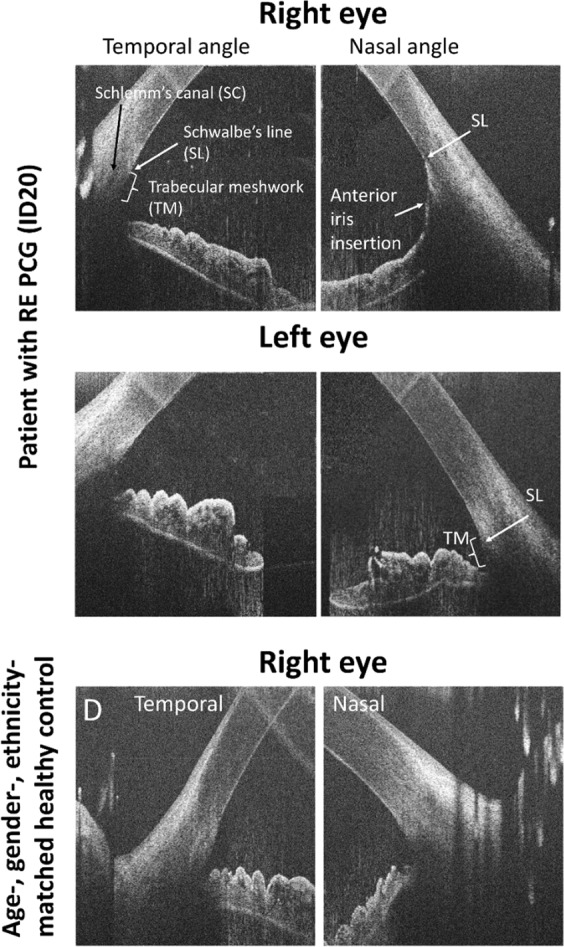
Results: Anterior HH-OCT in PCG visualised Haab's striae in 14.3%, uneven internal cornea in 9.5% and epithelial thickening in 11.9% of patients with central corneal thickening (CCT, p < 0.001). CCT was significantly correlated with the intraocular pressure (IOP, p < 0.001). The flat iris with a thin collarette zone was found in 59.5%, anterior iris insertion in 11.90% of eyes affected by PCG. Two independent examiners showed sensitivity and specificity of 87% and 77%, respectively, by instating iris thinning and flattening of the anterior profile.

Conclusions: Anterior HH-OCT has significant potential to improve diagnosis and management of PCG. Clinically relevant information can be obtained non-invasively and without sedation. High specificity makes anterior HH-OCT an important adjunct for management of PCG. Excellent visualisation of the iris insertion on OCT indicates potential for AS OCT to assist with surgical planning, including decision on the type of surgery and location of the incision.


Conflict of interest statement

The authors declare that they have no conflict of interest.

Figures

Fig. 1
Fig. 1
Optical coherence tomography (OCT) images of the anterior segment of the eye: a En face image showing the central position of the horizontal scan that was used for AS analysis (between dashed lines); b horizontal tomogram of the central cornea; c nasal and temporal high-resolution tomograms (1200 A scans and 12 B scans) indicating angle landmarks, including Schwalbe line (SL), trabecular meshwork (TM) and Schlemm’s canal (SC); d horizontal tomogram showing temporal and nasal irido-corneal angles; the iris area limited by anterior limiting membrane (ALM) and posterior epithelium (PE) was measured from the pupil to 2400 µm temporally from the pupil; the average iris thickness was measured between ALM and PE (e); standard deviation of the profile of ALM (f) was measured between 60 and 2400 µm temporally from the pupil
Fig. 2
Fig. 2
Corneal horizontal spectral domain–optical coherence tomography B-scan images of patients with PCG and healthy age-, gender- and ethnicity-matched controls. Patients with PCG showed a variety of changes, including epithelial thickening with a tortuous contour of the Bowman membrane (ID1, 6 and 13), uneven internal contour and the presence of Haab’s striae (ID3, 8)
Fig. 3
Fig. 3
Horizontal spectral domain–optical coherence tomography B-scan images showing iris changes in patients with PCG and healthy age-, gender- and ethnicity-matched controls
Fig. 4
Fig. 4
Distribution and mean ± SD of (a) iris thickness, (b) area and (c) deviation of the anterior limiting membrane in patients with primary congenital glaucoma (PCG) and healthy controls
Fig. 5
Fig. 5
Horizontal high-resolution spectral domain–optical coherence tomography B-scan images of the temporal and nasal irido-corneal angles in a patient with PCG in the right eye and a healthy age-, gender- and ethnicity-matched control. The image of the right affected eye of the patient shows abnormal anterior iris insertion in the nasal angle with the iris rout inserting at Schwalbe line (SL) covering the trabecular meshwork (TM). Normal configuration with a visible trabecular meshwork in the temporal angle of the affected eye, in the non-affected eye as well as in the control subject

References

    1. Tamm ER. [Development of the iridocorneal angle and congenital glaucoma] Der Ophthalmol: Z der Dtsch Ophthalmol Ges. 2011;108:610-4–616-7. doi: 10.1007/s00347-010-2294-5. - DOI - PubMed
    1. Ko F, Papadopoulos M, Khaw PT. Primary congenital glaucoma. In: Giacinto B, Carlo N editors. Progress in brain research, Chapter 9, Vol. 221. Elsevier; 2015. pp 177–89. - PubMed
    1. Todorova MG, Grieshaber MC, Camara RJ, Miny P, Palmowski-Wolfe AM. Anterior segment dysgenesis associated with Williams-Beuren syndrome: a case report and review of the literature. BMC Ophthalmol. 2014;14:70. doi: 10.1186/1471-2415-14-70. - DOI - PMC - PubMed
    1. Gould DB, John SWM. Anterior segment dysgenesis and the developmental glaucomas are complex traits. Hum Mol Genet. 2002;11:1185–93. doi: 10.1093/hmg/11.10.1185. - DOI - PubMed
    1. Hoguet A, Grajewski A, Hodapp E, Chang TC. A retrospective survey of childhood glaucoma prevalence according to Childhood Glaucoma Research Network classification. Indian J Ophthalmol. 2016;64:118–23. doi: 10.4103/0301-4738.179716. - DOI - PMC - PubMed

Publication types