Review
. 2019 May;4(5):734-747.
doi: 10.1038/s41564-019-0392-y. Epub 2019 Mar 18.

Antibody responses to viral infections: a structural perspective across three different enveloped viruses

Affiliations
Review

Antibody responses to viral infections: a structural perspective across three different enveloped viruses

Charles D Murin et al. Nat Microbiol. 2019 May.

Abstract

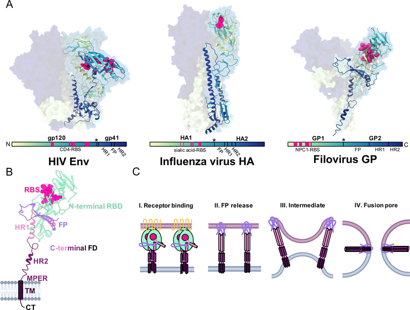
Antibodies serve as critical barriers to viral infection. Humoral immunity to a virus is achieved through the dual role of antibodies in communicating the presence of invading pathogens in infected cells to effector cells, and in interfering with processes essential to the viral life cycle (chiefly entry into the host cell). For individuals that successfully control infection, virus-elicited antibodies can provide lifelong surveillance and protection from future insults. One approach to understand the nature of a successful immune response has been to utilize structural biology to uncover the molecular details of antibodies derived from vaccines or natural infection and how they interact with their cognate microbial antigens. The ability to isolate antigen-specific B-cells and rapidly solve structures of functional, monoclonal antibodies in complex with viral glycoprotein surface antigens has greatly expanded our knowledge of the sites of vulnerability on viruses. In this Review, we compare the adaptive humoral immune responses to human immunodeficiency virus (HIV), influenza and filoviruses, with a particular focus on neutralizing antibodies. The pathogenesis of each of these viruses is quite different, providing an opportunity for comparison of immune responses: HIV causes a persistent, chronic infection; influenza, an acute infection with multiple exposures during a lifetime and annual vaccination; filoviruses, a virulent, acute infection. Neutralizing antibodies that develop under these different constraints are therefore sentinels that can provide insight into the underlying humoral immune responses, as well as important lessons to guide future development of vaccines and immunotherapeutics.


Figures

Figure 1.
Figure 1.. Points of antibody blockade to enveloped virus entry and egress.
Like a wrench thrown into the gears of a machine, antibodies are able to neutralize or protect against viruses by blocking one or more biological processes during viral entry or exit. The mechanism of neutralization depends largely on which epitope the antibody targets, and this subsequently determines which process is slowed or inhibited completely. For HIV, antibodies may block primary CD4 receptor (1) or secondary (2) CCR5/CXCR4 co-receptor binding. Filoviruses and influenza do not enter cells at the surface, but may be blocked by preventing attachment (3) or pinocytosis (4). For filoviruses, after endocytosis, antibodies must stay attached and survive acidification and subsequently prevent endosomal cleavage (5) or block endosomal receptor binding (6). For influenza, receptor binding can be blocked at the cell surface (7). In all cases, if receptor binding occurs, antibodies may directly block fusion through binding to fusion machinery. If viruses enter the cell, antibodies can potentially block egress of progeny virus (8). Non-neutralizing pathways to antibody-based protection include tagging cells for destruction by effector cells before viruses have a chance to exit (9), for example by antibody-dependent cellular cytotoxicity (ADCC) or by complement, or through agglutination of virions which can then be destroyed by effector cells (10). Additional abbreviations for human immunodeficiency virus 1, influenza virus and filovirus are HIV, flu and filov, respectively. This image was made using BioRender.
Figure 2.
Figure 2.. Antibody structure and domain topology.
When discussing antibodies in the context of viral immunity, we are generally referring to the IgG isotype, and particularly IgG1, which makes up the majority of serum antibodies and is primarily responsible for protection against infection. IgGs are heterodimers with two identical heavy chains (HC) and two identical light chains (LC) linked by disulfide bonds at a flexible hinge region that separates the Fab domains and Fc domain. The HC, named for its larger size, is a single polypeptide that contains four Ig domains, including the two Fab HC domains (VH and CH1), the hinge-region, and the Fc domain (CH2 and CH3), while the LC is composed of two LC Ig domains (VL and CL). The Fc region is responsible for linking antigens to effector cells and communicating their presence to the host through binding to FcγRs on effector cells, which exist in a variety of isotypes and are expressed at varying levels and compositions depending on the particular effector cell and activation state.
Figure 3.
Figure 3.. Shared structural features of type I glycoproteins.
A) The glycoproteins of HIV (PDB 5V7J), influenza (PDB 3KU3) and filoviruses (PDB 5JQ3) are widely divergent in sequence, size and shape but do share common structural features that are conserved in type I fusion mechanisms of enveloped virus entry. Here we show representative structures of glycoproteins from each of these viruses that are gradient colored from the N-terminus (light yellow) to the C-terminus (dark blue). This allows a comparison of where potential common sites of vulnerability exist on each of these structures in relation to divergent viruses, including the RBS, fusion loop, and stem. B) The sub-domain architecture of a type-I viral fusion protein is shown with this example of a GP1,2 subunit of Ebola virus GP (PDB 5JQ3). The N-terminal receptor binding domain (RBD, light green) is positioned above the C-terminal fusion domain (FD) and houses the receptor binding site (RBS, magenta). The FD contains a fusion peptide (FP, lavender), two heptad repeats (HR1 in pink and HR2 in purple), a hydrophobic membrane proximal external region (MPER, dark purple), and a transmembrane (TM, dark purple) anchor and C-terminal tail (CT, black). C) Type-I membrane fusion occurs in distinct stages. I) N-terminal RBDs bind to their cognate receptors, beginning the process of fusion. II) Receptor binding releases the FP, which pierces the host membrane and causes HR1 and HR2 to form an extended 3-helix bundle. III) During intermediate stages, it is thought that groups of viral spikes cluster to induce membrane buckling, allowing viral and host membranes to come close to each other. IV) Finally, HR2 collapses upon HR1, forming a 6-helix bundles that draws the TM domains together with the FPs, causing mixing of host and viral membranes and the formation of the fusion pore, which then permits the viral genome to exit into the host cytoplasm.
Figure 4.
Figure 4.. Examples of enveloped virus common and divergent sites of vulnerability targeted by neutralizing antibodies.
A) CD4 binding site (CD4bs) antibodies, such as VRC01 (PDB 5FYJ), bind in between HIV Env protomers using their HCs (top) and mimic the immunoglobulin fold (Ig) of the actual receptor (bottom). B) CDRH3 of F045–92 (PDB 4O58), and others like it, reach into the sialic acid binding pocket (top) and closely mimic the natural ligand (bottom). C) The antibody MR78 (PDB 5UQY) uses an extended hydrophobic CDRH3 (top) to bind to the NPC1 receptor binding site (RBS). D) The fusion peptide (FP) of Env sits near the base and is contacted largely by the HC of VRC34.01 (PDB 5I8H). E) The HA FP also sits near the base on the HA stalk and similarly is contacted largely by the HC of MEDI8852 (PDB 5JW3). F) For ADI-15878 (PDB 6DZL), contacts are made across HR1 with the HC and the FP is mostly contact by the LC. G) The HIV antibody 10E8 (Env PDB 5V7J and 10E8 PDB 5T85 fit into EMDB 3312) has evolved to partially contact portions of the viral lipid membrane. H) HA stalk antibodies typically have the broadest neutralizing paratopes and CR9114 (PDB 4FQI) contacts large portions of the extended HA2 alpha-helix with its HC. I) bNab ADI-16061 (GP PDB 5JQ3 and example Fab PDB 5HJ3 fit into EMD 8698) binds far below the base of GP, contacting conserved hydropbobic residues within HR2 and the MPER. J) The long-term exposure of the immune system to HIV allows for extensive SHM and antibody evolution, producing antibodies like PGT145 (PDB 5V8L), with an extended CDRH3 that is rigidified by a beta-hairpin structure with hydrophobic residues and sulfated tyrosines. This allows the antibody to reach deep into the apex pocket of Env. K) Ebolaviruses have two glycoproteins, the viral GP trimer and the soluble GP (sGP) dimer, which is expressed in large abundance during infection and is thought to be a type of immune decoy. The first 296 amino acids of GP and sGP are shared and the protective antibody 13C6 binds to both GP (left, PDB 5KEL) and sGP (right, PDB 5KEN) near a highly conserved residue (W275).
Figure 5.
Figure 5.. The immunogenic landscape of enveloped viruses illuminated by structural biology.
Overlaying low-pass filtered structures of selected nAbs bound to trimeric glycoproteins from A, B) HIV (blues and purples), C, D) influenza (reds and oranges) and E, F) filoviruses (yellows and greens) reveals the immunogenic landscape of these viral glycoproteins and the 90 degrees of approach angles that antibodies can utilize to probe the glycoprotein surface. Abs bound to a single protomer are shown for clarity. Stripping back these antibodies clarifies how these epitopes are focused into distinct regions of vulnerability, although nearly the entire surface is susceptible to nAb binding. Several of these sites of vulnerability are equivalent across these examples, including the apex, RBS, interface of RBS and fusion domains, FP and viral stem, which includes HR1, HR2 and the MPER domains.

References

    1. White JM & Whittaker GR Fusion of Enveloped Viruses in Endosomes. Traffic 17, 593–614, doi:10.1111/tra.12389 (2016). - DOI - PMC - PubMed
    1. Harrison SC Viral membrane fusion. Virology 479–480, 498–507, doi:10.1016/j.virol.2015.03.043 (2015). - DOI - PMC - PubMed
    1. Harris LJ, Larson SB, Hasel KW & McPherson A. Refined structure of an intact IgG2a monoclonal antibody. Biochemistry 36, 1581–1597, doi:10.1021/bi962514+ (1997). - DOI - PubMed
    1. Harris LJ, Skaletsky E. & McPherson A. Crystallographic structure of an intact IgG1 monoclonal antibody. J Mol Biol 275, 861–872, doi:10.1006/jmbi.1997.1508 (1998). - DOI - PubMed
    1. Saphire EO et al. Crystal structure of a neutralizing human IGG against HIV-1: a template for vaccine design. Science 293, 1155–1159, doi:10.1126/science.1061692 (2001). - DOI - PubMed

Publication types

Substances