. 2019 Aug;18(8):883-891.
doi: 10.1038/s41563-019-0307-6. Epub 2019 Mar 18.

Local nascent protein deposition and remodelling guide mesenchymal stromal cell mechanosensing and fate in three-dimensional hydrogels

Affiliations

Local nascent protein deposition and remodelling guide mesenchymal stromal cell mechanosensing and fate in three-dimensional hydrogels

Claudia Loebel et al. Nat Mater. 2019 Aug.

Abstract

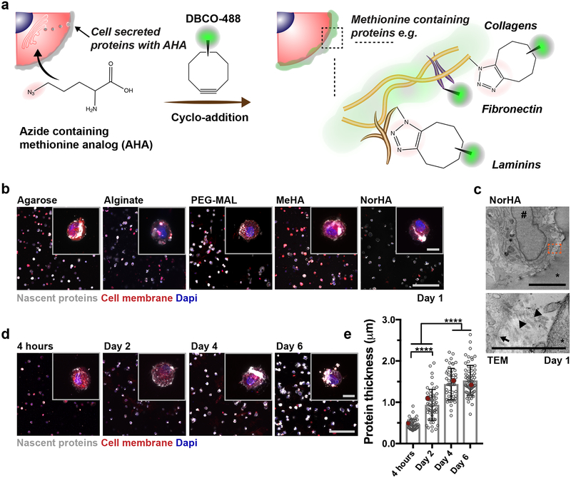
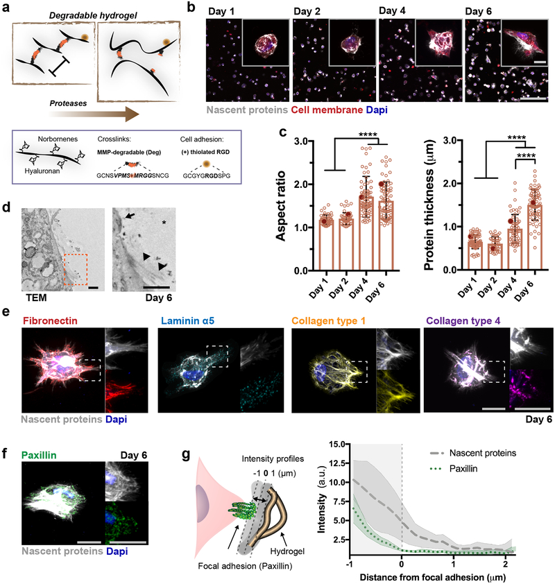
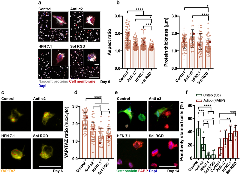
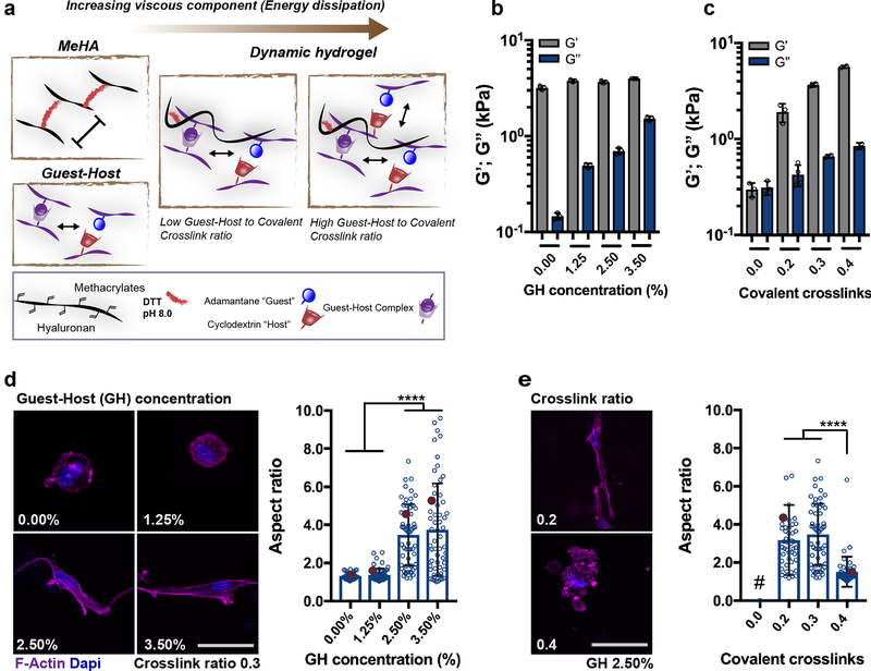
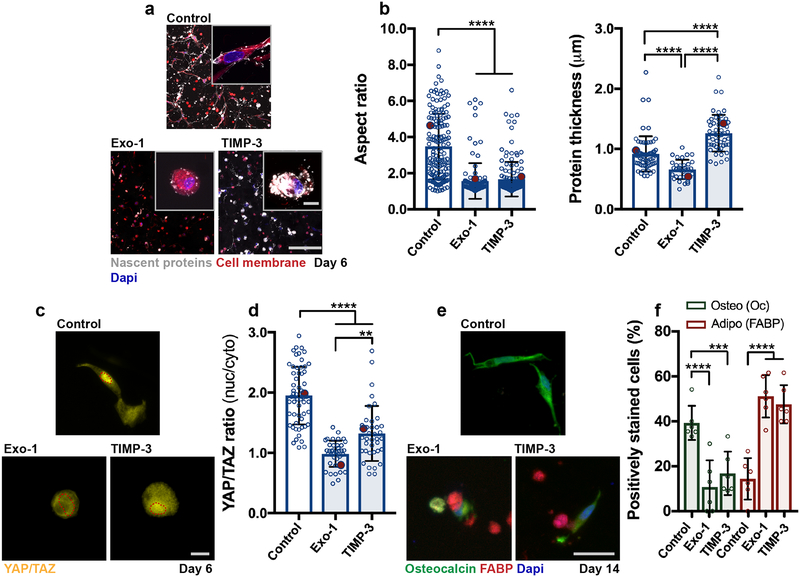
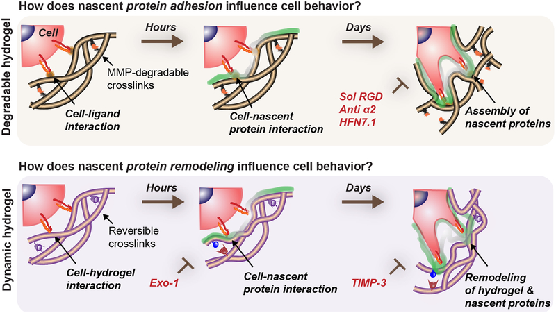
Hydrogels serve as valuable tools for studying cell-extracellular matrix interactions in three-dimensional environments that recapitulate aspects of native extracellular matrix. However, the impact of early protein deposition on cell behaviour within hydrogels has largely been overlooked. Using a bio-orthogonal labelling technique, we visualized nascent proteins within a day of culture across a range of hydrogels. In two engineered hydrogels of interest in three-dimensional mechanobiology studies-proteolytically degradable covalently crosslinked hyaluronic acid and dynamic viscoelastic hyaluronic acid hydrogels-mesenchymal stromal cell spreading, YAP/TAZ nuclear translocation and osteogenic differentiation were observed with culture. However, inhibition of cellular adhesion to nascent proteins or reduction in nascent protein remodelling reduced mesenchymal stromal cell spreading and nuclear translocation of YAP/TAZ, resulting in a shift towards adipogenic differentiation. Our findings emphasize the role of nascent proteins in the cellular perception of engineered materials and have implications for in vitro cell signalling studies and application to tissue repair.


Conflict of interest statement

Competing Financial Interests

The authors declare no competing financial interests.

Figures

Figure 1
Figure 1. Nascent protein deposition by encapsulated hMSCs occurs early, independent of hydrogel type.
a Schematic of nascent extracellular protein labeling. The methionine analog azidohomoalanine (AHA) is added to the culture media and incorporated into nascent proteins (e.g., fibronectin, collagens, laminins). The bio-orthogonal Cu(I)-free strain-promoted cyclo-addition between the azide and DBCO-modified fluorophore (DBCO-488) enables visualization of the nascent proteins. b Representative images of nascent proteins (white) deposited by hMSCs encapsulated in various hydrogels (alginate, agarose, maleimide modified poly(ethylene glycol) (PEG-MAL), methacrylated hyaluronic acid (MeHA), norbornene modified hyaluronic acid (NorHA)), E = ~9 kPa, scale bar 200 μm, inset 20 μm). c Representative transmission electron microscopy (TEM, * hydrogel, # nucleus; scale bar 5 μm left, 1 μm right) image of encapsulated hMSC after 1 day (24 h in culture). Orange box in top image indicates magnification in bottom image (arrow indicates cell membrane and arrowheads show collagen fibrils). d Representative images of nascent proteins (white) deposited by hMSCs encapsulated in non-degradable NorHA hydrogels (9.0 ± 0.7 kPa, mean ± SD, n = 3 independent measurements) and cultured in growth media (supplemented with AHA) up to 6 days (see Supplementary Figure 3 for daily changes up to 14 days, scale bar 200 μm, inset 20 μm). e Quantification of the accumulated nascent protein thickness deposited by hMSCs encapsulated in non-degradable NorHA hydrogels (n = 40 cells (4 hours), 55 cells (Day 2, Day 4) and 70 cells (Day 6) from 3 biologically independent experiments, mean ± SD, **** p ≤ 0.0001, one-way ANOVA with Bonferroni post hoc, red dots indicate measurements for magnified representative images in d).
Figure 2
Figure 2. Nascent ECM proteins create an adhesive layer at the cell-hydrogel interface.
a Schematic illustrating norbornene-modified hyaluronic acid (NorHA) hydrogels crosslinked via a thiol-ene reaction with MMP-degradable dithiol peptide crosslinkers and incorporating RGD for adhesion. b Representative images of nascent proteins (white, visualized via fluorescent DBCO labeling) deposited by hMSCs encapsulated in degradable NorHA hydrogels (9.0 ± 0.7 kPa, mean ± SD, n = 3 independent measurements) and cultured in growth media for up to 6 days (see Supplementary Figure 5 for daily changes up to 14 days, scale bar 200 μm, inset 20 μm). c Quantification of the accumulated nascent protein thickness deposited by hMSCs encapsulated in degradable NorHA hydrogels (n = 40 cells (4 hours), 68 cells (day 1, day 2, day 4) and 72 cells (day 6) from 3 biologically independent experiments), mean ± SD, **** p ≤ 0.0001, one-way ANOVA with Bonferroni post hoc, red dots indicate measurements for magnified representative images in b). d Representative transmission electron microscopy (TEM, * hydrogel) images of encapsulated hMSC after 6 days (scale bar 0.5 μm). Orange box in left image indicates magnification in right image (arrow indicates cell membrane and arrowheads show randomly aligned collagen fibrils). e Representative images (magnifications on right) of nascent proteins and fibronectin, laminin α5 and collagen type 1 and type 4 at 6 days (scale bars 20 μm). f Representative image (magnifications of single channels on right) of accumulated nascent proteins and focal adhesions stained for paxillin (scale bars 20 μm). g Schematic (left) illustrating the region used to generate intensity profiles (right) emanating from single FAs (representative image in f), n = 50 adhesions from 10 individual cells (2 biologically independent experiments), lines show median intensity profile, shaded areas demonstrate 95% confidence interval).
Figure 3
Figure 3. Adhesion to nascent proteins controls hMSC mechanosensing in degradable hydrogels.
a Representative images (scale bar 200 μm, inset 20 μm) of nascent protein deposition in MMP-degradable NorHA hydrogels after 6 days treatment without (control, note same image as in Fig. 2b), with monoclonal antibodies against integrin alpha 2 (anti α2, 20 μg/mL) or human fibronectin (HFN7.1, 5 μg/mL) or with soluble RGD (sol RGD, 0.5 mM). b Quantification of cell aspect ratio and accumulated nascent protein thickness deposited by hMSCs encapsulated in degradable NorHA hydrogels (n = 133 cells (control), n = 155 cells (anti α2), and n = 152 cells (HFN7.1), n = 124 cells (sol RGD) from 2 biologically independent experiments), mean ± SD, **** p ≤ 0.0001, *** p ≤ 0.001, * p ≤ 0.05, one-way ANOVA with Bonferroni post hoc, red dots indicate measurements for magnified images in a). c Representative images and d quantification of nuclear/cytoplasmic (nuc/cyto) YAP/TAZ ratios of hMSCs encapsulated in degradable NorHA hydrogels, cultured for 6 days in adipogenic-osteogenic media (scale bar 50 μm), quantifications: n = 60 cells (control), n = 51 cells (anti α2), and n = 40 cells (HFN7.1), n = 39 cells (sol RGD) from 2 biologically independent experiments), mean ± SD, **** p ≤ 0.0001, * p ≤ 0.05, one-way ANOVA with Bonferroni post hoc, red dots indicate measurements for magnified images in c). e Immunostaining for fatty-acid binding protein (FABP, adipogenic marker) and osteocalcin (Oc, osteogenic marker) after 14 days in adipogenic-osteogenic media (scale bar 50 μm). f Quantification of positively stained cells (percentage,%) towards osteogenesis (Oc positive) and adipogenesis (FABP positive) after 14 days, n = 6 samples from 2 biologically independent experiments), mean ± SD, **** p ≤ 0.0001, *** p ≤ 0.001, ** p ≤ 0.01, * p ≤ 0.05, two-way ANOVA with Bonferroni post hoc).
Figure 4
Figure 4. Dynamic hydrogel composition modulates viscoelastic properties and cell spreading.
a Schematic of guest-host double-network (DN) hydrogel formation, from the combination of (i) covalently crosslinked HA hydrogel (MeHA) without RGD peptide and crosslinked with 1,4-dithiothreitol (DTT) and (ii) guest-host (GH) hydrogel assembled through mixing of HA modified with cyclodextrin (host) or adamantane (guest). Rheological measurements (1 Hz, 0.5% strain) of storage modulus (G’, elastic component) and loss modulus (G”, viscous component) for DN hydrogels with b increasing GH polymer concentration at a given covalent crosslink ratio (0.3) and c increasing covalent crosslinking (ratio of thiols to methacrylates) at a given GH polymer concentration (2.50%, n = 3 independent measurements per group, mean ± SD). d Representative images of F-Actin immunostaining and quantification of aspect ratio of hMSCs encapsulated in DN hydrogels (G’ 3.5 ± 0.45 kPa, n = 3 independent measurements per group, mean ± SD) with different GH concentration but same covalent crosslinking ratio (0.3) (scale bar 20 μm), quantifications: n = 34 cells (0.00%), n = 58 cells (1.25%, 2.50%) and n = 56 cells (3.50%) from 2 biologically independent experiments), mean ± SD, **** p ≤ 0.0001, one-way ANOVA with Bonferroni post hoc, red dots indicate measurements for images on the left. e Representative images of F-Actin immunostaining and quantification of aspect ratio of hMSCs encapsulated in DN hydrogels with different covalent crosslinking ratios but same GH polymer concentration (2.50%). Note that image of 0.3 corresponds to 2.50% in d) (scale bar 20 μm), quantifications: n = 46 cells (0.2) and n = 49 (0.4) from 2 biologically independent experiments), mean ± SD, **** p ≤ 0.0001, one-way ANOVA with Bonferroni post hoc, red dots indicate measurements for images on the left, # GH only hydrogels (i.e. no covalent crosslinks) were not stable over 6 days in culture).
Figure 5
Figure 5. Nascent protein remodeling is required for cell spreading and osteogenesis in dynamic hydrogels.
a Representative images of nascent protein deposition of hMSCs encapsulated in DN hydrogels (G’ 3.6 ± 0.1 kPa, G” 0.7 ± 0.05 kPa, n = 3 independent measurements, mean ± SD) treated with an inhibitor of exocytosis and vesicular trafficking Exo-1 (120 nM) and a recombinant tissue inhibitor of metalloproteinases-3 (TIMP-3, 5 nM encapsulated) during 6 days in growth media (scale bar 200 μm, insets 20 μm). b Quantification of cell aspect ratio and accumulated nascent protein thickness deposited by hMSCs encapsulated in dynamic hydrogels (aspect ratio: n = 161 cells (control), n = 150 cells (TIMP-3), and n = 122 cells (Exo-1), protein thickness: n = 76 cells (control), n = 61 cells (TIMP-3), and n = 55 cells (Exo-1), from 3 biologically independent experiments), mean ± SD, **** p ≤ 0.0001, one-way ANOVA with Bonferroni post hoc, red dots indicate measurements for magnified images in a). c Representative images and d quantification of nuclear/cytoplasmic (nuc/cyto) YAP/TAZ ratios of hMSCs encapsulated in dynamic hydrogels and cultured for 6 days in adipogenic-osteogenic media (scale bar 50 μm, n = 59 cells (Control) and n = 39 cells (TIMP-3, Exo-1), from 2 biologically independent experiments), mean ± SD, **** p ≤ 0.0001, ** p ≤ 0.01, one-way ANOVA with Bonferroni post hoc, red dots indicate measurements for images in c). e Immunostaining for fatty-acid binding protein (FABP, adipogenic marker) and osteocalcin (Oc, osteogenic marker) after 14 days in adipogenic-osteogenic media (scale bar 50 μm). f Quantification of positively stained cells (percentage,%) towards osteogenesis (Oc positive) and adipogenesis (FABP positive) after 14 days, n = 6 samples from 2 biologically independent experiments), mean ± SD, **** p ≤ 0.0001, *** p ≤ 0.001, two-way ANOVA with Bonferroni post hoc).
Figure 6
Figure 6. Nascent protein adhesion and remodeling enhance cell spreading in degradable/dynamic hydrogels.
Cells interact with a 3D hydrogel and presented ligands for a short time period before depositing nascent proteins to form a pericellular matrix. The hydrogel properties determine if encapsulated cells can spread, but adhesion and active remodeling of the nascent proteins are required for spreading. For example, in hydrogels that cells can locally degrade (e.g., protease-sensitive crosslinkers), nascent proteins guide cell behavior as an assembled interfacial layer that cells adhere to. Perturbation of cell-ECM adhesion through inhibiting specific cell-nascent protein interactions (e.g., sol RGD, anti α2, HFN7.1) inhibits spreading and decreases its downstream cellular outcomes (YAP/TAZ nuclear translocation, osteogenic differentiation). Similarly, in dynamic microenvironments (e.g., viscoelastic hydrogels) where spreading is protease-independent, nascent protein deposition and remodeling are needed for mechanosensing (YAP/TAZ nuclear translocation, osteogenic differentiation) and are blocked by inhibiting nascent protein secretion (Exo-1) and remodeling (TIMP-3).

References

    1. Kim SH, Turnbull J & Guimond S Extracellular matrix and cell signalling: the dynamic cooperation of integrin, proteoglycan and growth factor receptor. J Endocrinol 209, 139–151, (2011). - PubMed
    1. Guvendiren M & Burdick JA Engineering synthetic hydrogel microenvironments to instruct stem cells. Curr Opin Biotechnol 24, 841–846, (2013). - PMC - PubMed
    1. Tibbitt MW & Anseth KS Hydrogels as extracellular matrix mimics for 3D cell culture. Biotechnol Bioeng 103, 655–663, (2009). - PMC - PubMed
    1. Drury JL & Mooney DJ Hydrogels for tissue engineering: scaffold design variables and applications. Biomaterials 24, 4337–4351, (2003). - PubMed
    1. Wells RG The role of matrix stiffness in regulating cell behavior. Hepatology 47, 1394–1400, (2008). - PubMed

Publication types