. 2019 Apr;26(4):322-330.
doi: 10.1038/s41594-019-0200-7. Epub 2019 Mar 18.

RNA structure maps across mammalian cellular compartments

Affiliations

RNA structure maps across mammalian cellular compartments

Lei Sun et al. Nat Struct Mol Biol. 2019 Apr.

Abstract

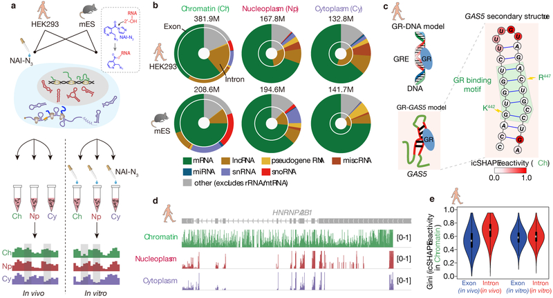
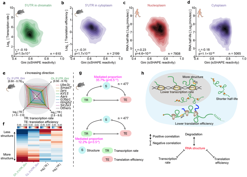
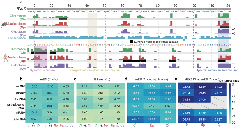
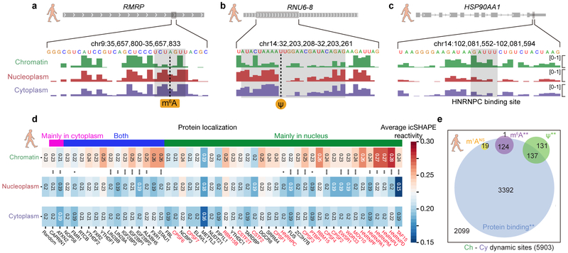
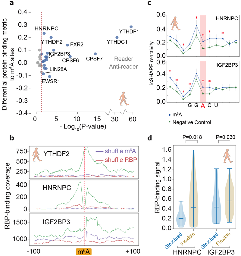
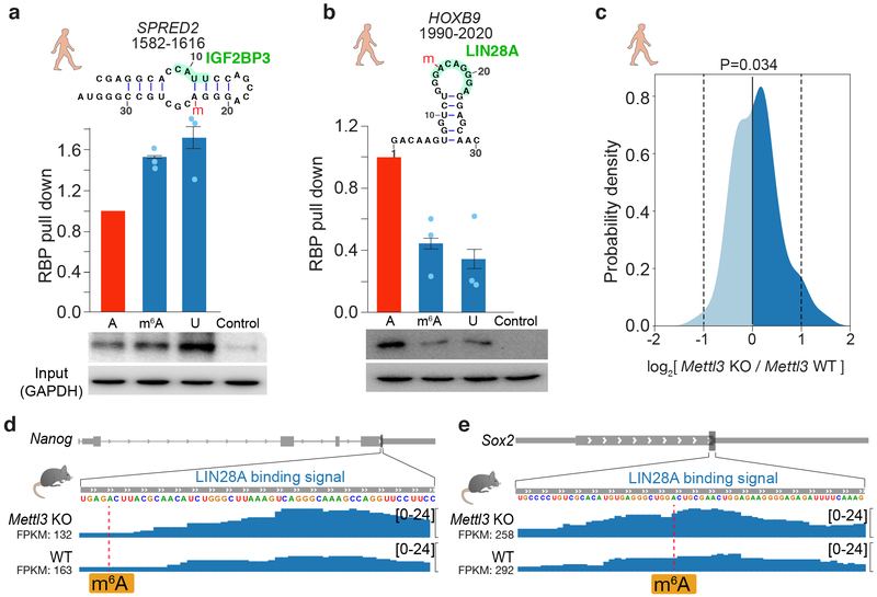
RNA structure is intimately connected to each step of gene expression. Recent advances have enabled transcriptome-wide maps of RNA secondary structure, called 'RNA structuromes'. However, previous whole-cell analyses lacked the resolution to unravel the landscape and also the regulatory mechanisms of RNA structural changes across subcellular compartments. Here we reveal the RNA structuromes in three compartments, chromatin, nucleoplasm and cytoplasm, in human and mouse cells. The cytotopic structuromes substantially expand RNA structural information and enable detailed investigation of the central role of RNA structure in linking transcription, translation and RNA decay. We develop a resource with which to visualize the interplay of RNA-protein interactions, RNA modifications and RNA structure and predict both direct and indirect reader proteins of RNA modifications. We also validate a novel role for the RNA-binding protein LIN28A as an N6-methyladenosine modification 'anti-reader'. Our results highlight the dynamic nature of RNA structures and its functional importance in gene regulation.


Figures

Fig 1 ∣
Fig 1 ∣. Chromatin fractions are enriched for pre-mRNA and lncRNA structures.
a, Experimental overview of the icSHAPE protocol. The dashed box highlights the chemical structure of NAI-N3 and its covalent bond with the 2'-OH group of RNA, which allows probing of RNA structures inside living cells. b, Donut charts showing read distributions of different RNA types in the three cellular compartments. The outer circles represent exon coverage while the inner circles represent intron coverage. c, GAS5 RNA secondary structure with icSHAPE reactivity scores shown in color. The nucleotides outlined in red interact with GR amino acids, shown in blue. d, UCSC tracks showing icSHAPE reactivity scores (y-axis), along the RNA sequence. 1 denotes unstructured (single-stranded) regions, and 0 denotes fully-structured regions. e, Violin plot of Gini index of icSHAPE data in exon versus in intron. The thick black bar in the center of the Violin plot represents the interquartile range, the thin black line extended from it represents the 95% confidence intervals, and the white dot is the median. The numbers of sliding windows (width = 20nt) on the respective regions from the left to the right are n=18930, n=5926, n=51409, n=82648.
Fig 2 ∣
Fig 2 ∣. RNA structure plays a central role in connecting transcription, translation and RNA degradation.
a-d, Scatter plots of (a) transcription rate versus 5'UTR RNA structure in chromatin, (b) translational efficiency versus 5'UTR RNA structure in cytoplasm, (c) RNA half-life versus full-length-transcript RNA structure in nucleoplasm, and (d) RNA half-life versus RNA structure in cytoplasm. The 2-tailed p-value was calculated by python package function scipy.stats.pearsonr. rp is the Pearson correlation efficient. e, Radar diagram showing 5'UTR RNA structure in chromatin, 5'UTR RNA structure in the cytoplasm, transcription rate, and translational efficiency. Grey lines show all genes, and the colored lines highlight representative transcripts. f, Heatmap of 5'UTR RNA structure in chromatin, 5'UTR RNA structure in cytoplasm, transcription rate, and translational efficiency. Each strip represents an average of a bin comprising 5% data, ranked by RNA-structure reactivity in the chromatin fraction, 477 common transcripts are shown. g, Mediator model (above) and cofounding model (bottom) of RNA structure in connecting transcription rate and translation efficiency. P-values were calculated by two-sided t-test. h, Schematic showing RNA structure connects transcription, translation and RNA degradation.
Fig 3 ∣
Fig 3 ∣. RNA structure differences in cellular context.
a, U12 small nuclear RNA (snRNA) structural change across cellular compartments, and the structural divergence in two species. Tracks show the icSHAPE score plotted along the RNA sequence. The black bars highlight RNA structural change regions. b-e, Heatmaps showing fractions of structurally-different regions across cellular compartments (b) in vivo, (c) in vitro, (d) between in vivo and in vitro, and (e), between human and mouse. Dashed lines represent insufficient data.
Fig 4 ∣
Fig 4 ∣. RNA modification and RBP binding underlie RNA structural changes.
a-c, RNA structural change at (a) an m6A-modified site, (b) a Ψ-modified site, and (c) an HNRNPC-binding site. Tracks show the icSHAPE score plotted along the RNA sequence. d, Heatmap of average icSHAPE scores in RBP binding regions in different cellular compartments, ranked by increasing structural change (from left to right) between the chromatin and the nucleoplasmic fractions. Proteins are annotated by their known localizations, with chromatin-associated RBPs shown in red. P-values were calculated by single-sided Mann-Whitney U test and corrected by the Bonferroni method. Source data for panel d are available online. e, The number and overlap of different types of RNA modification sites and RBP binding sites in regions with RNA structural change. P-values were calculated by a permutation test for 1,000 times. * p-value < 0.05; ** p-value < 1e-3; *** p-value < 1e-5.
Fig 5 ∣
Fig 5 ∣. Structural analysis dissects different types of m6A readers.
a, Differential RBP binding to m6A sites and control sites containing an m6A motif. P-values are calculated to show the statistical significance of the binding differences by single-sided Mann-Whitney U test and corrected by the Benjamini/Hochberg method. Source data for panel a are available online. b, Metagene profiles of protein binding in m6A-flanking regions. c, Metagene profiles showing that RNA structures are different between known m6A-modified sites and unmodified sites (negative control), at m6A motifs overlapping a binding site of IGF2BP3 and HNRNPC. P-values were calculated by single-sided Mann-Whitney U test, red asterisks, p-values less than 0.01. The error bars represent the standard error of the mean. The numbers of HNRNPC and IGF2BP3 binding regions are 86 and 56, respectively. d, Violin plots of RBP-binding strengths of HNRNPC and IGF2BP3 in structured and flexible regions containing a m6A motif. Structured and flexible regions are defined as the RBP-binding regions at the bottom 30% or top 30% of average icSHAPE scores, respectively. P-values were calculated by single-sided Mann-Whitney U test. The numbers of HNRNPC and IGF2BP3 binding regions in comparison are 137 and 320, respectively.
Fig 6 ∣
Fig 6 ∣. Validation of IGF2BP3 as an indirect m6A reader and LIN28A as an anti-reader.
a-b, RNA pull-down assays and western blots for (a) IGF2BP3 and (b) LIN28A, using RNA probes that contain unmodified A, m6A, and U, respectively, derived from the indicated positions in the transcripts. m6A sites are marked with a red “m”. Histograms show mean of RNA pull-down from three independent replicates. The error bars represent standard error of mean (s.e.m.).Uncropped blots are shown in Supplementary Data Set 1. c. Density plot of LIN28A binding strength (log ratio) at m6A sites in Mettl3 knockout (KO) versus wild-type mES cells. P-value is calculated by two-sided t-test. The number of transcripts is 145. d-e, Signal tracks of Nanog and Sox2 showing LIN28A binding at specific loci in Mettl3 KO and wildtype mES cells.

Similar articles

Cited by

References

    1. Sharp PA The centrality of RNA. Cell 136, 577–580, doi:10.1016/j.cell.2009.02.007 (2009). - DOI - PubMed
    1. Pan T & Sosnick T RNA folding during transcription. Annu Rev Biophys Biomol Struct 35, 161–175, doi:10.1146/annurev.biophys.35.040405.102053 (2006). - DOI - PubMed
    1. Warf MB & Berglund JA Role of RNA structure in regulating pre-mRNA splicing. Trends in biochemical sciences 35, 169–178, doi:10.1016/j.tibs.2009.10.004 (2010). - DOI - PMC - PubMed
    1. Martin KC & Ephrussi A mRNA localization: gene expression in the spatial dimension. Cell 136, 719–730, doi:10.1016/j.cell.2009.01.044 (2009). - DOI - PMC - PubMed
    1. Kozak M Regulation of translation via mRNA structure in prokaryotes and eukaryotes. Gene 361, 13–37, doi:10.1016/j.gene.2005.06.037 (2005). - DOI - PubMed

Publication types