. 2019 May;22(5):828-839.
doi: 10.1038/s41593-019-0358-7. Epub 2019 Mar 18.

Multiplexed peroxidase-based electron microscopy labeling enables simultaneous visualization of multiple cell types

Affiliations

Multiplexed peroxidase-based electron microscopy labeling enables simultaneous visualization of multiple cell types

Qiyu Zhang et al. Nat Neurosci. 2019 May.

Abstract

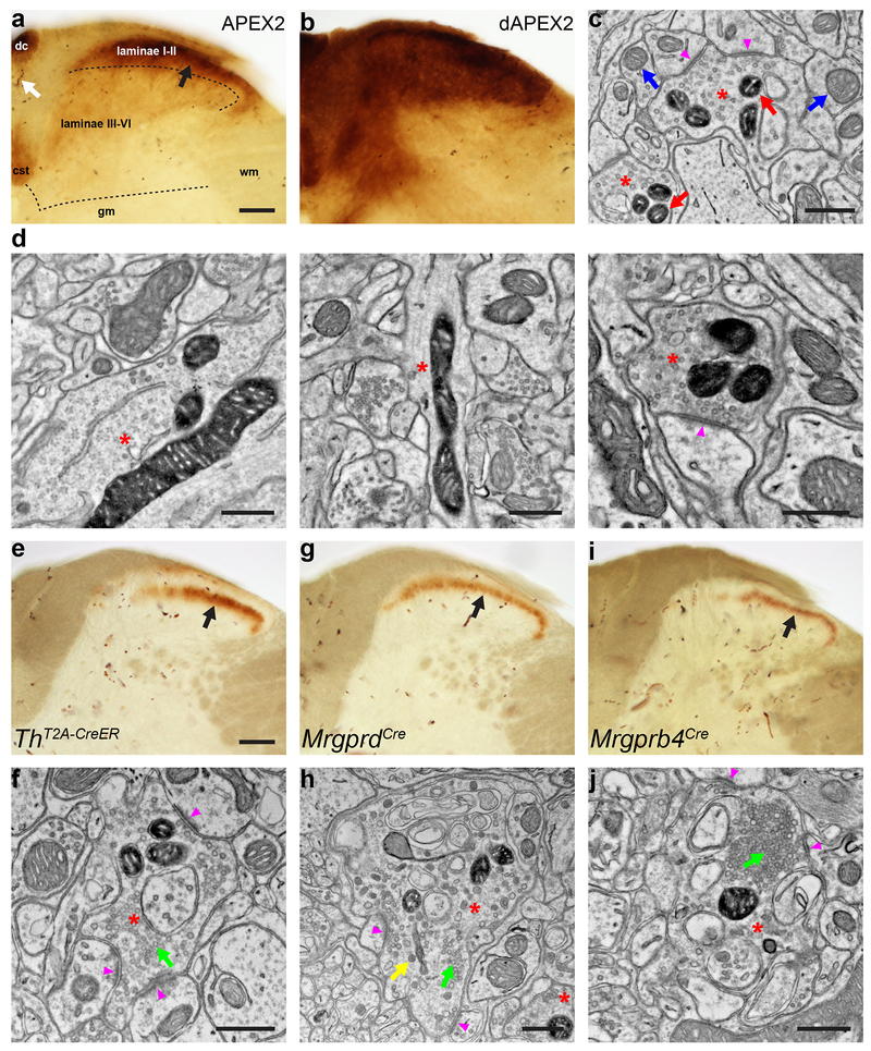
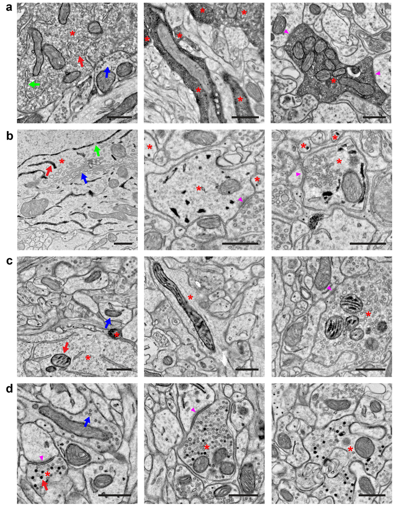
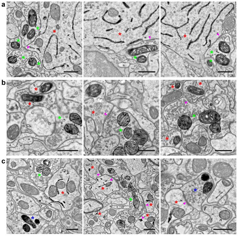
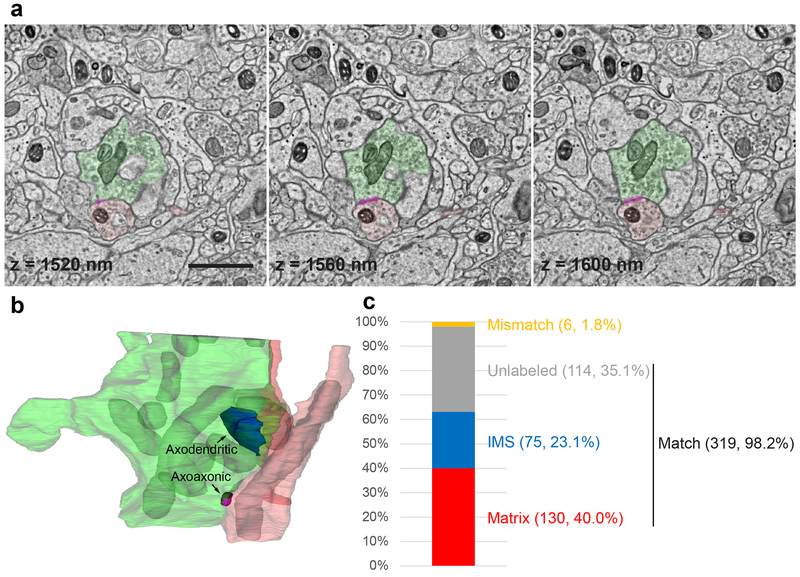
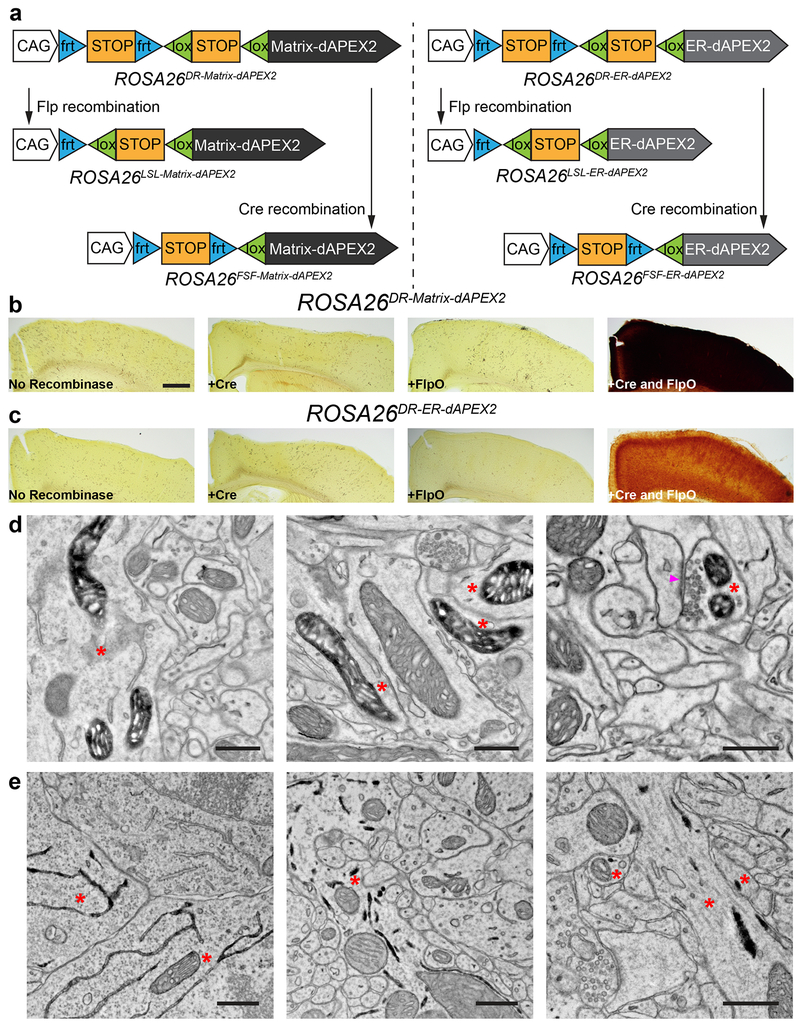
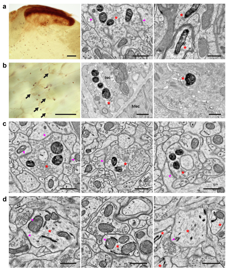
Electron microscopy (EM) is a powerful tool for circuit mapping, but identifying specific cell types in EM datasets remains a major challenge. Here we describe a technique enabling simultaneous visualization of multiple genetically identified neuronal populations so that synaptic interactions between them can be unequivocally defined. We present 15 adeno-associated virus constructs and 6 mouse reporter lines for multiplexed EM labeling in the mammalian nervous system. These reporters feature dAPEX2, which exhibits dramatically improved signal compared with previously described ascorbate peroxidases. By targeting this enhanced peroxidase to different subcellular compartments, multiple orthogonal reporters can be simultaneously visualized and distinguished under EM using a protocol compatible with existing EM pipelines. Proof-of-principle double and triple EM labeling experiments demonstrated synaptic connections between primary afferents, descending cortical inputs, and inhibitory interneurons in the spinal cord dorsal horn. Our multiplexed peroxidase-based EM labeling system should therefore greatly facilitate analysis of connectivity in the nervous system.


Conflict of interest statement

Competing Interests

The authors declare no competing interests.

Figures

Figure 1.
Figure 1.. dAPEX2 is a sensitive reporter for visualizing axons of long projection neurons with EM
(a) LM image of the spinal cord dorsal horn after systemic transduction of AAV9-mito-V5-APEX2. Black arrow: staining from mito-V5-APEX2. Laminae I-II has much stronger staining than laminae III-VI. White arrow: endogenous peroxidase activity from erythrocytes. dc: dorsal column, cst: corticospinal tract, gm: grey matter, wm: white matter. n = 4 animals and experiments. (b) LM image of the spinal cord dorsal horn after systemic transduction of AAV9-Matrix-dAPEX2. Note the robust staining in laminae III-VI with Matrix-dAPEX2 and generally increased staining levels compared to mito-V5-APEX2. n = 6 animals and experiments. (c) EM image of the spinal cord dorsal horn after systemic transduction of AAV9-Matrix-dAPEX2. Asterisks: labeled axon terminals. Red arrows: labeled mitochondria. Blue arrows: unlabeled mitochondria. Arrowheads: synapses made by the labeled axon terminals. Note that ultrastructural details of the labeled neurons are not obscured by staining products. n = 3 animals and experiments. (d) EM images of a cortical layer 5 neuron labeled using Tg(Rbp4-Cre)KL100 and AAV1-DIO-Matrix-dAPEX2 (asterisks). (Left) Soma of a labeled neuron. (Middle) Dendrite of a labeled neuron. (Right) Corticospinal axon in the spinal cord dorsal horn of a cortical layer 5 neuron labeled using Tg(Rbp4-Cre)KL100. Arrowhead: synapse made by the labeled neuron. n = 2 animals and experiments. (e) LM image of spinal cord dorsal horn from a ThT2A-CreER animal transduced with AAV9-DIO-Matrix-dAPEX2 and treated with tamoxifen from P14-21 to label C-LTMRs. Arrow: staining from labeled C-LTMR axons. n = 6 animals and experiments. (f) EM image from the same animal in e. Asterisk: labeled C-LTMR axon terminal with round, clear vesicles (green arrow). Most C-LTMR axon terminals have only clear vesicles. Arrowheads: synapses made by the labeled C-LTMR terminal. n = 4 animals and experiments. (g) LM image of spinal cord dorsal horn from an MrgprdCre animal transduced with AAV9-DIO-Matrix-dAPEX2. Arrow: staining from labeled Mrgprd+ polymodal nociceptor afferents. n = 4 animals and experiments. (h) EM image from the same animal in g. Asterisk: labeled Mrgprd+ polymodal nociceptor axon terminal. Unlike C-LTMRs, these neurons can exhibit both round, clear vesicles (green arrow) and large, variably shaped dense-core vesicles (yellow arrow). Arrowheads: synapses made by the labeled Mrgprd+ polymodal nociceptor axon terminal. n = 3 animals and experiments. (i) LM image of spinal cord dorsal horn from an Mrgprb4Cre animal transduced with AAV9-DIO-Matrix-dAPEX2. Arrow: staining from labeled Mrgprb4+ afferents. n = 4 animals and experiments. (j) EM image from the same animal in i. Asterisk: labeled Mrgprb4+ afferent axon terminal with a dense cluster of vesicles (green arrow), a configuration rarely seen in C-LTMRs, but often present in both Mrgprb4+ and Mrgprd+ afferents. Arrowheads: synapses made by the labeled Mrgprb4+ afferent axon terminal. n = 3 animals and experiments. Scale bars: a, b: 100 μm, c, d: 0.5 μm, e, g, i: 100 μm, f, h, j: 0.5 μm.
Figure 2.
Figure 2.. Peroxidase constructs targeted to different subcellular compartments for multiplexed EM labeling
(a) EM images showing localization of dAPEX2. Asterisks: labeled neurons. Staining in the cytoplasm is often not uniform and can appear granular. (Left) Soma of a cortical layer 5 neuron labeled using Tg(Rbp4-Cre)KL100. Red arrow: labeled cytoplasm. Blue arrow: unlabeled cytoplasm. Note that membrane-limited organelles, such as ER (green arrow), mitochondria, and Golgi apparatus, can usually be distinguished in stained cells. (Middle) Dendrites of cortical layer 5 neurons labeled using Tg(Rbp4-Cre)KL100. (Right) Axon of a primary sensory neuron in the spinal cord dorsal horn after AAV9 systemic transduction. Arrowheads: synapses made by the labeled neuron. n = 2 animals and experiments for each condition. (b) EM images showing localization of ER-dAPEX2. Asterisks: labeled neurons. (Left) Soma of a cortical layer 5 neuron labeled using Tg(Rbp4-Cre)KL100. Red arrow: labeled ER. Blue arrow: unlabeled Golgi apparatus. Note that nuclear envelope is labeled as expected and nuclear pores (green arrows) are clearly visible, unobscured by the reaction product. (Middle) Inhibitory interneurons in the dorsal horn labeled using Slc32a1IRES-Cre. Arrowhead: a synapse received by an inhibitory interneuron. (Right) Inhibitory interneurons in the spinal cord dorsal horn labeled using Slc32a1IRES-Cre. Arrowhead: a synapse made by an inhibitory interneuron. Note that identification of small ER profiles can be difficult and only clearly identified profiles are marked. n = 2 animals and experiments for each condition. (c) EM images showing localization of IMS-dAPEX2. Asterisks: labeled neurons. (Left) Soma of a cortical layer 5 neuron labeled using Tg(Rbp4-Cre)KL100. Red arrow: labeled mitochondrion. Blue arrow: unlabeled mitochondrion. Preservation of the full extent of IMS staining is not always achieved, potentially due to difficulty in sectioning dense heavy metal labeling, however this usually does not hinder identification of stained mitochondria. (Middle) Dendrite of cortical layer 5 neuron labeled using Tg(Rbp4-Cre)KL100. (Right) Axon in the spinal cord dorsal horn after AAV9 systemic transduction. Arrowhead: synapse made by the labeled neuron. n = 2 animals and experiments for each condition. (d) EM images showing localization of SV-HRP. Asterisks: labeled neurons. Not every vesicle in transduced cells is stained. (Left) Corticocortical axon of a cortical layer 5 neuron labeled using Tg(Rbp4-Cre)KL100. Red arrow: labeled vesicle. Blue arrow: unlabeled vesicle. Arrowhead: synapse made by the labeled neuron. (Middle) Corticospinal axon in the spinal cord dorsal horn of a cortical layer 5 neuron labeled using Tg(Rbp4-Cre)KL100. Arrowhead: synapse made by the labeled neuron. (Right) Axon in the spinal cord dorsal horn after AAV9 systemic transduction. n = 2 animals and experiments for each condition. Scale bars: 0.5 μm.
Figure 3.
Figure 3.. Double and triple EM labeling using orthogonal peroxidase reporter constructs
(a) EM images showing double labeling of cortical layer 5 pyramidal neurons (ER) using Tg(Rbp4-Cre)KL100 and AAV1-DIO-ER-dAPEX2 (red asterisks), and fast-spiking GABAergic interneurons (mitochondrial matrix) using PvalbT2A-FlpO and AAV1-FDIO-Matrix-dAPEX2 (green asterisks). Note the symmetric perisomatic synapses made by fast-spiking interneurons onto layer 5 pyramidal neurons (arrowheads). n = 4 animals and experiments. (b) EM images showing double labeling of spinal cord dorsal horn inhibitory interneurons (mitochondrial matrix) using Slc32a1IRES-Cre and AAV1-DIO-Matrix-dAPEX2 (red asterisks), and primary somatosensory afferents (mitochondrial IMS) using AvilFlpO and AAV9-FDIO-IMS-dAPEX2 (green asterisks). Arrowheads: an axodendritic synapse from a primary somatosensory afferent to an inhibitory interneuron (Middle) and an axoaxonic synapse from an inhibitory interneuron to a primary somatosensory afferent (Right). n = 3 animals and experiments. (c) EM images showing triple labeling of dorsal horn inhibitory interneurons (ER) using Slc32a1IRES-Cre and AAV1-DIO-ER-dAPEX2 (red asterisks), primary somatosensory afferents (mitochondrial IMS) using AvilFlpO and AAV9-FDIO-IMS-dAPEX2 (green asterisks), and corticospinal inputs (mitochondrial matrix) using cortical injections of AAV1-Matrix-dAPEX2 (blue asterisks). (Left) All three stains can be clearly visualized and distinguished in the same field of view. (Middle) A primary somatosensory afferent making axodendritic synaptic contacts onto inhibitory interneurons (arrowheads). Note the numerous synaptic contacts made by the primary somatosensory afferent, which is characteristic of the central axons of glomeruli. (Right) A corticospinal axon making an axodendritic synapse onto an inhibitory interneuron (arrowhead). This type of simple synaptic arrangement is typical of corticospinal inputs. n = 2 animals and experiments. Scale bars: 0.5 μm.
Figure 4.
Figure 4.. Multiplexed peroxidase labeling in volume EM
(a) Three consecutive sections from one of the samples shown in Fig. 3b in which spinal cord dorsal horn inhibitory interneurons (mitochondrial matrix) were labeled using Slc32a1IRES-Cre and AAV1-DIO-Matrix-dAPEX2 (axon in light red and dendrite in dark red), and primary somatosensory afferents (mitochondrial IMS) were labeled using AvilFlpO and AAV9-FDIO-IMS-dAPEX2 (green). Magenta overlay: axoaxonic synapse between an inhibitory interneuron and the primary afferent. The z coordinates from the top of the volume are noted on each image. Scale bar: 1 μm. See also Supplementary Video 1. (b) The 3D reconstruction of the same primary afferent and inhibitory interneuron profiles. Labeled mitochondria (grey) and an axodendritic synapse between the primary afferent and an inhibitory interneuron (blue) are additionally reconstructed. See also Supplementary Video 2. (c) Level of concordance between independent annotations of mitochondria (matrix-labeled, IMS-labeled, or unlabeled) in a volume of 12 × 8 × 2 μm by two annotators. The numbers of each category as well as their proportions of the total number of mitochondria are indicated in parentheses. The three categories Matrix, IMS, and Unlabeled all contain matching annotations, while the Mismatch category contains mismatching annotations.
Figure 5.
Figure 5.. Generation of recombinase-dependent mouse dAPEX2 reporter lines
(a) Schematics showing overviews of the six mouse reporter lines. Single-recombinase-dependent lines were generated by germline deletion of one of the STOP cassettes. (b, c) LM images showing cortical sections after injections of AAVs encoding various recombinases into dual-recombinase-dependent ROSA26DR-Matrix-dAPEX2 (b) and ROSA26DR-ER-dAPEX2 animals (c). Only endogenous peroxidase activity was observed when no recombinase, Cre alone, or FlpO alone was transduced (left three panels). dAPEX2 peroxidase staining was observed only following co-injection of Cre and FlpO viruses (rightmost panels). (d) EM images from the cortex of a ROSA26DR-Matrix-dAPEX2 animal co-transduced with Cre and FlpO. Asterisks: labeled neurons. Labeled mitochondria can be seen in soma, dendrites, and axons, consistent with results using AAVs to express peroxidase constructs. Arrowhead: synapse made by the labeled neuron. n = 4 animals and experiments. (e) EM images of the cortex of a ROSA26DR-ER-dAPEX2 animal co-transduced with Cre and FlpO. Asterisks: labeled neurons. Labeled ER can be seen in somata and dendrites, as expected. n = 4 animals and experiments. Scale bars: b, c: 500 μm, d, e: 0.5 μm.
Figure 6.
Figure 6.. Mouse dAPEX2 reporter lines exhibit robust EM staining
(a) Spinal cord dorsal horn images from an Scn10aCre; AvilFlpO; ROSA26DR-Matrix-dAPEX2 animal. (Left) LM image showing the expected pattern of heavy labeling in superficial laminae and lighter labeling in deep laminae expected from the expression of NaV1.8 in both small- and a subset of large-diameter neurons. (Middle) EM image showing labeling in superficial laminae. Asterisk: labeled C-fiber axon terminal. Arrowheads: synapses made by the labeled C-fiber. (Right) EM image showing labeling in deep laminae. Asterisks: labeled axons. Note the myelination around one of the profiles. n = 2 animals and experiments. (b) Skin images from an Scn10aCre; AvilFlpO; ROSA26DR-Matrix-dAPEX2 animal. (Left) LM image showing labeled lanceolate endings (arrows). Free nerve endings are also labeled but not visible in this focal plane. (Middle) EM image showing a labeled lanceolate ending (asterisk) around a hair follicle. tsc: terminal Schwann cell, hfec: hair follicle epithelial cell. (Right) EM image showing a labeled free nerve ending (asterisk) in the epidermis. n = 2 animals and experiments. (c) EM images from a ThT2A-CreER; AvilFlpO; ROSA26DR-Matrix-dAPEX2 animal treated with tamoxifen at P14 to label C-LTMRs. Asterisks: labeled C-LTMR terminals. Arrowheads: synapses made by labeled C-LTMRs. n = 2 animals and experiments. (d) EM images from an Slc32a1IRES-Cre; ROSA26LSL-ER-dAPEX2 animal. Asterisks: labeled neurons. (Left) A dendrite of a cortical inhibitory interneuron. Arrowheads: synapses received by the labeled inhibitory interneuron. (Middle) A dendrite of a spinal cord dorsal horn inhibitory interneuron. Arrowhead: synapse received by the labeled inhibitory interneuron. (Right) Dendrites of striatal inhibitory neurons. Arrowhead: synapse received by a labeled inhibitory neuron. n = 2 animals and experiments. Scale bars: a: (Left) 100 μm, (Middle and Right) 0.5 μm, b: (Left) 100 μm, (Middle and Right) 0.5 μm, c, d: 0.5 μm.

References

    1. Daigle TL, et al. A Suite of Transgenic Driver and Reporter Mouse Lines with Enhanced Brain-Cell-Type Targeting and Functionality. Cell 174, 465–480 e422 (2018). - PMC - PubMed
    1. Zhu F, et al. Architecture of the Mouse Brain Synaptome. Neuron 99, 781–799 e710 (2018). - PMC - PubMed
    1. Feinberg EH, et al. GFP Reconstitution Across Synaptic Partners (GRASP) defines cell contacts and synapses in living nervous systems. Neuron 57, 353–363 (2008). - PubMed
    1. Martell JD, et al. A split horseradish peroxidase for the detection of intercellular protein-protein interactions and sensitive visualization of synapses. Nat. Biotechnol 34, 774–780 (2016). - PMC - PubMed
    1. Dani A, Huang B, Bergan J, Dulac C & Zhuang X Superresolution imaging of chemical synapses in the brain. Neuron 68, 843–856 (2010). - PMC - PubMed

Method-only References

    1. Deverman BE, et al. Cre-dependent selection yields AAV variants for widespread gene transfer to the adult brain. Nat. Biotechnol 34, 204–209 (2016). - PMC - PubMed
    1. Lakso M, et al. Efficient in vivo manipulation of mouse genomic sequences at the zygote stage. Proc. Natl. Acad. Sci. U. S. A 93, 5860–5865 (1996). - PMC - PubMed
    1. Rodriguez CI, et al. High-efficiency deleter mice show that FLPe is an alternative to Cre-loxP. Nat. Genet 25, 139–140 (2000). - PubMed
    1. Martell JD, Deerinck TJ, Lam SS, Ellisman MH & Ting AY Electron microscopy using the genetically encoded APEX2 tag in cultured mammalian cells. Nat. Protoc 12, 1792–1816 (2017). - PMC - PubMed
    1. Hua Y, Laserstein P & Helmstaedter M Large-volume en-bloc staining for electron microscopy-based connectomics. Nat. Commun 6, 7923 (2015). - PMC - PubMed

Publication types