Review
. 2019 Apr;122(4):278-285.
doi: 10.1007/s00113-019-0626-8.

Surgical applications of three-dimensional printing in the pelvis and acetabulum: from models and tools to implants

Affiliations
Review

Surgical applications of three-dimensional printing in the pelvis and acetabulum: from models and tools to implants

Christian Fang et al. Unfallchirurg. 2019 Apr.

Abstract

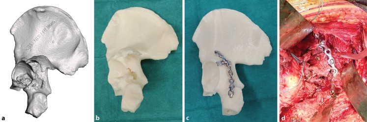
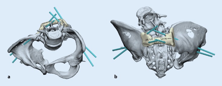
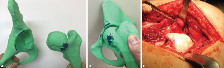
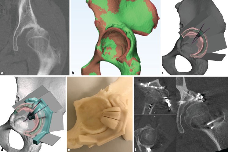
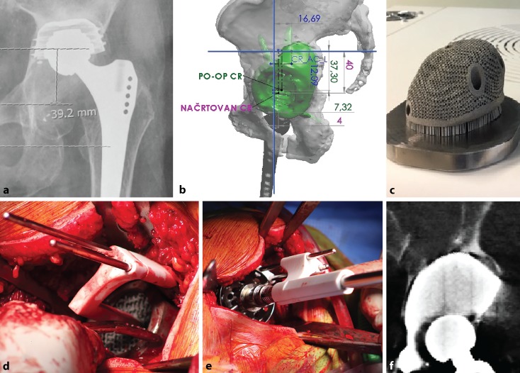
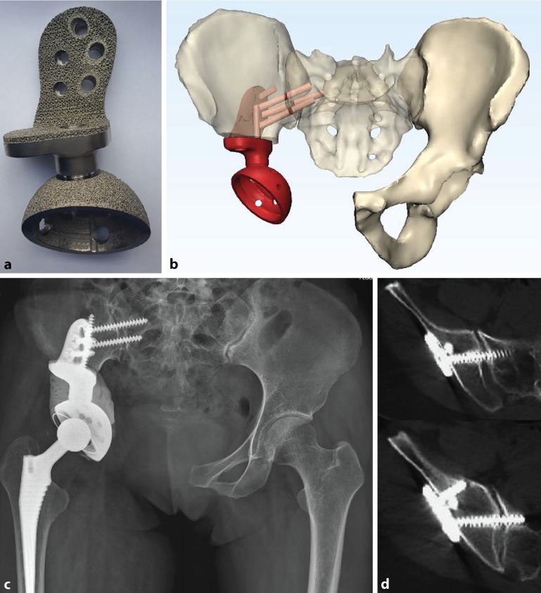
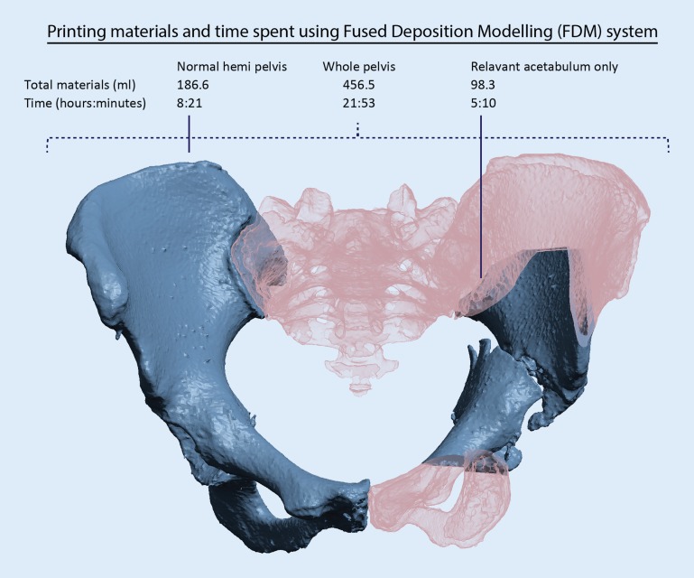
There are numerous orthopaedic applications of three-dimensional (3D) printing for the pelvis and acetabulum. The authors reviewed recently published articles and summarized their experience. 3D printed anatomical models are particularly useful in pelvic and acetabular fracture surgery for planning, implant templating and for anatomical assessment of pathologies such as CAM-type femoroacetabular impingement and rare deformities. Custom-made metal 3D printed patient-specific implants and instruments are increasingly being studied for pelvic oncologic resection and reconstruction of resected defects as well as for revision hip arthroplasties with favourable results. This article also discusses cost-effectiveness considerations when preparing pelvic 3D printed models from a hospital 3D printing centre.

Es gibt zahlreiche orthopädische Anwendungen des 3‑dimensionalen (3‑D) Drucks für Becken und Acetabulum. Im vorliegenden Beitrag werden in jüngerer Zeit publizierte Beiträge und eigene Erfahrungen zusammengefasst. Mittels 3‑D-Druck erstellte anatomische Modelle sind in der operativen Versorgung von Becken- und Acetabulumfrakturen besonders nützlich für die Planung, das Implantat-Templating und die anatomische Abklärung von Krankheitsbildern wie dem femoroacetabulären Cam-Impingement und seltenen Deformitäten. Mittels 3‑D-Druck maßgeschneiderte, patientenspezifische Metallimplantate und Instrumente werden vermehrt im Rahmen der onkologischen Beckenresektion und nachfolgenden Rekonstruktion von Defekten sowie in der Revisionsendoprothetik an der Hüfte getestet; die Ergebnisse sind positiv. Im vorliegenden Beitrag wird auch die Wirtschaftlichkeit diskutiert, wenn Beckenmodelle in einem innerklinischen Zentrum für 3‑D-Druck hergestellt werden.

Keywords: 3D printing; Hip replacement; Orthopaedics; Pelvic tumour; Pelvis.


Conflict of interest statement

C. Fang is co-founder of Lifespans Ltd. H. Cai serves as a paid consultant to AK Medical and Koln 3D Medical Technology. E. Kuong, E. Chui, Y.C. Siu, T. Ji and I. Drstvenšek declare that they have no competing interests.

Figures

Fig. 1
Fig. 1
An example of a digital three-dimensional (3D) model of a posterior wall acetabular fracture in stereolithography format (a). The 1:1-sized 3D printed model allows the surgeon to accurately appreciate fracture morphology (b). A mirrored model is printed using the opposite intact hemi-pelvis for easy and accurate plate contouring (c). Surgical plan after fracture fixation (d). The implants are placed according to the surgical plan after fracture fixation reduction
Fig. 2
Fig. 2
Design of a patient-specific drill guide for the placement of bilateral dual iliac screws for a patient with osteogenesis imperfecta undergoing posterior spinal fusion and instrumentation surgery in inlet (a) and posterior (b) orientations
Fig. 3
Fig. 3
An example of showing the location and simulating mechanical femoroacetabular impingement in a 12-year-old patient. The offending CAM-type lesion is accurately located before osteochondroplasty, marked (ab) and seen in surgery (c)
Fig. 4
Fig. 4
A 15-year-old patient with a malunited acetabulum posterior wall with hip subluxation (a). Digital model (green) compared to the mirrored opposite (red) (b). A closing-wedge volume-reducing osteotomy is planned (c) with 3D printed cutting jig (de). Postoperative computed tomography showing satisfactory restoration of hip congruency (f)
Fig. 5
Fig. 5
Patient with Paprosky IIIa defect and acetabular component loosening (a). Planning is performed using a digital three-dimensional model (b). Custom-made porous titanium alloy acetabular component printed using direct metal laser sintering (c). Intraoperative use of patient-specific instrumentation placed Schanz screws as positional references for implant (d) and reamer (e) placement. Postoperative computed tomography scan confirming satisfactory positioning (f)
Fig. 6
Fig. 6
A 39-year-old patient with right acetabulum chondrosarcoma resection, reconstructed with an electron-beam melting three-dimensionally printed porous modular hemi-pelvic endoprosthesis (a) with preoperative virtual planning (b). Radiograph at 2 years (c), with computed tomography showing osteointegration (d)
Fig. 7
Fig. 7
Using a fused deposition modelling (FDM) system at a 0.01-in. layer thickness (Fortus 450mc, Stratasys, Eden Prairie, MN, USA). Printing only the relevant fractured acetabulum results in 78% savings in materials and 76% savings in printing time compared to the whole pelvis

Similar articles

Cited by

References

    1. Mumith A, Thomas M, Shah Z, Coathup M, Blunn G. Additive manufacturing. Bone Jt J. 2018;100–B(4):455–460. doi: 10.1302/0301-620X.100B4.BJJ-2017-0662.R2. - DOI - PubMed
    1. Wong TM, Jin J, Lau TW, Fang C, Yan CH, Yeung K, et al. The use of three-dimensional printing technology in orthopaedic surgery. J Orthop Surg (hong kong) 2017;25(1):2309499016684077. doi: 10.1177/2309499016684077. - DOI - PubMed
    1. Fadero PE, Shah M. Three dimensional (3D) modelling and surgical planning in trauma and orthopaedics. surgeon. 2014;12(6):328–333. doi: 10.1016/j.surge.2014.03.008. - DOI - PubMed
    1. Boudissa M, Courvoisier A, Chabanas M, Tonetti J. Computer assisted surgery in preoperative planning of acetabular fracture surgery: state of the art. Expert Rev Med Devices. 2018;15(1):81–89. doi: 10.1080/17434440.2017.1413347. - DOI - PubMed
    1. Cromeens BP, Ray WC, Hoehne B, Abayneh F, Adler B, Besner GE. Facilitating surgeon understanding of complex anatomy using a three-dimensional printed model. J Surg Res. 2017;216:18–25. doi: 10.1016/j.jss.2017.04.003. - DOI - PubMed

LinkOut - more resources