Review
. 2019 Apr;28(4):332-344.
doi: 10.1111/exd.13913.

An updated classification of hair follicle morphogenesis

Affiliations
Review

An updated classification of hair follicle morphogenesis

Nivedita Saxena et al. Exp Dermatol. 2019 Apr.

Abstract

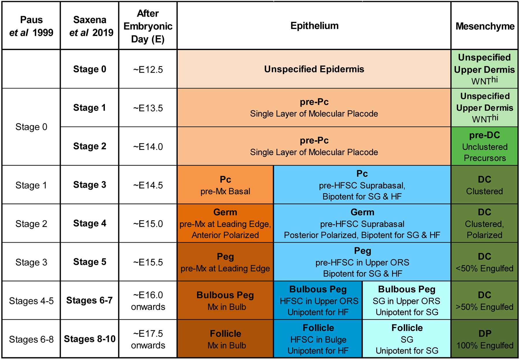
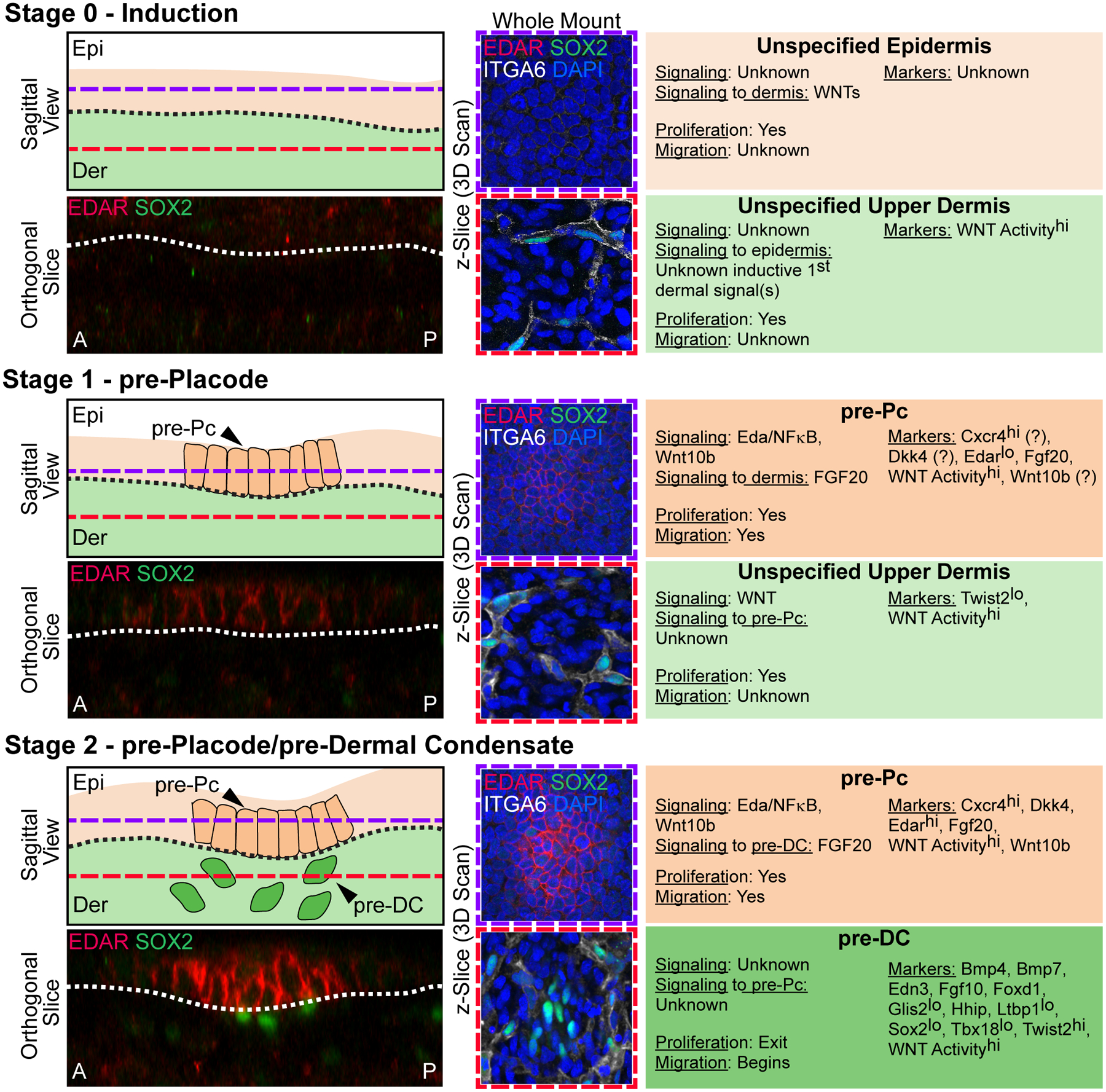
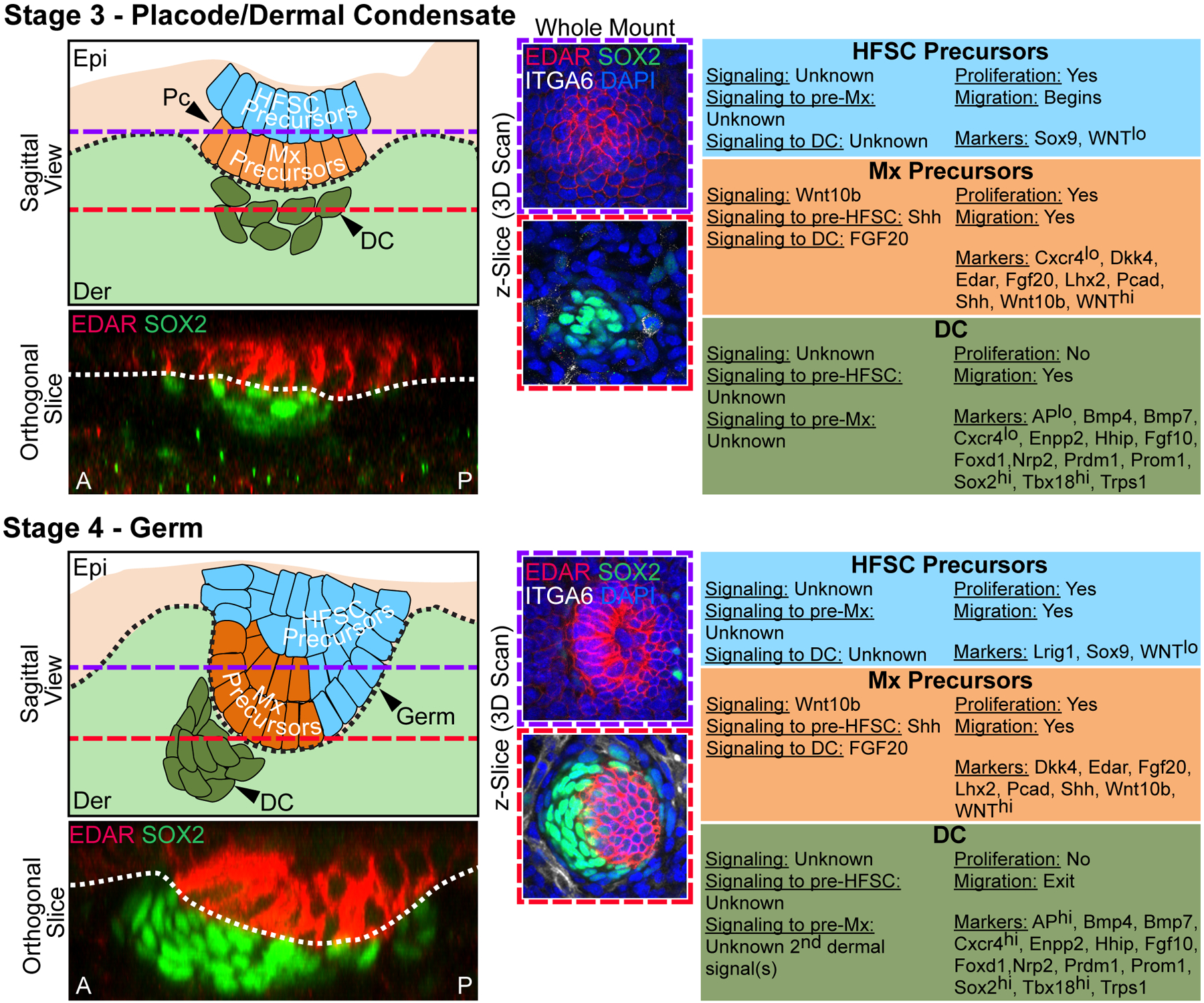
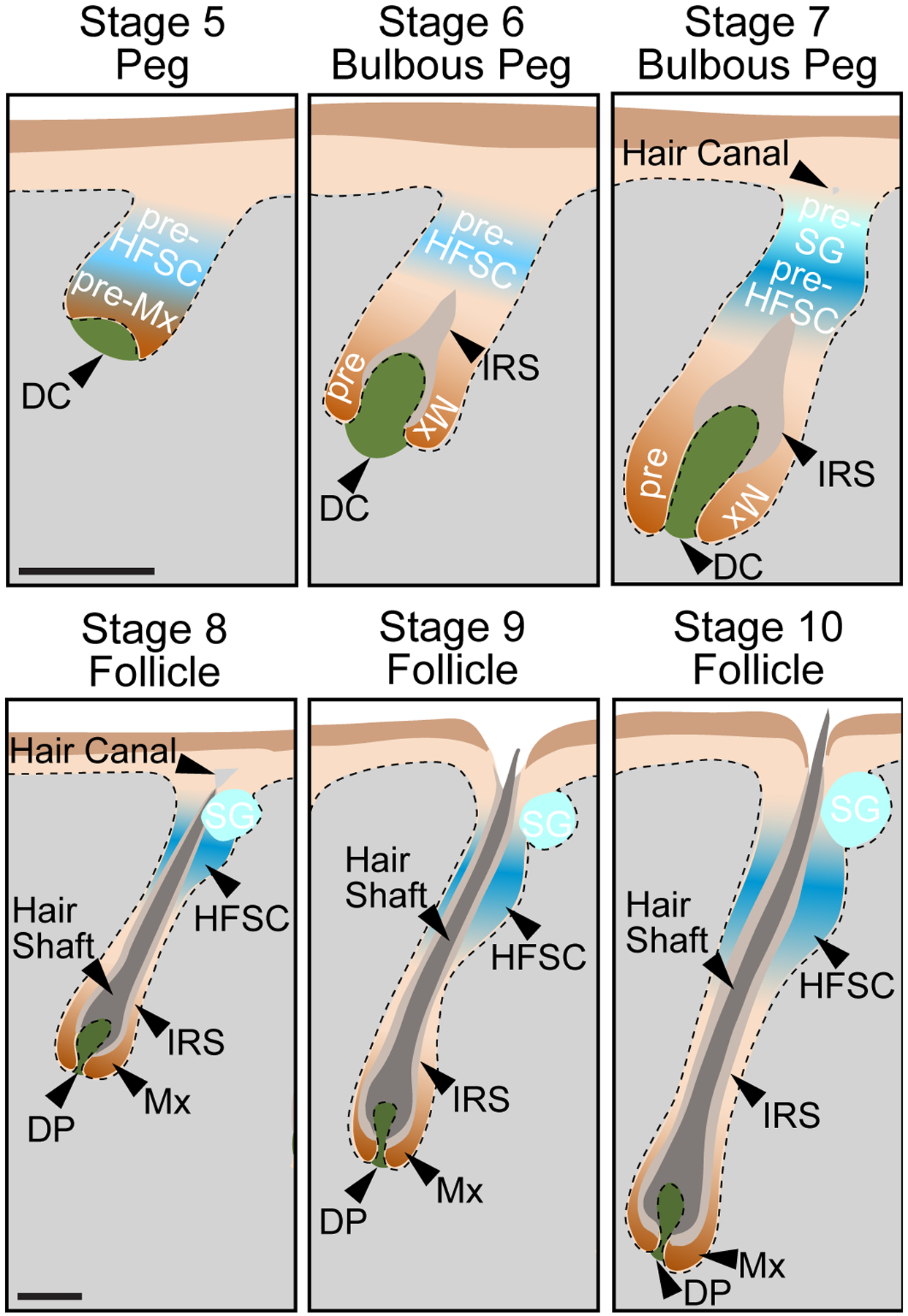
Hair follicle (HF) formation in developing embryonic skin requires stepwise signalling between the epithelial epidermis and mesenchymal dermis, and their specialized derivatives, the placode/germ/peg and dermal condensate/papilla, respectively. Classically, distinct stages of HF morphogenesis have been defined, in the mouse model, based on (a) changes in cell morphology and aggregation; (b) expression of few known molecular markers; (c) the extent of follicle downgrowth; and (d) the presence of differentiating cell types. Refined genetic strategies and recent emerging technologies, such as live imaging and transcriptome analyses of isolated cell populations or single cells, have enabled a closer dissection of the signalling requirements at different stages of HF formation, particularly early on. They have also led to the discovery of precursor cells for placode, dermal condensate and future bulge stem cells that, combined with molecular insights into their fate specification and subsequent formation, serve as novel landmarks for early HF morphogenetic events and studies of the signalling networks mediating these processes. In this review, we integrate the emergence of HF precursor cell states and novel molecular markers of fate and formation to update the widely used 20-year-old seminal classification guide of HF morphogenetic stages by Paus et al. We then temporally describe the latest insights into the early cellular and molecular events and signalling requirements for HF morphogenesis in relation to one another in a holistic manner.

Keywords: classification; dermal condensate; dermal papilla; guide; hair follicle morphogenesis; placode progenitors; stem cell niche.


Conflict of interest statement

CONFLICT OF INTEREST

The authors have declared no conflicting interests.

Figures

Figure 1.
Figure 1.. Updated Classification of Hair Follicle Morphogenesis Stages
Updated hair follicle morphogenetic classification including new stages for emergence and spatial localization of recently discovered precursor cell states in the epithelial and mesenchymal compartments, including pre-Pc, pre-DC and HFSC precursors. Differentiation of the pre-Pc into the Pc, then into Mx (matrix), HFSC, and SG (sebaceous gland), as well as of the pre-DC into the DP serve as hallmarks for stage identification. The previous classification system by Paus et al, 1999 is outlined. The approximate gestational ages are provided for HF stages as they first appear during the first wave of primary guard hair formation.
Figure 2.
Figure 2.. Stages 0–2 of Hair Follicle Morphogenesis
Left top: Sagittal view schematic of HF morphogenesis stages. Purple dashed lines mark the epidermal Z-plane, and red dashed lines mark the dermal Z-plane of 3D-imaged confocal scans of whole mount immunofluorescence of E15.0 back skin. A and P denote anterior and posterior orientation of embryonic skin (head is left). Left bottom: Orthogonal slice from a 3D reconstruction of whole mount immunofluorescence for EDAR and SOX2. Middle: Whole mount immunofluorescence for EDAR, SOX2, and ITGA6 in the epithelial plane (top, purple frame), and dermal plane (bottom, red frame). DAPI marks all nuclei. Right: Description of autocrine and paracrine signaling, proliferation and migration status, and markers of the relevant epithelial and mesenchymal populations at each stage. Stage 0 – Induction. The uniform unspecified multipotent epidermis resides over an unspecified dermis. EDAR is not expressed in the epidermal compartment and only SOX2+/ITGA6+ Schwann cells are present in the dermis. Widespread Wnt signaling activity in the upper dermis sets up HF induction by the critical, but still unknown “first dermal signal(s)”. Stage 1 – pre-Placode. Emergence of placode precursors (pre-Pc), the fated “molecular placode”, in the epidermis over an unspecified dermis. The pre-Pc in the epidermal plane expresses EDAR. Only SOX2+/ ITGA6+ Schwann cells are present in the dermal compartment. Markers with question marks refer to known expression by in situ hybridization and/or protein staining at E13.5, in which the presence of recently discovered pre-DC (stage 2) cannot be ruled out. Stage 2 – pre-Placode/pre-Dermal Condensate. pre-Pc in the epidermis over dermal condensate precursors (pre-DC) in the dermis. The pre-Pc in the epidermal plane expresses EDAR more strongly. Pre-DC emerge as low-level SOX2+/ITGA6 unclustered cells underneath pre-Pc.
Figure 3.
Figure 3.. Stages 3–4 of Hair Follicle Morphogenesis
Left top: Sagittal view schematic of HF morphogenesis stages. Purple dashed lines mark the epidermal Z-plane, and red dashed lines mark the dermal Z-plane of 3D-imaged confocal scans of whole mount immunofluorescence of E15.0 back skin. A and P denote anterior and posterior orientation of embryonic skin (head is left). Left bottom: Orthogonal slice from a 3D reconstruction of whole mount immunofluorescence for EDAR and SOX2. Middle: Whole mount immunofluorescence for EDAR, SOX2, and ITGA6 in the epithelial plane (top, purple frame), and dermal plane (bottom, red frame). DAPI marks all nuclei. Right: Description of autocrine and paracrine signaling, proliferation and migration status, and markers of the relevant epithelial and mesenchymal populations at each stage. Stage 3 – Placode/Dermal Condensate. Pc in the epithelium, with matrix (Mx) precursors at the basal layer and HFSC precursors in the suprabasal layer, above an aggregating DC in the dermis. Pronounced EDAR expression is detectable in maturing placodes. Pronounced SOX2 expression is present in the maturing DC. Stage 4 – Germ. Downgrown germ in the epithelium, with matrix (Mx) precursors at the basal layer and HFSC precursors in the suprabasal layers, above an aggregating and polarized DC in the dermis. At this stage, the DC has more cells and is further condensed. Note anterior polarization (towards the head) of the downgrowing germ with the DC at the leading edge. EDAR continues to be highly expressed at the germ stage. SOX2 is highly expressed in the DC.
Figure 4.
Figure 4.. Stages 5–10 of Hair Follicle Morphogenesis
Sagittal view schematics for the late stages of HF morphogenesis. Pseudoscale bars represent the relative scale for stages 5–7 and stages 8–10. Stage 5 – Peg. Downgrowth of the hair peg, comprised of Mx precursors at the leading edge, and HFSC precursors above them. The DC is beginning the be engulfed. Stage 6 – Bulbous Peg. At the bulbous peg stage, Mx precursors engulf the DC by > 50% in the bulb region. HFSC precursors remain in the upper ORS. The IRS is beginning to form. Stage 7 – Bulbous Peg. At this later bulbous peg stage, Mx precursors almost entirely engulf the DC. Bipotent HFSC precursors bifurcate into unipotent HFSC precursors, which reside in the presumptive bulge area, and sebocytes that will make up the sebaceous gland (pre-SG), which reside in the upper part of the follicle. The hair canal is first becoming visible. Stage 8 – Follicle. At the follicle stage, the Mx now completely engulfs the DP. The IRS reaches the hair canal, and the lineages of the hair shaft are being produced. HFSC reside in the bulge. The sebaceous gland (SG) is formed. Stage 9 – Follicle. The follicle further elongates into the dermis. The tip of the hair shaft leaves the IRS and enters the hair canal. The DP is even more fully narrowed. Stage 10 – Follicle. The hair shaft emerges from the epidermis. The follicle has reached its maximum length expanding into the subcutaneous adipose layers.
Figure 5.
Figure 5.. Molecular Markers for Early Hair Follicle Morphogenesis Cell States and Types
Marker expression for cell types at the key stages of early hair follicle morphogenesis. Genes are color-coded for expression in respective compartments; high (hi) and low (lo) expression is noted where it is known. Grey boxes with question marks refer to unknown expression by in situ hybridization and/or protein staining, at related neighboring stages. Color boxes with question markers (Cxcr4, Dkk4, Wnt10b) in Stage 1 pre-Pc refers to known expression by in situ hybridization or protein staining at E13.5, where the presence of recently discovered pre-DC (stage 2) cannot be ruled out. References for each marker are on the right.

Similar articles

Cited by

References

    1. Sperling LC Hair anatomy for the clinician. J Am Acad Dermatol 1991: 25: 1–17. - PubMed
    1. Cotsarelis G, Sun TT, Lavker RM. Label-retaining cells reside in the bulge area of pilosebaceous unit: implications for follicular stem cells, hair cycle, and skin carcinogenesis. Cell 1990: 61: 1329–37. - PubMed
    1. Oshima H, Rochat A, Kedzia C et al. Morphogenesis and renewal of hair follicles from adult multipotent stem cells. Cell 2001: 104: 233–45. - PubMed
    1. Taylor G, Lehrer MS, Jensen PJ et al. Involvement of follicular stem cells in forming not only the follicle but also the epidermis. Cell 2000: 102: 451–61. - PubMed
    1. Tumbar T, Guasch G, Greco V et al. Defining the epithelial stem cell niche in skin. Science 2004: 303: 359–63. - PMC - PubMed

Publication types