

Non-invasive assessment of glioma microstructure using VERDICT MRI: correlation with histology
- PMID: 30888488
- PMCID: PMC6719328
- DOI: 10.1007/s00330-019-6011-8
Non-invasive assessment of glioma microstructure using VERDICT MRI: correlation with histology
Abstract
Purpose: This prospective study evaluated the use of vascular, extracellular and restricted diffusion for cytometry in tumours (VERDICT) MRI to investigate the tissue microstructure in glioma. VERDICT-derived parameters were correlated with both histological features and tumour subtype and were also used to explore the peritumoural region.
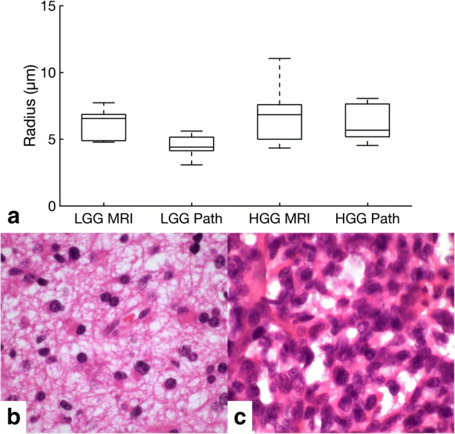
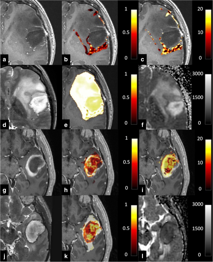
Methods: Fourteen consecutive treatment-naïve patients (43.5 years ± 15.1 years, six males, eight females) with suspected glioma underwent diffusion-weighted imaging including VERDICT modelling. Tumour cell radius and intracellular and combined extracellular/vascular volumes were estimated using a framework based on linearisation and convex optimisation. An experienced neuroradiologist outlined the peritumoural oedema, enhancing tumour and necrosis on T2-weighted imaging and contrast-enhanced T1-weighted imaging. The same regions of interest were applied to the co-registered VERDICT maps to calculate the microstructure parameters. Pathology sections were analysed with semi-automated software to measure cellularity and cell size.
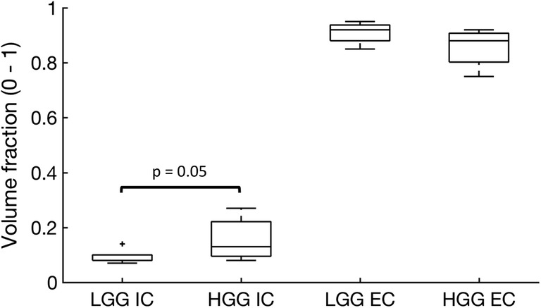
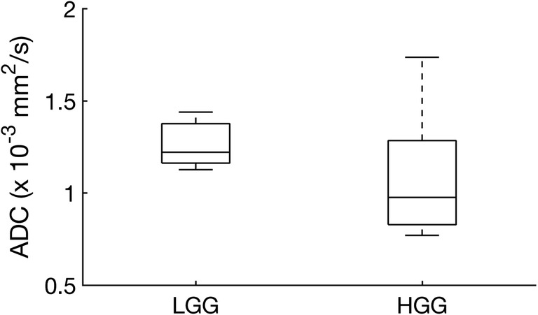
Results: VERDICT parameters were successfully calculated in all patients. The imaging-derived results showed a larger intracellular volume fraction in high-grade glioma compared to low-grade glioma (0.13 ± 0.07 vs. 0.08 ± 0.02, respectively; p = 0.05) and a trend towards a smaller extracellular/vascular volume fraction (0.88 ± 0.07 vs. 0.92 ± 0.04, respectively; p = 0.10). The conventional apparent diffusion coefficient was higher in low-grade gliomas compared to high-grade gliomas, but this difference was not statistically significant (1.22 ± 0.13 × 10⁻³ mm2/s vs. 0.98 ± 0.38 × 10⁻³ mm2/s, respectively; p = 0.18).
Conclusion: This feasibility study demonstrated that VERDICT MRI can be used to explore the tissue microstructure of glioma using an abbreviated protocol. The VERDICT parameters of tissue structure correlated with those derived on histology. The method shows promise as a potential test for diagnostic stratification and treatment response monitoring in the future.
Key points: • VERDICT MRI is an advanced diffusion technique which has been correlated with histopathological findings obtained at surgery from patients with glioma in this study. • The intracellular volume fraction measured with VERDICT was larger in high-grade tumours compared to that in low-grade tumours. • The results were complementary to measurements from conventional diffusion-weighted imaging, and the technique could be performed in a clinically feasible timescale.
Keywords: Brain neoplasms; Cancer; Diagnostic imaging; Diffusion magnetic resonance imaging; Glioma.
Conflict of interest statement
The authors declare that they have no conflict of interest.
Figures
Similar articles
-
Noninvasive diffusion magnetic resonance imaging of brain tumour cell size for the early detection of therapeutic response.Sci Rep. 2020 Jun 8;10(1):9223. doi: 10.1038/s41598-020-65956-4. Sci Rep. 2020. PMID: 32514049 Free PMC article.
-
Microstructural characterization of normal and malignant human prostate tissue with vascular, extracellular, and restricted diffusion for cytometry in tumours magnetic resonance imaging.Invest Radiol. 2015 Apr;50(4):218-27. doi: 10.1097/RLI.0000000000000115. Invest Radiol. 2015. PMID: 25426656
-
Comprehensive Brain Tumour Characterisation with VERDICT-MRI: Evaluation of Cellular and Vascular Measures Validated by Histology.Cancers (Basel). 2023 Apr 27;15(9):2490. doi: 10.3390/cancers15092490. Cancers (Basel). 2023. PMID: 37173965 Free PMC article.
-
The role of imaging in the management of adults with diffuse low grade glioma: A systematic review and evidence-based clinical practice guideline.J Neurooncol. 2015 Dec;125(3):457-79. doi: 10.1007/s11060-015-1908-9. Epub 2015 Nov 3. J Neurooncol. 2015. PMID: 26530262
-
High-Grade Glioma Treatment Response Monitoring Biomarkers: A Position Statement on the Evidence Supporting the Use of Advanced MRI Techniques in the Clinic, and the Latest Bench-to-Bedside Developments. Part 1: Perfusion and Diffusion Techniques.Front Oncol. 2022 Mar 3;12:810263. doi: 10.3389/fonc.2022.810263. eCollection 2022. Front Oncol. 2022. PMID: 35359414 Free PMC article. Review.
Cited by
-
Quantitative Analysis With Time-Dependent Diffusion MRI for Assessing WHO/ISUP Tumor Grade in Clear Cell Renal Cell Carcinoma.Korean J Radiol. 2025 Jul;26(7):678-687. doi: 10.3348/kjr.2024.1202. Epub 2025 Jun 13. Korean J Radiol. 2025. PMID: 40527738 Free PMC article.
-
Predicting Survival in Patients with Brain Tumors: Current State-of-the-Art of AI Methods Applied to MRI.Diagnostics (Basel). 2022 Sep 1;12(9):2125. doi: 10.3390/diagnostics12092125. Diagnostics (Basel). 2022. PMID: 36140526 Free PMC article. Review.
-
Emerging methods for prostate cancer imaging: evaluating cancer structure and metabolic alterations more clearly.Mol Oncol. 2021 Oct;15(10):2565-2579. doi: 10.1002/1878-0261.13071. Epub 2021 Aug 30. Mol Oncol. 2021. PMID: 34328279 Free PMC article. Review.
-
Magnetic Fields and Cancer: Epidemiology, Cellular Biology, and Theranostics.Int J Mol Sci. 2022 Jan 25;23(3):1339. doi: 10.3390/ijms23031339. Int J Mol Sci. 2022. PMID: 35163262 Free PMC article. Review.
-
Separating Glioma Hyperintensities From White Matter by Diffusion-Weighted Imaging With Spherical Tensor Encoding.Front Neurosci. 2022 Apr 21;16:842242. doi: 10.3389/fnins.2022.842242. eCollection 2022. Front Neurosci. 2022. PMID: 35527815 Free PMC article.
References
Publication types
MeSH terms
Grants and funding
LinkOut - more resources
Full Text Sources
Medical
