. 2019 Aug;34(8):1387-1392.
doi: 10.1002/jbmr.3719. Epub 2019 May 6.

Growth Plate Borderline Chondrocytes Behave as Transient Mesenchymal Precursor Cells

Affiliations

Growth Plate Borderline Chondrocytes Behave as Transient Mesenchymal Precursor Cells

Koji Mizuhashi et al. J Bone Miner Res. 2019 Aug.

Abstract

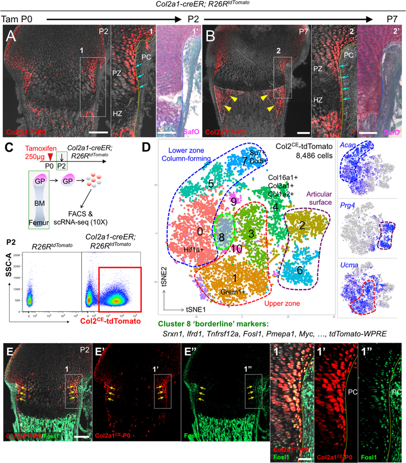
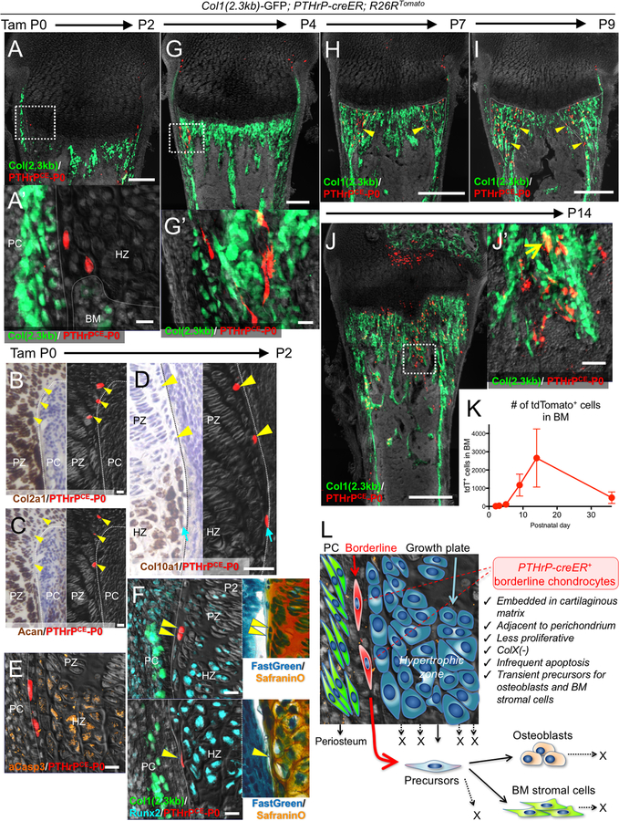
The growth plate provides a substantial source of mesenchymal cells in the endosteal marrow space during endochondral ossification. The current model postulates that a group of chondrocytes in the hypertrophic zone can escape from apoptosis and transform into cells that eventually become osteoblasts in an area beneath the growth plate. The growth plate is composed of cells with various morphologies; particularly at the periphery of the growth plate immediately adjacent to the perichondrium are "borderline" chondrocytes, which align perpendicularly to other chondrocytes. However, in vivo cell fates of these special chondrocytes have not been revealed. Here we show that borderline chondrocytes in growth plates behave as transient mesenchymal precursor cells for osteoblasts and marrow stromal cells. A single-cell RNA-seq analysis revealed subpopulations of Col2a1-creER-marked neonatal chondrocytes and their cell type-specific markers. A tamoxifen pulse to Pthrp-creER mice in the neonatal stage (before the resting zone was formed) preferentially marked borderline chondrocytes. Following the chase, these cells marched into the nascent marrow space, expanded in the metaphyseal marrow, and became Col(2.3 kb)-GFP+ osteoblasts and Cxcl12-GFPhigh reticular stromal "CAR" cells. Interestingly, these borderline chondrocyte-derived marrow cells were short-lived, as they were significantly reduced during adulthood. These findings demonstrate based on in vivo lineage-tracing experiments that borderline chondrocytes in the peripheral growth plate are a particularly important route for producing osteoblasts and marrow stromal cells in growing murine endochondral bones. A special microenvironment neighboring the osteogenic perichondrium might endow these chondrocytes with an enhanced potential to differentiate into marrow mesenchymal cells. © 2019 American Society for Bone and Mineral Research.

Keywords: DEVELOPMENTAL MODELING; GENETIC ANIMAL MODELS; GROWTH PLATE; OSTEOBLASTS; STROMAL/STEM CELLS.


Conflict of interest statement

The authors declare no conflict of interest.

Figures

Figure 1.
Figure 1.. Single cell RNA-seq characterization of neonatal growth plate chondrocytes.
(A,B) Cell-fate analysis of Col2a1-creER+ neonatal growth plate chondrocytes (P0-pulsed) at P2 (A) and P7 (B). PC: perichondrium, PZ: proliferating zone, HZ: hypertrophic zone. Right panels: SafraninO & FastGreen staining of identical sections. Arrows: tdTomato+ borderline chondrocytes, arrowheads: tdTomato+ cells in peripheral metaphyseal marrow space. Scale bars: 200μm (left panels), 100μm (right panels). n=3 mice for each experiment. (C,D) Single cell RNA-seq analysis of Col2a1-creER+ neonatal growth plate chondrocytes. (D) t-SNE-based visualization of major classes of FACS-sorted Col2a1CE-tdTomato+ single cells at P2 (Cluster 0–10). Cluster 0,5,7: lower zone column-forming chondrocytes, Cluster1,3: upper zone chondrocytes, Cluster 2,6: articular surface chondrocytes, Cluster 4: collagen-rich articular cells, Cluster 8: borderline chondrocytes, Cluster 9,10: cells in cell cycle. Dots: individual cells, color: cell type. Green contour: borderline chondrocytes. Right panels: feature plots of Acan, Prg4 and Ucma. Blue: high expression, grey: no expression. n=8,486 cells. (E) In situ validation of the identified marker, Fosl1. Col2a1-creER; R26RtdTomato distal femurs at P2 (P0-pulsed). (E’,E”): tdTomato, Fosl1-Alexa488 single channel, (1–1”): magnified views of the dotted area. Scale bars: 200μm (E-E”), 50μm (1–1”). n=3 mice.
Figure 2.
Figure 2.. Pthrp-creER+ borderline chondrocytes in the neonatal stage behave as transient mesenchymal precursor cells.
Cell-fate analysis of Pthrp-creER+ borderline chondrocytes (P0-pulsed) at P2 (A-F), P4 (G), P7 (H), P9 (I) and P14 (J). (A,F-J): Col1(2.3kb)-GFP; Pthrp-creER; R26RtdTomato distal femurs, (B-E): Pthrp-creER; R26RtdTomato distal femurs. (A’,G’,J’): magnified views of the dotted areas. In (B-D), left panels (bright field) and right panels (dark field) represent the identical sections. Right panels in (F): SafraninO & FastGreen staining of left panels. Arrowheads in (B-F): tdTomato+ borderline chondrocytes denoting identical cells. PC: perichondrium, PZ: proliferating zone, HZ: hypertrophic zone. Arrowheads in (H,I): tdTomato+ cells in peripheral metaphyseal marrow space, arrow in (J): GFP+tdTomato+ osteoblasts. Scale bars: 200μm (A,G), 500μm (H-J), 20μm (A-F), 50μm (J’). n=3 mice for each experiment. (K): Quantification of tdTomato+ marrow cells at each time point. n=6 (P2), n=4 (P4), n=3 (P5, P9, P14, P36) mice, data are presented as mean ± S.D. (L): Concluding diagram. Borderline chondrocytes in growth plates can translocate into marrow space and behave as transient precursors for osteoblasts and stromal cells by escaping hypertrophy and apoptosis.

Comment in

References

    1. Yang L, Tsang KY, Tang HC, Chan D, Cheah KS. Hypertrophic chondrocytes can become osteoblasts and osteocytes in endochondral bone formation. Proc Natl Acad Sci U S A. August 2014;111(33):12097–102. Epub 2014/08/04. - PMC - PubMed
    1. Ono N, Ono W, Nagasawa T, Kronenberg HM. A subset of chondrogenic cells provides early mesenchymal progenitors in growing bones. Nat Cell Biol. December 2014;16(12):1157–67. - PMC - PubMed
    1. Zhou X, von der Mark K, Henry S, Norton W, Adams H, de Crombrugghe B. Chondrocytes transdifferentiate into osteoblasts in endochondral bone during development, postnatal growth and fracture healing in mice. PLoS Genet. December 2014;10(12):e1004820 Epub 2014/12/04. - PMC - PubMed
    1. Mizuhashi K, Ono W, Matsushita Y, Sakagami N, Takahashi A, Saunders TL, et al. Resting zone of the growth plate houses a unique class of skeletal stem cells. Nature. November 2018;563(7730):254–8. Epub 2018/10/31. - PMC - PubMed
    1. Bianco P, Cancedda FD, Riminucci M, Cancedda R. Bone formation via cartilage models: the “borderline” chondrocyte. Matrix Biol. July 1998;17(3):185–92. - PubMed

Publication types