. 2019 May 1;199(9):1076-1085.
doi: 10.1164/rccm.201810-1869OC.

Extracellular DNA, Neutrophil Extracellular Traps, and Inflammasome Activation in Severe Asthma

Affiliations

Extracellular DNA, Neutrophil Extracellular Traps, and Inflammasome Activation in Severe Asthma

Marrah E Lachowicz-Scroggins et al. Am J Respir Crit Care Med. .

Abstract

Rationale: Extracellular DNA (eDNA) and neutrophil extracellular traps (NETs) are implicated in multiple inflammatory diseases. NETs mediate inflammasome activation and IL-1β secretion from monocytes and cause airway epithelial cell injury, but the role of eDNA, NETs, and IL-1β in asthma is uncertain. Objectives: To characterize the role of activated neutrophils in severe asthma through measurement of NETs and inflammasome activation. Methods: We measured sputum eDNA in induced sputum from 399 patients with asthma in the Severe Asthma Research Program-3 and in 94 healthy control subjects. We subdivided subjects with asthma into eDNA-low and -high subgroups to compare outcomes of asthma severity and of neutrophil and inflammasome activation. We also examined if NETs cause airway epithelial cell damage that can be prevented by DNase. Measurements and Main Results: We found that 13% of the Severe Asthma Research Program-3 cohort is "eDNA-high," as defined by sputum eDNA concentrations above the upper 95th percentile value in health. Compared with eDNA-low patients with asthma, eDNA-high patients had lower Asthma Control Test scores, frequent history of chronic mucus hypersecretion, and frequent use of oral corticosteroids for maintenance of asthma control (all P values <0.05). Sputum eDNA in asthma was associated with airway neutrophilic inflammation, increases in soluble NET components, and increases in caspase 1 activity and IL-1β (all P values <0.001). In in vitro studies, NETs caused cytotoxicity in airway epithelial cells that was prevented by disruption of NETs with DNase. Conclusions: High extracellular DNA concentrations in sputum mark a subset of patients with more severe asthma who have NETs and markers of inflammasome activation in their airways.

Keywords: IL-1β; asthma; caspase 1; extracellular DNA; neutrophil extracellular traps.


Figures

Figure 1.
Figure 1.
Increased extracellular DNA (eDNA) in sputum from a subset of patients with asthma is associated with increased neutrophil numbers and neutrophil activation. (A) A subset of patients with asthma has sputum eDNA concentrations above the 95th percentile value in healthy control subjects. (B) Sputum eDNA concentrations are not significantly correlated with sputum eosinophils. (C) Sputum eDNA concentrations are not significantly correlated with the T-helper cell type 2 (Th2) gene mean. (D) Sputum eDNA concentrations are significantly correlated with sputum neutrophils. (E) Sputum eDNA concentrations are significantly correlated with myeloperoxidase (MPO) concentrations. (F) Sputum MPO concentrations are significantly higher in eDNA-high asthma than in eDNA-low asthma. ***P < 0.001. Circles represent individual data points.
Figure 2.
Figure 2.
Extracellular DNA (eDNA)-high asthma is associated with poor asthma control and symptoms of chronic mucus hypersecretion but not with airway mucus plugging. (A) The Asthma Control Test (ACT) score is significantly lower in eDNA-high asthma than in eDNA-low asthma. (B) Chronic mucus hypersecretion (also called “chronic bronchitis”) is more prevalent in eDNA-high asthma than in eDNA-low asthma. Chronic mucus hypersecretion data were available for 297 DNA-low patients and 40 DNA-high patients. *P < 0.05 and ***P < 0.001. Circles represent individual data points.
Figure 3.
Figure 3.
Soluble neutrophil extracellular trap complexes are higher in extracellular DNA (eDNA)-high asthma than in eDNA-low asthma. (A) Neutrophil elastase (NE)–DNA complexes are significantly higher in eDNA-high asthma than in eDNA-low asthma. (B) Citrullinated histone H3 (H3Cit)–DNA complexes are significantly higher in eDNA-high asthma than in eDNA-low asthma. Healthy, n = 35; eDNA-low, n = 42; eDNA-high, n = 44. *P < 0.05, **P < 0.01, and ***P < 0.001. Circles represent individual data points. RLU = relative luminometer units.
Figure 4.
Figure 4.
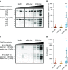
Caspase-1 activity and IL-1β concentrations are higher in extracellular DNA (eDNA)-high asthma than in eDNA-low asthma. (A) Western blot data for caspase 1 in sputum from three healthy control subjects, three patients with eDNA-low asthma, and three patients with eDNA-high asthma. The bands show caspase 1 zymogen and cleavage at Asp297 yielding the activated protease bands p20 and p10. (B) Bioluminescence data for caspase 1 in sputum showing significantly higher concentrations in eDNA-high asthma (n = 44) than in eDNA-low asthma (n = 42) or healthy control subjects (n = 35). (C) Western blot data for IL-1β in sputum from three healthy control subjects, three patients with eDNA-low asthma, and three patients with eDNA-high asthma. The data show cleavage of IL-1β at Asp116 in eDNA asthma. (D) ELISA data for IL-1β in sputum showing significantly higher concentrations in eDNA-high asthma (n = 44) than eDNA-low asthma (n = 42) or healthy control subjects (n = 35). ***P < 0.001. Circles represent individual data points. RLU = relative luminometer units.
Figure 5.
Figure 5.
Neutrophil extracellular trap (NET)-mediated injury to airway epithelial cells (AECs). (A) Apical exposure of AECs grown at air–liquid interface (ALI) to NETs (6 μg/ml) for 24 hours causes upregulation of gene expression for IL-6 and IL-8 (data are from epithelial cells from seven donors). (B) Apical exposure of AECs grown at ALI to NETs (6 μg/ml) for 24 hours causes release of IL-6 and IL-8 protein into the conditioned media (data are from epithelial cells from seven donors). (C) Glucose-6-phosphate dehydrogenase (G6PD) release into conditioned media of AECs in ALI culture after exposure to NETs (6 μg/ml) at the apical surface for 24 hours in the presence or absence of DNase I cotreatment (data are from epithelial cells from five donors). Error bars represent SEM. *P < 0.05 and ***P < 0.0001. Circles represent individual data points. RFU = relative fluorescence units.

Comment in

References

    1. Moore WC, Hastie AT, Li X, Li H, Busse WW, Jarjour NN, et al. National Heart, Lung, and Blood Institute’s Severe Asthma Research Program. Sputum neutrophil counts are associated with more severe asthma phenotypes using cluster analysis. J Allergy Clin Immunol. 2014;133:1557–1563.e5. - PMC - PubMed
    1. Krishnamoorthy N, Douda DN, Brüggemann TR, Ricklefs I, Duvall MG, Abdulnour RE, et al. National Heart, Lung, and Blood Institute Severe Asthma Research Program-3 Investigators. Neutrophil cytoplasts induce TH17 differentiation and skew inflammation toward neutrophilia in severe asthma. Sci Immunol. 2018;3:eaao4747. - PMC - PubMed
    1. Wright TK, Gibson PG, Simpson JL, McDonald VM, Wood LG, Baines KJ. Neutrophil extracellular traps are associated with inflammation in chronic airway disease. Respirology. 2016;21:467–475. - PubMed
    1. Kim RY, Pinkerton JW, Essilfie AT, Robertson AAB, Baines KJ, Brown AC, et al. Role for NLRP3 inflammasome-mediated, IL-1β-dependent responses in severe, steroid-resistant asthma. Am J Respir Crit Care Med. 2017;196:283–297. - PubMed
    1. Rossios C, Pavlidis S, Hoda U, Kuo CH, Wiegman C, Russell K, et al. Unbiased Biomarkers for the Prediction of Respiratory Diseases Outcomes (U-BIOPRED) Consortia Project Team. Sputum transcriptomics reveal upregulation of IL-1 receptor family members in patients with severe asthma. J Allergy Clin Immunol. 2018;141:560–570. - PubMed

Publication types