

Detection of breast cancer lymph node metastases in frozen sections with a point-of-care low-cost microscope scanner
- PMID: 30889174
- PMCID: PMC6424449
- DOI: 10.1371/journal.pone.0208366
Detection of breast cancer lymph node metastases in frozen sections with a point-of-care low-cost microscope scanner
Abstract
Background: Detection of lymph node metastases is essential in breast cancer diagnostics and staging, affecting treatment and prognosis. Intraoperative microscopy analysis of sentinel lymph node frozen sections is standard for detection of axillary metastases but requires access to a pathologist for sample analysis. Remote analysis of digitized samples is an alternative solution but is limited by the requirement for high-end slide scanning equipment.
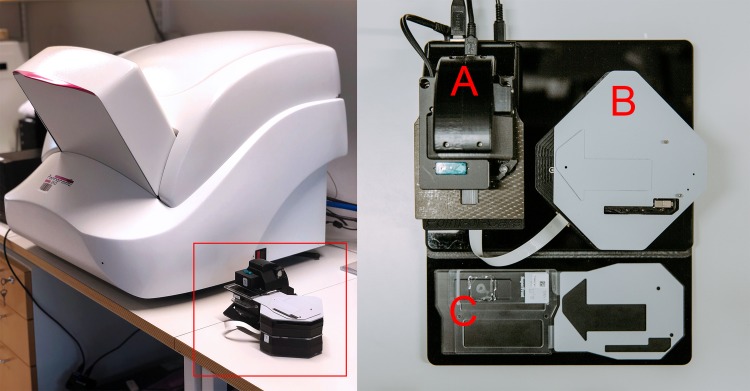
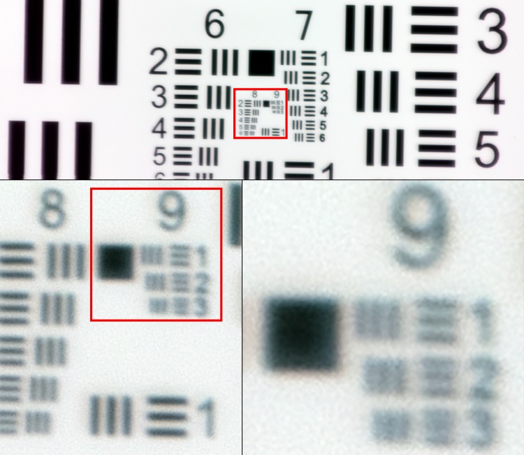
Objective: To determine whether the image quality achievable with a low-cost, miniature digital microscope scanner is sufficient for detection of metastases in breast cancer lymph node frozen sections.
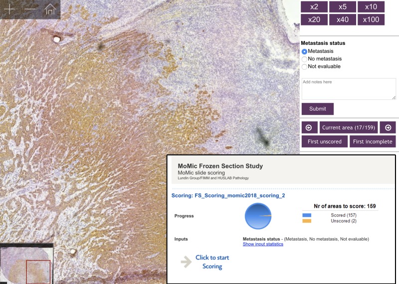
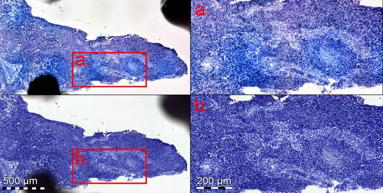
Methods: Lymph node frozen sections from 79 breast cancer patients were digitized using a prototype miniature microscope scanner and a high-end slide scanner. Images were independently reviewed by two pathologists and results compared between devices with conventional light microscopy analysis as ground truth.
Results: Detection of metastases in the images acquired with the miniature scanner yielded an overall sensitivity of 91% and specificity of 99% and showed strong agreement when compared to light microscopy (k = 0.91). Strong agreement was also observed when results were compared to results from the high-end slide scanner (k = 0.94). A majority of discrepant cases were micrometastases and sections of which no anticytokeratin staining was available.
Conclusion: Accuracy of detection of metastatic cells in breast cancer sentinel lymph node frozen sections by visual analysis of samples digitized using low-cost, point-of-care microscopy is comparable to analysis of digital samples scanned using a high-end, whole slide scanner. This technique could potentially provide a workflow for digital diagnostics in resource-limited settings, facilitate sample analysis at the point-of-care and reduce the need for trained experts on-site during surgical procedures.
Conflict of interest statement
I have read the journal's policy and the authors of this manuscript have the following competing interests: Johan Lundin and Mikael Lundin are founders and co-owners of Fimmic Oy, Helsinki, Finland. This does not alter our adherence to PLOS ONE policies on sharing data and materials.
Figures
Similar articles
-
Sentinel lymph node as a new marker for therapeutic planning in breast cancer patients.J Surg Oncol. 2004 Mar;85(3):102-11. doi: 10.1002/jso.20022. J Surg Oncol. 2004. PMID: 14991881 Review.
-
Application of automated image analysis reduces the workload of manual screening of sentinel lymph node biopsies in breast cancer.Histopathology. 2017 Dec;71(6):866-873. doi: 10.1111/his.13305. Epub 2017 Sep 22. Histopathology. 2017. PMID: 28677240
-
Touch preparation or frozen section for intraoperative detection of sentinel lymph node metastases from breast cancer.Ann Surg Oncol. 2003 Dec;10(10):1166-70. doi: 10.1245/aso.2003.04.023. Ann Surg Oncol. 2003. PMID: 14654472
-
Prospective and Retrospective Analysis of Whole-Slide Images of Sentinel and Targeted Lymph Node Frozen Sections in Breast Cancer.Mod Pathol. 2025 Apr;38(4):100708. doi: 10.1016/j.modpat.2025.100708. Epub 2025 Jan 7. Mod Pathol. 2025. PMID: 39788205
-
Evaluation of intraoperative frozen section diagnosis of sentinel lymph nodes in breast cancer.Jpn J Clin Oncol. 2004 Mar;34(3):113-7. doi: 10.1093/jjco/hyh023. Jpn J Clin Oncol. 2004. PMID: 15078905 Review.
Cited by
-
Fine-Tuned DenseNet-169 for Breast Cancer Metastasis Prediction Using FastAI and 1-Cycle Policy.Sensors (Basel). 2022 Apr 13;22(8):2988. doi: 10.3390/s22082988. Sensors (Basel). 2022. PMID: 35458972 Free PMC article.
-
The value and significance of nucleolar organizer region proteins as markers of malignancy in breast cancer patients.Saudi Med J. 2024 Oct;45(10):1028-1033. doi: 10.15537/smj.2024.45.10.20240483. Saudi Med J. 2024. PMID: 39379117 Free PMC article.
-
Utility of a Low-Cost 3-D Printed Microscope for Evaluating Esophageal Biopsies.Ann 3D Print Med. 2024 Feb;13:100145. doi: 10.1016/j.stlm.2024.100145. Epub 2024 Jan 10. Ann 3D Print Med. 2024. PMID: 38405263 Free PMC article.
-
A novel deep learning-based point-of-care diagnostic method for detecting Plasmodium falciparum with fluorescence digital microscopy.PLoS One. 2020 Nov 17;15(11):e0242355. doi: 10.1371/journal.pone.0242355. eCollection 2020. PLoS One. 2020. PMID: 33201905 Free PMC article.
References
-
- Siegel RL, Miller KD, Jemal A. Cancer statistics, 2018. CA: a Cancer Journal for Clinicians 2018. January;68(1):7–30. - PubMed
-
- Fisher B, Bauer M, Wickerham DL, Redmond CK, Fisher ER, Cruz AB, et al. Relation of number of positive axillary nodes to the prognosis of patients with primary breast cancer. An NSABP update. Cancer 1983. November 01;52(9):1551–1557. - PubMed
-
- Weiser MR, Montgomery LL, Susnik B, Tan LK, Borgen PI, Cody HS. Is routine intraoperative frozen-section examination of sentinel lymph nodes in breast cancer worthwhile?. Annals of Surgical Oncology 2000. October;7(9):651–655. - PubMed
Publication types
MeSH terms
LinkOut - more resources
Full Text Sources
Medical
