. 2019 Mar 19;14(3):e0212888.
doi: 10.1371/journal.pone.0212888. eCollection 2019.

Defects in assembly explain reduced antiviral activity of the G249D polymorphism in human TRIM5α

Affiliations

Defects in assembly explain reduced antiviral activity of the G249D polymorphism in human TRIM5α

Sevnur Kömürlü et al. PLoS One. .

Abstract

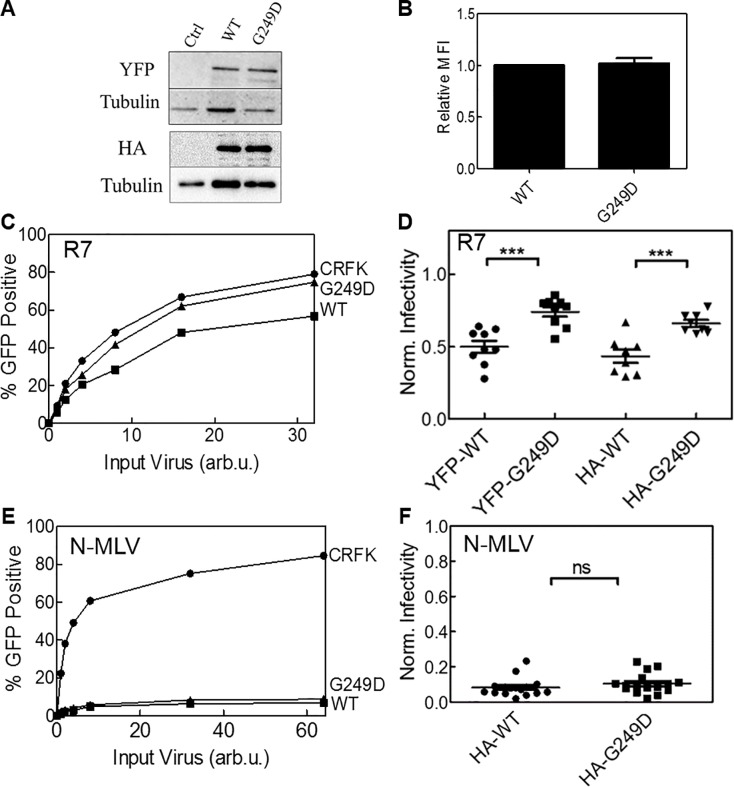
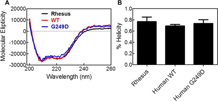
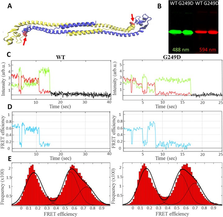
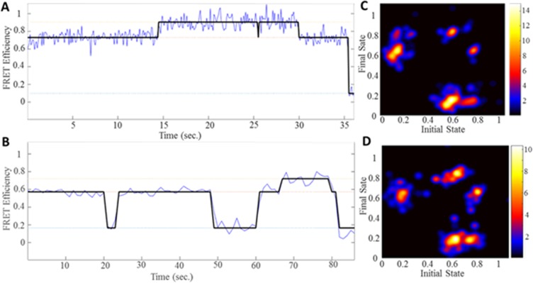
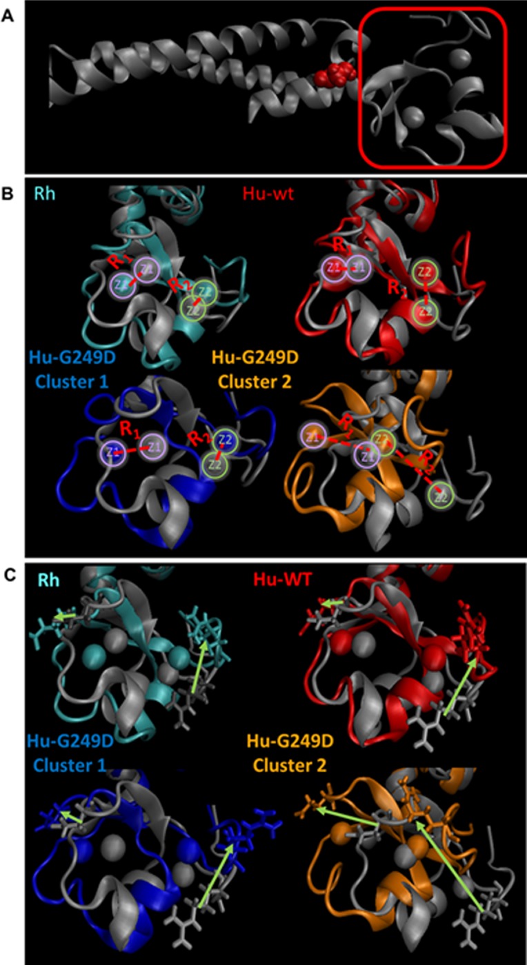
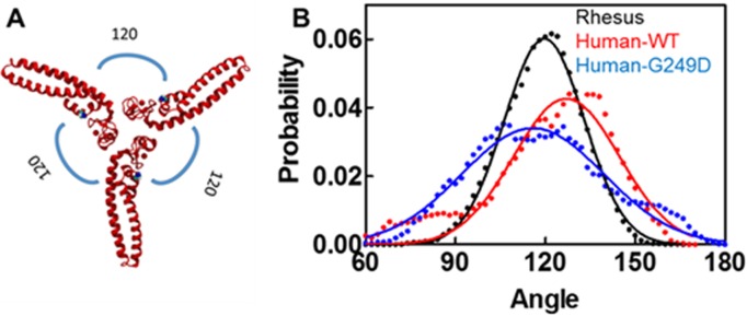
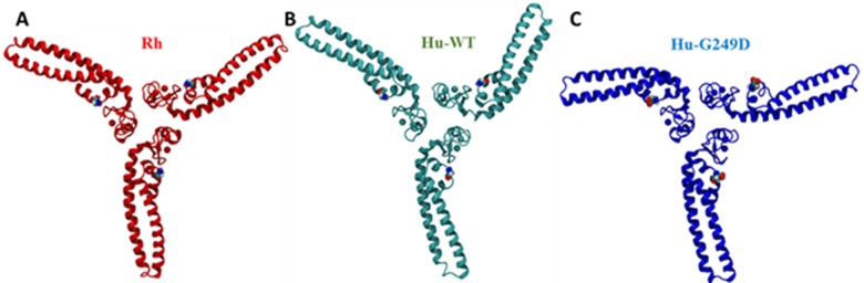
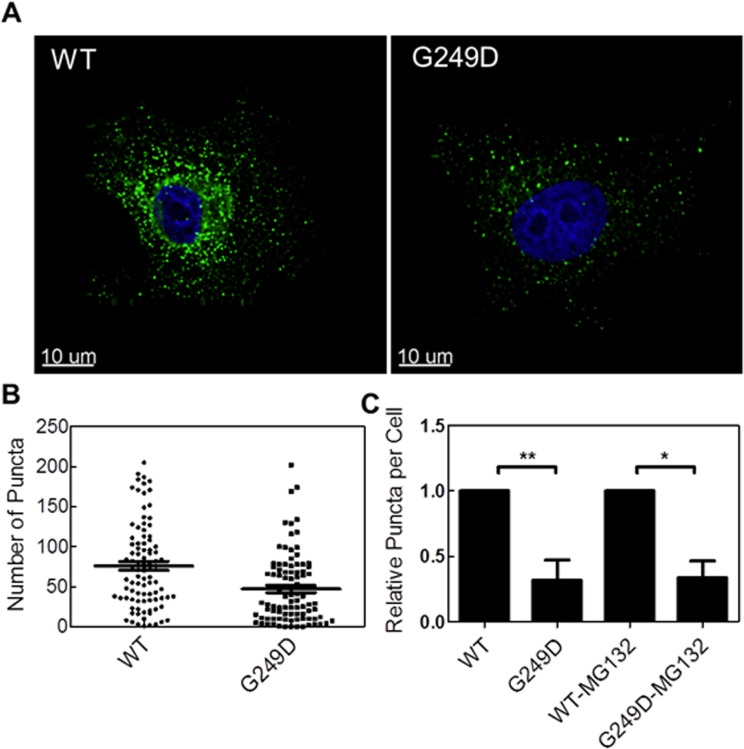
TRIM5α is an interferon inducible restriction factor which contributes to intrinsic defense against HIV infection by targeting the HIV capsid protein CA. Although human TRIM5α (huTRIM5α) does not potently inhibit HIV-1 infection, the ability of huTRIM5α to exhibit some control of HIV-1 infection is evidenced by a single nucleotide polymorphism in huTRIM5α which substitutes aspartic acid to glycine at position 249 (G249D) in the L2 region and is associated with higher susceptibility to HIV-1 infection. To understand the mechanistic basis for the reduced antiviral activity, we employed biophysical and cell biological methods coupled with molecular dynamics simulations to compare WT and the G249D polymorphism of huTRIM5α. We investigated the differences in conformational dynamics of rhesus and huTRIM5α Coiled Coil-Linker 2 (CC-L2) dimers utilizing circular dichroism and single molecule-Fluorescence Energy Transfer (sm-FRET). These methods revealed that the G249D dimer exhibits secondary structure and conformational dynamics similar to WT huTRIM5α. Homology modelling revealed that G249 was present on the hairpin of the antiparallel dimer, in a position which may act to stabilize the adjacent BBox2 domain which mediates the inter-dimeric contacts required for the formation of TRIM5 assemblies. We therefore asked if the G249D mutant forms assemblies in cells with the same efficiency as WT protein by expressing these proteins as YFP fusions and quantifying the number of assemblies in cells. In cells expressing comparable amounts of protein, the G249D mutant formed fewer assemblies than WT protein, in agreement with our homology modeling predictions and molecular dynamics simulations of dimers and higher oligomers of TRIM5α, providing a mechanistic explanation of the reduced antiviral activity of the G249D polymorphism.


Conflict of interest statement

The authors’ declare that they have no competing interests.

Figures

Fig 1
Fig 1. The G249D polymorphism of huTRIM5α exhibits reduced restriction of HIV-1 but not N-MLV.
CRFK cells transduced to stably express YFP and HA tagged human TRIM5α. (A) Protein expression of YFP and HA tagged human WT, G249D TRIM5α and untransduced (control) cell lines by immunoblot. Tubulin used as a loading control. (B) Relative mean fluorescence intensities of WT and G249D TRIM5α by flow cytometry. Error bars represent SEM of 5 biological replicates. (C) Equivalent number of CRFK cells infected by R7ΔEnvGFP or N-MLV (E) by a serial of dilutions. Percent GFP positive cells quantified by flow cytometry 48 hours post infection. (D and F) Normalized infectivity for cell lines expressing YFP and HA tagged human WT, G249D TRIM5α. Normalization done by using percent GFP positive cells of untransduced cell line for virus concentrations resulting less than 35% infectivity (p<0.05 compared to the WT).
Fig 2
Fig 2. G249D polymorphism in human Trim5α does not affect α-helical content.
(A) Circular dichroism of purified CC-L2 peptides of rhesus (in black), human WT (in red) and G249D (in green) TRIM5α given in molecular elipticity units. (B) Helical content is calculated by using Selcon II software. Error bars represent SEM of 4 biological replicates.
Fig 3
Fig 3. Three different conformational states are observed for human WT and G249D CCL2 dimers.
Dually labelled human wt and G249D Trim5α for sm-FRET measurements. (A) Cartoon representation of human wt CC-L2 dimers (yellow and blue) depicting location of fluorophore (Alexa 488 by green and Alexa 594 by red pentagon) conjugation and G249 residue (red arrows). (B) Gel electrophoresis of purified and dually labelled CC-L2 peptides imaged by using green (526 nm) or red (610 nm) emission filters. (C) Representative fluorescence intensity changes over time (donor shown in green, acceptor in red) obtained by TIRF microscopy for WT (left panel) and G249D (right panel) mutant respectively. (D) Calculated FRET efficiencies for CCL2 Dimers for WT (left panel) and G249D mutant (right panel) respectively. (E) Composite histogram of FRET efficiencies compiled from individual traces and fit to three Gaussian distributions for WT (left panel) and G249D mutant (right panel).
Fig 4
Fig 4. Human WT and G249D TRIM5α exhibits similar conformational dynamics by Hidden Markov Model analysis.
Idealized FRET traces were obtained by fitting each trace to a three state Markov Model (in black) for WT (A) and G249D (B) CCL2 dimer (in blue). Transition density plots were generated from the modelled FRET trajectories for WT (C) and G249D (D) CCL2 dimer (68 and 52 traces with 240 and 185 transitions were used for generation of TDP plots for WT and G249D respectively).
Fig 5
Fig 5. Human G249D CCL2 dimer has distorted placement of BBox-2 domain compared crystal structure.
(A) Crystal structure of rhesus CCL2 given in silver. Trajectories from MD simulations classified into clusters according to their RMSD (threshold value of 2.25 Å). (B) Rhesus CCL2 dimer major cluster (in cyan, 64%), human WT CCL2 dimer major cluster (in red, 89%), human G249D CCL2 dimer cluster 1 (in blue, 64%), human G249D CCL2 dimer cluster 2 (in orange, 10%). R1 and R2 represents the distances between zinc ions and zinc ions are color coded (purple for the first and green for the second zinc ion). (C) Representative side chains are depicted for each cluster. Arrows represent how the side chains displaced compared to rhesus crystal structure.
Fig 6
Fig 6. Human WT and G249D Bcc miniTRIM oligomers exhibit wider angle distributions than rhesus miniTRIM.
(A) Rhesus Bcc miniTRIM structure with representative angles. (B) Probability distribution of angles for rhesus (in black), human WT (in red) and G249D (in blue) Bcc miniTRIMs obtained from MD simulations of our homology models (circles) and Gaussian fits (solid lines). The angles are defined by the angle between Cα atoms of residues 117 and 174 for rhesus and 115 and 172 for human oligomers and gated for 2 degrees to obtain frequency distribution.
Fig 7
Fig 7. Representative structures of Human WT and G249D Bcc miniTRIM oligomers by MD simulations showing distortions in G249D miniTRIM.
We used Bcc miniTRIM structures starting geometries for our homology models and obtained MD simulations with trajectories showing distortions in three-fold symmetry. (A) Rhesus Bcc miniTRIM (in red) (B) Human WT Bcc miniTRIM (in cyan) (C) Human G249D Bcc miniTRIM (in blue).
Fig 8
Fig 8. Cytoplasmic body formation is reduced by G249D polymorphism in human Trim5α.
(A) Representative cell images for CRFK cells stably expressing WT or G249D mutant Trim5α (green) with DAPI stain (blue). (B) Number of cytoplasmic bodies per cell for CRFK stably expression YFP tagged WT or G249D mutant Trim5α were determined from surface analysis (C) Relative puncta is computed by normalization to average number of cytoplasmic bodies per cell for cells expressing WT Trim5α and computed for five biological replicates. Error bars represent SEM for five biological replicates (p<0.05 compared to the WT for no treatment and treatment cases).

Similar articles

References

    1. Stremlau M, Owens CM, Perron MJ, Kiessling M, Autissier P, Sodroski J. The cytoplasmic body component TRIM5α restricts HIV-1 infection in Old World monkeys. Nature. 2004;427:848 10.1038/nature02343 https://www.nature.com/articles/nature02343#supplementary-information. - DOI - PubMed
    1. Stremlau M, Perron M, Lee M, Li Y, Song B, Javanbakht H, et al. Specific recognition and accelerated uncoating of retroviral capsids by the TRIM5alpha restriction factor. Proc Natl Acad Sci U S A. 2006;103 10.1073/pnas.0509996103 - DOI - PMC - PubMed
    1. Pertel T, Hausmann S, Morger D, Zuger S, Guerra J, Lascano J, et al. TRIM5 is an innate immune sensor for the retrovirus capsid lattice. Nature. 2011;472(7343):361–5. Epub 2011/04/23. 10.1038/nature09976 - DOI - PMC - PubMed
    1. Grutter MG, Luban J. TRIM5 structure, HIV-1 capsid recognition, and innate immune signaling. Current opinion in virology. 2012;2(2):142–50. Epub 2012/04/10. 10.1016/j.coviro.2012.02.003 - DOI - PMC - PubMed
    1. Ozato K, Shin D-M, Chang T-H, Morse Iii HC. TRIM family proteins and their emerging roles in innate immunity. Nature Reviews Immunology. 2008;8:849 10.1038/nri2413 https://www.nature.com/articles/nri2413#supplementary-information. - DOI - PMC - PubMed

Publication types

MeSH terms