. 2019 Mar 19;15(3):e1007873.
doi: 10.1371/journal.pgen.1007873. eCollection 2019 Mar.

An ABCA4 loss-of-function mutation causes a canine form of Stargardt disease

Affiliations

An ABCA4 loss-of-function mutation causes a canine form of Stargardt disease

Suvi Mäkeläinen et al. PLoS Genet. .

Abstract

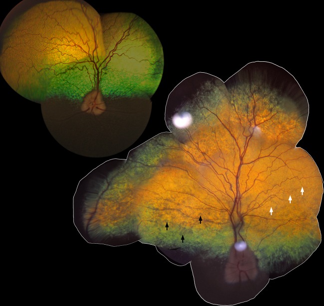
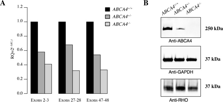
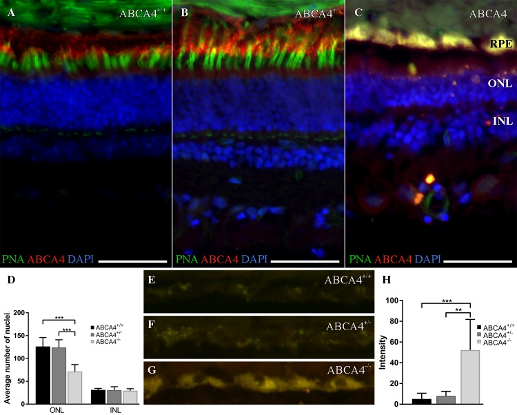
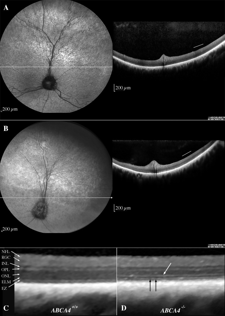
Autosomal recessive retinal degenerative diseases cause visual impairment and blindness in both humans and dogs. Currently, no standard treatment is available, but pioneering gene therapy-based canine models have been instrumental for clinical trials in humans. To study a novel form of retinal degeneration in Labrador retriever dogs with clinical signs indicating cone and rod degeneration, we used whole-genome sequencing of an affected sib-pair and their unaffected parents. A frameshift insertion in the ATP binding cassette subfamily A member 4 (ABCA4) gene (c.4176insC), leading to a premature stop codon in exon 28 (p.F1393Lfs*1395), was identified. In contrast to unaffected dogs, no full-length ABCA4 protein was detected in the retina of an affected dog. The ABCA4 gene encodes a membrane transporter protein localized in the outer segments of rod and cone photoreceptors. In humans, the ABCA4 gene is associated with Stargardt disease (STGD), an autosomal recessive retinal degeneration leading to central visual impairment. A hallmark of STGD is the accumulation of lipofuscin deposits in the retinal pigment epithelium (RPE). The discovery of a canine homozygous ABCA4 loss-of-function mutation may advance the development of dog as a large animal model for human STGD.


Conflict of interest statement

I have read the journal's policy and the authors of this manuscript have the following competing interests: A patent application (US No. 62/662,362) has been filed by the following authors and inventors, TB, GA, BE and SM. CM and SR are affiliated with a diagnostic lab marketing genetic tests for dogs. TB, GA and SM are affiliated with a university department that provides genetic testing for animals.

Figures

Fig 1
Fig 1. Retinal morphology in vivo in canine Stargardt disease.
The tapetal fundus of the right eye from an 11-year-old unaffected Labrador retriever (upper left; LAB27) and a 10-year-old affected dog (lower right; LAB4). Black arrows show areas with abnormal, grayish, hyporeflective appearance and white arrows indicate attenuation of the retinal blood vessels.
Fig 2
Fig 2. Loss-of-function mutation in the canine ABCA4 gene.
(A) Sanger sequencing traces spanning positions Chr6:55,146,545–55,146,564 (CanFam3.1) in exon 28 of the ABCA4 gene of a wild-type, unaffected (ABCA4+/+) dog, a heterozygous (ABCA4+/-) dog, and a homozygous (ABCA4-/-) affected dog. (B) Predicted domain structure of canine full-length ABCA4 protein, based on the proposed human structure [28], and the putative truncated product as a result of the premature stop codon at amino acid residue 1,395. The inferred canine exon numbers are indicated. (C) Schematic representation of the region where the insertion of cytosine (C) is found showing the nucleotide and amino acid sequences of a full-length (top) and truncated (bottom) protein. (D) Predicted topological organization of ABCA4 [29, 30] with the insertion leading to a premature stop codon marked with an arrow. ECD1 = first extracellular domain; TMD1 = first membrane-spanning region; NBD1 = first nucleotide-binding domain; ECD2 = second extracellular domain; TMD2 = second membrane-spanning region; NBD2 = second nucleotide-binding domain.
Fig 3
Fig 3. Characterization of ABCA4 mRNA expression and western blot analyses of ABCA4 protein levels in the canine retina.
(A) Relative ABCA4 mRNA expression levels by quantitative RT-PCR in three different regions in three dogs with different genotypes (ABCA4+/+, ABCA4+/-, and ABCA4-/-), normalized to GAPDH expression. (B) Western blot analyses of ABCA4 (above), GAPDH (middle), and RHO (below) protein levels in retinal tissue of dogs with the three different genotypes.
Fig 4
Fig 4. Fluorescence histochemistry of ABCA4, cone photoreceptors, and autofluorescence in the canine retina.
(A-C) Fluorescence micrographs showing ABCA4 expression (red), FITC-conjugated peanut agglutinin (PNA, green), and DAPI nuclear staining (blue) in wild-type (ABCA4+/+), heterozygous (ABCA4+/-), and affected (ABCA4-/-) retinas. PNA labels cone photoreceptors. Autofluorescence, indicative of lipofuscin accumulation, was seen in the ABCA4-/- RPE. (D) Bar graph with the average number of DAPI-stained nuclei within a given region of the ONL and the INL. (E-G) Fluorescence micrographs of RPE without immunohistochemistry show autofluorescence. (H) Bar graph with background-corrected mean autofluorescence-intensity in the RPE. Note the reduction of ABCA4-immunoreactivity and PNA binding, higher autofluorescence, and fewer nuclei in the ONL in the ABCA4-/- compared to ABCA4+/+ or ABCA4+/- retinas. All scale bars = 50 μm; RPE = retinal pigment epithelium; ONL = outer nuclear layer; INL = inner nuclear layer; Because there was only one individual per genotype, the statistics are valid for the technical replicates. ANOVA with Tukey’s post hoc test, n = 6; **P < 0.01; ***P < 0.001; mean ± S.D.
Fig 5
Fig 5. Histopathology.
Light microscopic histology of a 12-year-old affected (LAB4) dog (A, B), 12-year old heterozygote (LAB6) dog (C, D) and 10-year old unaffected (German spaniel) dog (E, F) taken at comparable locations in the superior central retina (0.5–1.5 cm dorsal to the optic nerve on the sagittal plane). Cone photoreceptors in the retina of the affected dog (A) were scarce (cone nuclei indicated with black arrow) compared with the retinas of the heterozygote (C) and wild-type (E) dogs. In the affected dog (B), accumulation of lipofuscin was abundant in retinal epithelial cells (thick white arrow), compared to heterozygote (D) and wild-type (F) dogs. Photoreceptor outer segment disruption is artifactual. All scale bars = 20 microns; ONL = outer nuclear layer.
Fig 6
Fig 6. Flash-electroretinography (FERG) were used to assess retinal function in vivo.
The green, blue, grey and black tracings indicate wild-type (LAB22; ABCA4+/+) unaffected dog, a heterozygous (LAB6; ABCA4+/-) dog, and two homozygous (LAB4 and LAB3; ABCA4-/-) affected dogs, respectively. Black arrows in A, B and C indicate 3 cd/m2/s-flash stimuli and the red line in D indicate 0.02 cd/m2/s-flash stimuli and scales show amplitude on the y-axis (μV) and time in ms on the x-axis for each type of response. (A) A dark-adapted, mixed rod-cone response. (B) Light-adapted cone transient responses (C) and cone flicker response at 30 Hz. Note that the affected dog had a delayed response to the stimuli. (D) The dark-adapted rod responses monitored during one hour in an affected (LAB3) and a wild-type female (LAB22).
Fig 7
Fig 7. In vivo retinal morphology assessed with cSLO and OCT.
cSLO-images (left) and OCTs (right) (A) from the left eye of a 12-year-old unaffected dog (LAB22) along the visual streak and (B) from the left eye of a 10-year-old affected dog (LAB10), where the horizontal extension of the visual streak is indicated by the black arrows. (C) A magnification of the temporal retina (corresponding to the area below the white bar in the OCTs) of the unaffected dog (LAB22) and accordingly (D) of the affected dog (LAB10), with thickened and hyperreflective ELM (white arrow) and fragmented EZ (black arrows). cSLO = confocal scanning laser ophthalmoscopy; OCT = optical coherence tomography; ELM = external limiting membrane; EZ = ellipsoid zone (inner-to-outer segment junction).
Fig 8
Fig 8. Analysis of retinal layer thickness using OCT.
Graphs showing the mean thickness of retinal layers from OCTs of two wild-type dogs at the age of 10- and 12-years (green dots; LAB23 and LAB22, respectively) and two affected dogs at the age of 10- and 12-years (blue squares; LAB10 and LAB16, respectively). The thickness is measured every 0.5 mm from the rim of the optic nerve head (0) along the visual streak. The solid lines indicate 95% confidence intervals. Measurement of the (A) total retinal thickness, (B) inner retina (C) Rec+, (D) ONL and (E) EZ+RPE. (F) The distances presented in the five graphs are shown on the magnified OCT image from a wild-type Labrador retriever dog. OCT = optical coherence tomography; EZ = ellipsoid zone (inner-to-outer segment junction); Rec+ = total photoreceptor length; RPE = retinal pigment epithelium; ONL = outer nuclear layer.

References

    1. Berger W, Kloeckener-Gruissem B, Neidhardt J. The molecular basis of human retinal and vitreoretinal diseases. Progress in Retinal and Eye Research. 2010;29(5):335–75. 10.1016/j.preteyeres.2010.03.004 - DOI - PubMed
    1. den Hollander AI, Black A, Bennett J, Cremers FPM. Lighting a candle in the dark: advances in genetics and gene therapy of recessive retinal dystrophies. The Journal of Clinical Investigation. 2010;120(9):3042–53. 10.1172/JCI42258 PMC2929718. - DOI - PMC - PubMed
    1. Miyadera K, Kato K, Aguirre-Hernandez J, Tokuriki T, Morimoto K, Busse C, et al. Phenotypic variation and genotype-phenotype discordance in canine cone-rod dystrophy with an RPGRIP1 mutation. Molecular vision. 2009;15:2287–305. Epub 2009/11/26. - PMC - PubMed
    1. Downs LM, Wallin-Håkansson B, Bergström T, Mellersh CS. A novel mutation in TTC8 is associated with progressive retinal atrophy in the golden retriever. Canine Genetics and Epidemiology. 2014;1:4 10.1186/2052-6687-1-4 PMC4574394. - DOI - PMC - PubMed
    1. Downs LM, Hitti R, Pregnolato S, Mellersh CS. Genetic screening for PRA-associated mutations in multiple dog breeds shows that PRA is heterogeneous within and between breeds. Vet Ophthalmol. 2014;17(2):126–30. Epub 2013/11/22. 10.1111/vop.12122 . - DOI - PubMed

Publication types

MeSH terms

Substances