. 2019 Mar 19;14(3):e0213553.
doi: 10.1371/journal.pone.0213553. eCollection 2019.

Aicardi-Goutières Syndrome associated mutations of RNase H2B impair its interaction with ZMYM3 and the CoREST histone-modifying complex

Affiliations

Aicardi-Goutières Syndrome associated mutations of RNase H2B impair its interaction with ZMYM3 and the CoREST histone-modifying complex

Alexander Shapson-Coe et al. PLoS One. .

Abstract

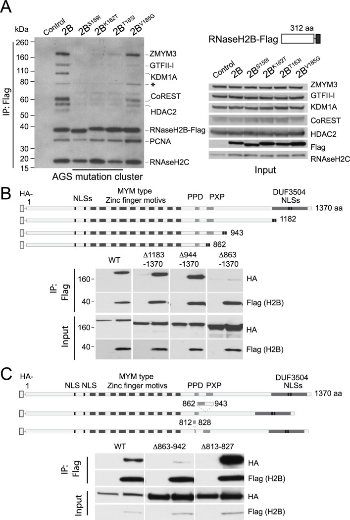
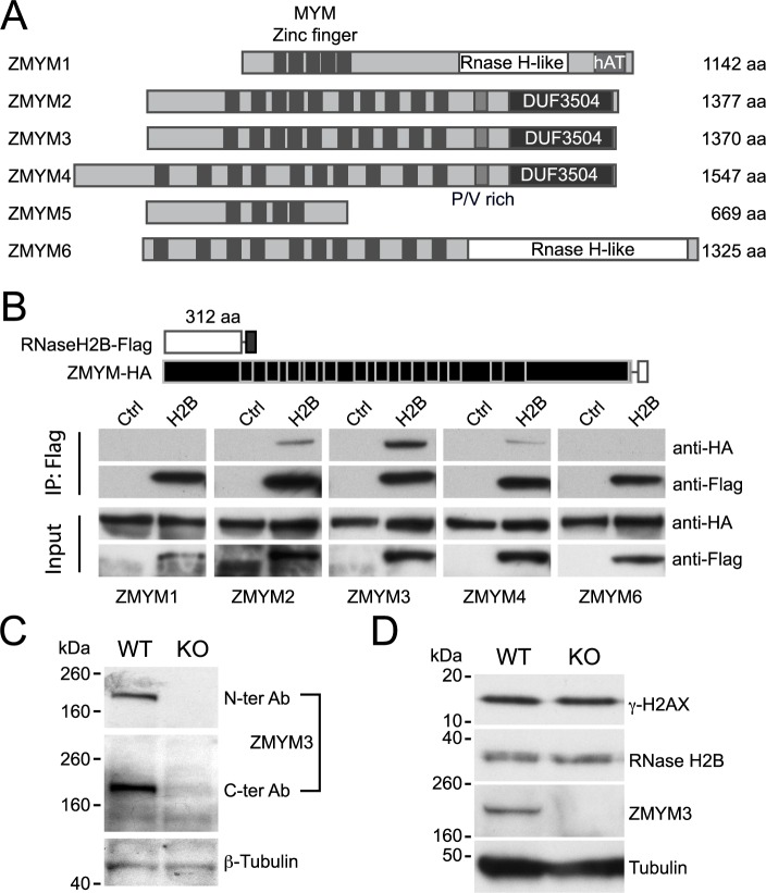
DNA-RNA hybrids arise in all cell types, and are removed by multiple enzymes, including the trimeric ribonuclease, RNase H2. Mutations in human RNase H2 result in Aicardi-Goutières syndrome (AGS), an inflammatory brain disorder notable for being a Mendelian mimic of congenital viral infection. Previous studies have shown that several AGS-associated mutations of the RNase H2B subunit do not affect trimer stability or catalytic activity and are clustered on the surface of the complex, leading us to speculate that these mutations might impair important interactions of RNase H2 with so far unidentified proteins. In this study, we show that AGS mutations in this cluster impair the interaction of RNase H2 with several members of the CoREST chromatin-silencing complex that include the histone deacetylase HDAC2 and the demethylase KDM1A, the transcriptional regulators RCOR1 and GTFII-I as well as ZMYM3, an MYM-type zinc finger protein. We also show that the interaction is mediated by the zinc finger protein ZMYM3, suggesting that ZMYM3 acts as a novel type of scaffold protein coordinating interactions between deacetylase, demethylase and RNase H type enzymes, raising the question of whether coordination between histone modifications and the degradation of RNA-DNA hybrids may be required to prevent inflammation in humans.


Conflict of interest statement

The authors have declared that no competing interests exist.

Figures

Fig 1
Fig 1. Identification of a novel RNase H2 binding partner ZMYM3.
(A) Surface representation of human RNase H2 and location of mutations on the surface of the B subunit. The A, C and B subunits are in red, pink and blue respectively with residues mutated in Aicardi–Goutières syndrome shown in yellow and the catalytic site boxed in black. The location of the S159I, K162T and T163I cluster of mutations are indicated on the top view of the structure. Protein Data Bank accession code: 3PUF [14]. (B) Protein complexes associated with wild-type RNase H2B are disrupted by single amino acid substitutions. Pull down of protein complexes associated with RNase H2B-Flag are shown by silver-stained SDS-Page. Lanes correspond to the individual AGS mutations indicated as well wild type (2B) RNase H2B and nonspecific proteins (Ctrl). The closed arrows indicate the RNase H2B subunits. The open arrow indicates a band that is lost in mutant forms of RNase H2. The same result was observed in three independent pull-downs. (C) Peptides identified by mass spectrometric analysis of the regions corresponding to the arrows indicated in (B), ranked by number of peptides identified. Results for the region corresponding to band 4 from two independent pulldowns are listed separately. The most abundant peptides correspond to the RNase H2 subunits and known associated proteins such as PCNA (in black). The common protein identified in band 4 corresponds to the zinc finger MYM-type protein 3 (bold). (D) Endogenous untagged ZMYM3 is associated with endogenous RNase H2B. The schematic representation indicates the protein size in amino acids (aa). Rabbit anti-RNase H2B immunoprecipitaes from HEK293T cell lysates compared control rabbit IgG from serum. The same result was observed in three independent pull-downs. The input corresponds to ~6% of the lysates used for IP. Rabbit anti-ZMYM3 was developed with goat anti-Rabbit Peroxidase, and the rabbit anti-RNase H2B with TrueBlot anti-rabbit IgG Peroxidase. (Uncropped files are included in S7 Fig).
Fig 2
Fig 2. RNase H2 interaction with ZMYM3 and CoREST is mediated the proline rich region.
(A) ZMYM3 and RNase H2B are associated as part of the larger CoREST complex. Transfected FLAG-RNase H2B pull downs in HEK293T cell extracts brings down ZMYM3 and CoREST, but both are lost or partially lost in mutants S159I, K162T and T163. On the left, individual developed films are shown as an overlap to allow comparison of the different protein sizes. The input on the right (~3% of the lysates) is shown for the endogenous RNase H and members of the CoREST complex as well as the Flag-RNase H2B transfected protein. Antibodies used for western blotting were Rabbit anti-ZMYM3, Mouse anti-FLAG M2 Peroxidase, Rabbit anti-RNase H2C, Rabbit anti-GFII-I, Rabbit anti-CoREST, Rabbit anti-HDAC2, Goat anti-Rabbit IgG Peroxidase and Goat anti-Mouse IgG Peroxidase. The * indicates an isoform of GTFII-I. The same result was observed in four independent pull-downs. (B) HA-tagged ZMYM3 protein fragments in HEK293T cell extracts co-immunoprecipitated with FLAG-tagged RNase H2B. The schematic shows the domain architecture of ZMYM3 and the C-terminally truncated fragments, including the nuclear localisation region from the DUF3504 domain (black). The first and last amino acids of full and truncation proteins are indicated according to full-length human ZMYM3 (UniProtKB: Q14202.2). PPD: Proline-rich region. NLS: Nuclear localisation signal. Each pull down is shown next to lysates transfected with HA-tagged ZMYM3 fragments and Flag-vector controls. The input equals 3% of the total lysate. Antibodies used to visualise the proteins were Mouse anti-FLAG M2 Peroxidase and Rat anti-HA Peroxidase. (C) Small internal deletions of the conserved proline rich region of motif ZMYM3 prevent its interaction with FLAG-tagged RNase H2B. As B, amino acids shown in the schematic on either side of the deleted segment are according to full-length human ZMYM3 (UniProtKB: Q14202.2). Each pull down is shown next to lysates transfected with HA-tagged ZMYM3 fragments and Flag-vector controls. The input equals 3% of the total lysate. Antibodies used to visualise the proteins were Mouse anti-FLAG M2 Peroxidase and Rat anti-HA Peroxidase. The same results were observed for B and C in three independent pull-downs each.
Fig 3
Fig 3. The ZMYM3 zinc finger domains mediate its interactions with the CoREST complex.
(A) Pulldown of members of the CoREST complex by FLAG-tagged fragments of ZMYM3 zinc finger domains. The schematic shows the domain architecture of ZMYM3 and the Flag tagged fragments. Lysates from cells transfected with empty vector are show as controls. The input equals 3% of the total lysate. Levels of the endogenous KDM1A, HDAC2 and RCOR1/CoREST proteins relative to each other were consistent in all experiments and are shown as control of each other in input lysates. The expression of fragment 620–678 (corresponding to zinc finger 8) was reproducibly lower than that of the rest of the fragments, including the fragment corresponding to zinc finger 9, as shown. Antibodies used to visualise the proteins were Rabbit anti-CoREST, Rabbit anti-HDAC2, Rabbit anti-KDM1A (LSD1), Goat anti-Rabbit IgG Peroxidase and Mouse anti-FLAG M2 Peroxidase. The size of the molecular standards is indicated in kDa. (B) Pulldown of GFII-I co-immunoprecipitated with HA-tagged fragments of ZMYM3 zinc finger domains. As in A, the schematic shows the fragments identified by the corresponding amino acids. Antibodies used were Rabbit anti-GFII-I, Goat anti-Rabbit IgG-HRP and Rat anti-HA Peroxidase. The same results were observed for A and B in three independent pull-downs each.
Fig 4
Fig 4. ZMYM3 and RNase H2B interaction is redundant with other ZMYM proteins.
(A) Schematic representation of the conserved members of the human ZMYM protein family. Conserved domains are aligned based on homology. The MYM type zinc fingers are indicated as is the proline rich (PV) region and the domain of unknown function DUF3504. ZMYM 1 and 6 contain a domain with structural homology to the RNase H fold, but have no known RNase activity. (B) Pull down of transfected FLAG-tagged RNase H2B and HA-tagged human ZMYM proteins. Empty Flag-vector transfected HEK293T cells lysates are shown as control next to each co-expressing HA- tagged ZMYM 1, 2, 3, 4 and 6 lysates. Antibodies used were Mouse anti-FLAG M2 Peroxidase and Rat anti-HA Peroxidase. The input equals 3% of the total lysate. The same results were observed in three independent pull-downs. (C) Protein expression of mouse ZMYM3 in control and Zmym3-/ ES cells. Two different monoclonal antibodies directed against the N terminus and C terminus of the protein were used to detect ZMYM3 in C57Bl/6 derived ES cells. Targeted Zmym3-/ lacking Exon3 (KO) do not express either portion of the protein. The relative size of molecular standards is shown in kDa. β-tubulin levels are shown as control. (D) Expression of the damage response marker γ-H2AX is not affected in Zmym3-/ ES cells. Protein lysates from the parental C57Bl/6 derived ES cells (WT) compared with the Zmym3-/ ES cell lysates (KO). The relative size of molecular standards is shown in kDa. α-tubulin levels are shown as control. Antibodies used were Goat anti-ZMYM3, Mouse anti-γ-H2AX, Mouse anti-α-Tubulin, Rabbit anti-RNase H2B, Goat anti-Rabbit IgG Peroxidase, Donkey anti-Goat IgG Peroxidase and Goat anti-Mouse IgG Peroxidase. The same results in C and D were observed in three independent clones of targeted C57Bl/6-derived ES cells.

Similar articles

Cited by

References

    1. Aicardi J, Goutieres F. A Progressive familial encephalopathy in infancy with calcifications of the basal ganglia and chronic cerebrospinal fluid lymphocytosis. Annals of Neurology. Wiley Subscription Services, Inc., A Wiley Company; 1984;15: 49–54. 10.1002/ana.410150109 - DOI - PubMed
    1. Crow YJ. Aicardi-Goutières Syndrome. Pagon RA, Adam MP, Ardinger HH, Wallace SE, Amemiya A, Bean LJ, et al., editors. Seattle (WA): University of Washington, Seattle; 2005. - PubMed
    1. Campbell IL, Krucker T, Steffensen S, Akwa Y, Powell HC, Lane T, et al. Structural and functional neuropathology in transgenic mice with CNS expression of IFN-alpha. Brain Res. 1999;835: 46–61. - PubMed
    1. Rice GI, del Toro Duany Y, Jenkinson EM, Forte GMA, Anderson BH, Ariaudo G, et al. Gain-of-function mutations in IFIH1 cause a spectrum of human disease phenotypes associated with upregulated type I interferon signaling. Nat Genet. 2014;46: 503–509. 10.1038/ng.2933 - DOI - PMC - PubMed
    1. Crow YJ, Hayward BE, Parmar R, Robins P, Leitch A, Ali M, et al. Mutations in the gene encoding the 3“-5” DNA exonuclease TREX1 cause Aicardi-Goutières syndrome at the AGS1 locus. Nat Genet. 2006;38: 917–920. 10.1038/ng1845 - DOI - PubMed

Publication types

MeSH terms

Supplementary concepts