Review
. 2019 Mar 18;35(3):347-367.
doi: 10.1016/j.ccell.2019.01.007.

Integrin Signaling in Cancer: Mechanotransduction, Stemness, Epithelial Plasticity, and Therapeutic Resistance

Affiliations
Review

Integrin Signaling in Cancer: Mechanotransduction, Stemness, Epithelial Plasticity, and Therapeutic Resistance

Jonathan Cooper et al. Cancer Cell. .

Abstract

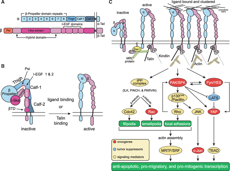
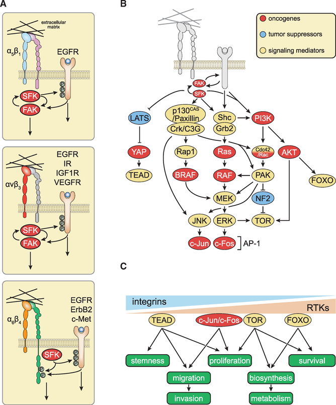
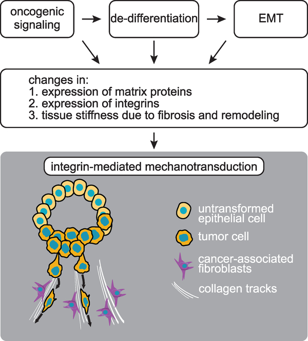
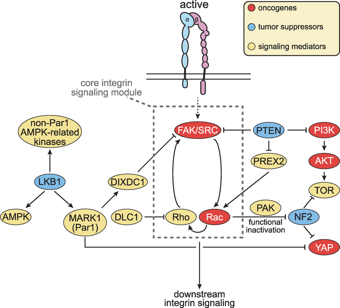
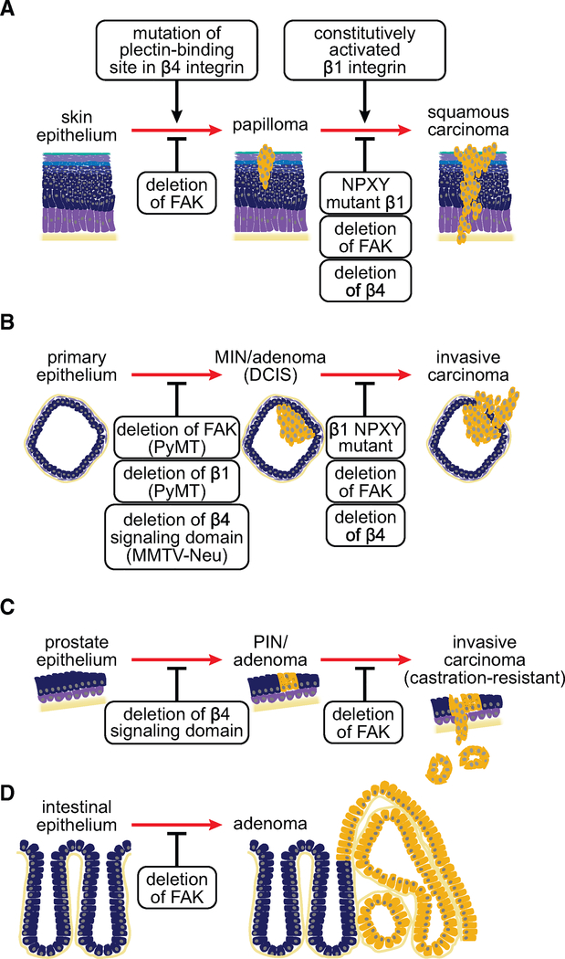
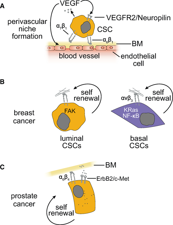
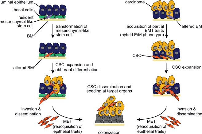
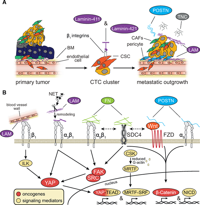
Integrins mediate cell adhesion and transmit mechanical and chemical signals to the cell interior. Various mechanisms deregulate integrin signaling in cancer, empowering tumor cells with the ability to proliferate without restraint, to invade through tissue boundaries, and to survive in foreign microenvironments. Recent studies have revealed that integrin signaling drives multiple stem cell functions, including tumor initiation, epithelial plasticity, metastatic reactivation, and resistance to oncogene- and immune-targeted therapies. Here, we discuss the mechanisms leading to the deregulation of integrin signaling in cancer and its various consequences. We place emphasis on novel functions, determinants of context dependency, and mechanism-based therapeutic opportunities.

Keywords: FAK signaling; cancer stem cell; extracellular niche; integrins; mechanotransduction; metastasis and invasion; receptor tyrosine kinase; therapeutic resistance; therapeutic targeting of integrins; tumor microenvironment.


Figures

Figure 1.
Figure 1.. Integrin-Mediated Signal Transduction
(A) Domain organization and structure of a generic integrin. The α and β subunits have large extracellular domains and short cytoplasmic domains. Exceptions to this generic domain structure include the a subunits of leukocyte integrins (αL, αM, and αX) and those of collagen-binding β1 integrins, which have an I domain inserted between β propeller domains 2 and 3. When present, the I domain participates in ligand binding together with the I-like domain in the extracellular portion of the β subunit. In addition, the β4 integrin is also structurally variant as it possesses a large and unique cytoplasmic domain with two pairs of type III fibronectin-like repeats and connects with the keratin, not the actin, cytoskeleton at hemidesmosomes. (B) Allostery-driven bidirectional signaling. The β propeller in the N-terminal portion of the α subunit combines with the I-like and hybrid domain in the corresponding portion of the β subunit to form the ligand binding pocket and the head piece of the integrin. Inactive integrins exhibit a closed conformation (“are bent at their knees”): the ligand binding pocket possesses low affinity for ligand and faces toward the plasma membrane and the “legs” (α subunits Calf-1 and −2; β subunit I-EGF3, I-EGF4 and the membrane-proximal tail domain βTD), transmembrane and cytoplasmic domains are adjoined (left). Talin binding to the β subunit cytoplasmic domain triggers large conformational changes that include an extension of the legs and a separation of the heterodimeric subunits at the level of the transmembrane and cytoplasmic domains. Ligand binding to partially active integrins can induce the same conformational changes, reinforcing integrin activation. Active integrins are extended and possess a ligand binding-competent headpiece exposed toward the extracellular space and a β cytotail bound to talin (right). These mechanisms ensure that talin binding induces integrin activation and, conversely, ligand binding induces talin binding and hence association with the actin cytoskeleton. (C) Integrin activation, clustering, and autonomous signaling. Current models suggest that GTP-Rap1 induces MRL proteins, such as RIAM and lamellipodin, to activate talin, which in turn binds to the integrin β subunit cytoplasmic domain, separating it from the corresponding portion of the α subunit. Talin binding thus triggers the large conformational changes that lead to integrin activation. Both talin and kindlin can connect to the actin cytoskeleton and promote integrin clustering, initiating robust intracellular signaling. In addition to organizing the cytoskeleton and thereby impinging on myocardin-related transcription factor (MRTF)/serum response factor (SRF), integrin signaling contributes to the activation of AP-1, inhibition of FOXO, and activation of YAP/TEAD. While the integrins impinge on these transcription factors predominantly through activation of MRTF, c-Jun, and YAP, receptor tyrosine kinases potently activate c-Fos and inhibit FOXO through the Ras-ERK and PI3K-AKT pathways (see Figure 2). Oncogenes are shown in red, tumor suppressors in blue, and mediators of integrin signaling in yellow.
Figure 2.
Figure 2.. Joint Integrin-Receptor Tyrosine Kinase Signaling and Mechanotransduction
(A) Membrane-proximal mechanisms underlying joint integrin-RTK signaling. Integrins buttress mitogenic signaling by RTKs at multiple levels. At or near the plasma membrane, integrin-activated SRC family kinases (SFKs) induce the phosphorylation of the P loop of RTKs, priming them for ligand-induced activation. The RTKs in turn induce phosphorylation of focal adhesion kinase (FAK) or the signaling domain of the β4 integrin. These elements recruit distinct subsets of signaling enzymes and adaptors, refining the specificity of individual partner RTKs. (B) Major membrane-distal interconnections between integrin and RTK signaling pathways. Integrins and RTKs cooperate to activate FAK, SFKs, and PI3K. The FAK-SFK complex phosphorylates and activates p130CAS and paxillin, thereby mediating activation of JNK and––in cells expressing Rap1––of BRAF. Several mechanisms ensure joint regulation of Ras and PI3K by integrins and RTKs. (C) Integrins and RTKs regulate multiple cellular functions of normal and transformed cells through additive, co-dependent, or synergistic mechanisms.
Figure 3.
Figure 3.. Origin and Consequences of Integrin-Mediated Mechanotransduction in Cancer
Oncogenic signaling and changes in cell fate contribute to deregulate integrin expression and function in cancer. Changes in matrix composition and structure enable integrins to transmit integrated biochemical and mechanical signals to the interior of cancer cells.
Figure 4.
Figure 4.. Rewiring of the Integrin Signaling Network in Cancer
Alterations in oncogenes and tumor suppressors deregulate integrin signaling in cancer. Oncogenic receptor tyrosine kinases and protumorigenic integrins collaborate to activate FAK-SFK signaling. Mutations in LKB1, MARK1, and DIXDC1 remove an inhibitory constraint. Mutations in PTEN exert the same effect by activating FAK as well as Rac. In addition to reinforcing activation of Rho and hence FAK, Rac activates PAK and functionally suppresses NF2. DLC1 opposes activation of Rho. Additional mechanisms enable specific integrins to promote or suppress tumorigenesis, often in a context-dependent manner.
Figure 5.
Figure 5.. Genetics Underlying Integral Roles of Integrins and FAK in Cancer Development and Progression
Genetic studies in mice revealed integrins, in particular β4, and FAK are necessary for tumor initiation and progression in models of skin (A), breast (B), prostate (C), and intestinal (D) cancers, yet the mechanisms underlying these dependencies vary between the models. DCIS, ductal carcinoma in situ; MIN, mammary intraepithelial neoplasia; PIN, prostatic intraepithelial neoplasia.
Figure 6.
Figure 6.. Specific Integrin Signaling Mechanisms Contribute to the Self-Renewal of Cancer Stem Cells in Different Tumor Types
(A) The cancer stem cells in several cancer types adhere to the endothelial basement membrane (BM) at perivascular niches via α6β1 or α7β1 integrins and undergo self-renewal in response to activation of FAK. (B and C) Shown are major integrin signaling mechanisms that enable epithelial BMs to support breast (B) and prostate (C) cancer stem cells.
Figure 7.
Figure 7.. EMT and Stemness
Models for the role of the EMT in stemness and metastatic colonization of carcinomas. Cancer stem cells may possess mesenchymal traits because they originate from the transformation of mesenchymal-like stem cells or the de-differentiation of neoplastic epithelial progenitors. Cancer stem cells with mesenchymal traits produce aberrantly differentiated epithelial progeny at the primary site and, because of their elevated invasive capacity, disseminate through the bloodstream and seed target organs (left). In a non-mutually exclusive model, the cancer stem cells possess epithelial traits but undergo a “partial” EMT in response to micro-environmental cues and thereby acquire the capacity to invade and disseminate (right). It is currently unresolved whether cancer stem cells with mesenchymal traits have to re-acquire epithelial traits (mesenchymal-epithelial transition [MET]) in order to outgrow into macroscopic metastases (bottom).
Figure 8.
Figure 8.. Integrin Signaling in Tumor Initiation and Re-initiation at Metastatic Sites
(A) Secretion of specific laminin (LAM) isoforms facilitates carcinoma initiation and re-initiation at metastatic sites by activating β1 integrin signaling. Tenascin-C (TNC) and periostin (POSTN) are also key stromal elements promoting metastatic reactivation. (B) Stromal elements and cancer cells cooperate to deposit the extracellular matrix of metastatic niches through several mechanisms including neutrophil extracellular trap (NET) remodeling. LAM, fibronectin (FN), POSTN, and TNC––acting through integrins, Syndecan-4 (SDC4), and the Wnt receptor Frizzled (FZD)––activate signaling pathways involved in metastatic outgrowth.

References

    1. Aceto N, Bardia A, Miyamoto DT, Donaldson MC, Wittner BS, Spencer JA, Yu M, Pely A, Engstrom A, Zhu H, et al. (2014). Circulating tumor cell clusters are oligoclonal precursors of breast cancer metastasis. Cell 158, 1110–1122. - PMC - PubMed
    1. Adams D, Gonzalez-Duarte A, O’Riordan WD, Yang CC, Ueda M, Kristen AV, Tournev I, Schmidt HH, Coelho T, Berk JL, et al. (2018). Patisiran, an RNAi therapeutic, for hereditary transthyretin amyloidosis. N. Engl. J. Med 379, 11–21. - PubMed
    1. Ahmed AA, Mills AD, Ibrahim AE, Temple J, Blenkiron C, Vias M, Massie CE, Iyer NG, McGeoch A, Crawford R, et al. (2007). The extracellular matrix protein TGFBI induces microtubule stabilization and sensitizes ovarian cancers to paclitaxel. Cancer Cell 12, 514–527. - PMC - PubMed
    1. Ahmed KM, Pandita RK, Singh DK, Hunt CR, and Pandita TK (2018). β1-Integrin impacts Rad51 stability and DNA double-strand break repair by homologous recombination. Mol. Cell. Biol 38, 10.1128/MCB.00672-17. - DOI - PMC - PubMed
    1. Alanko J, and Ivaska J (2016). Endosomes: emerging platforms for integrin-mediated FAK signalling. Trends Cell Biol 26, 391–398. - PubMed

Publication types