Review
. 2019 May:79:89-97.
doi: 10.1016/j.ceca.2019.02.006. Epub 2019 Feb 16.

Redox regulation of ER and mitochondrial Ca2+ signaling in cell survival and death

Affiliations
Review

Redox regulation of ER and mitochondrial Ca2+ signaling in cell survival and death

Suresh K Joseph et al. Cell Calcium. 2019 May.

Abstract

Physiological signaling by reactive oxygen species (ROS) and their pathophysiological role in cell death are well recognized. This review focuses on two ROS targets that are key to local Ca2+ signaling at the ER/mitochondrial interface - notably, inositol trisphosphate receptors (IP3Rs) and the mitochondrial calcium uniporter (MCU). Both transport systems are central to molecular mechanisms in cell survival and death. Methods for the measurement of the redox state of these proteins and for the detection of ROS nanodomains are described. Recent results on the redox regulation of these proteins are reviewed.

Keywords: Ca(2+); IP(3); IP(3) receptor; Mitochondrial calcium uniporter; Reactive oxygen species; Redox regulation.


Figures

Figure 1
Figure 1. Model of redox regulation of Ca2+signaling at the ER-mitochondrial interface.
IP3Rs are shown as undergoing reduction or oxidation. The binding of the ER luminal chaperone ERp44 favors the reduced species and inhibits channel function. Oxidative conditions induced by Ero1α stimulate channel function by altering the redox state of IP3R intraluminal thiols and/or by binding to ERp44. Oxidation of critical thiols in the cytosolic domain of the IP3R makes a major contribution to stimulation of channel activity. Enhanced efflux from the ER is directed into the mitochondria facilitated by MCU which itself is redox sensitive by a mechanism involving the cysteine-97 residue. Depending on the magnitude of the mitochondrial Ca2+uptake and the concomitant ROS production, the mitochondria can generate more ATP or open the PTP as a prelude to cell death.
Figure 2
Figure 2. Methods for measuring the redox state of IP3Rs in situ
Panel A. Work-flow diagram showing the steps used for measuring the redox state of IP3Rs by gel-shift assay or LC-MS/MS. Panel B. Representative gel-shift assays of HEK293 cells transfected with IP3R1 and treated for 10min with H2O2 (0.5mM) or thimerosal (50μM). TCA precipitated lysates were processed as described in Panel A and reacted with for 1h with 0.5mM MPEG-2 or MPEG-5. A control is also shown in which the cells were pretreated for 30min with DTT (10mM) before lysis. Panel C. Cartoon showing the redox state of cysteines in IP3R1 determined by LC-MS/MS. Redox lysates were prepared from transfected HEK293 cells treated in the presence and absence of thimerosal (50μM) and processed sequentially with iodoacetamide (IAM), DTT and biotin maleimides as depicted in Panel A. The lysates were processed on a sephadex-G25 column to exchange the sample into a buffer suitable for immunoprecipitation which was carried out with a C-terminal IP3R1 Ab. The immunoprecipitates were processed on 5% SDS-PAGE and the silver-stained IP3R1 was excised and processed for LC-MS/MS. Spectra were analyzed with the SEQUEST search engine for tryptic peptides containing cysteines modified with IAM (reduced) or biotin maleimide (oxidized). Underlined residues were identified in each of 3 independent trials. Oxidized residues observed in H2O2 (0.5mM) treated cells are indicated by arrows. Open circles are residues that were not detected in the analysis. For additional details see Ref. [68].
Figure 3
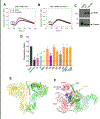
Figure 3. Location of functionally relevant redox-sensitive cysteines in IP3R1
Panel A & B. Representative traces illustrating the approach to measuring redox potentiation described in Section 2.3 are shown. HEK293 cells in which all 3 IP3R isoforms have been deleted by CRISPR were transfected with wild-type IP3R1 or a mutant in which the first 13 N-terminal cysteines are substituted with serine (cys-less)[28]. The cells were loaded with Fluo-8 AM and the cytosolic Ca2+ response to a subsaturating dose of carbachol (0.25μM) was measured in the absence of extracellular Ca2+ with and without a 2min preincubation with 10μM Thimerosal. The data were obtained with a flex station plate reader and are the mean + S.D. of 4 wells for each condition. Responses to maximal doses of carbachol were also quantified and were not significantly different between wild-type and cys-less mutant (not shown). Panel C. The expression of the cys-less mutant is shown by immunoblotting with IP3R1 Ab. Panel D The thimerosal-induced potentiation of the individual cysteines corresponding to the cys-less mutant were measured as described in panels A &. The amplitude of the Ca2+ response to 0.25μM carbachol in the presence or absence of thimerosal treatment was quantified as a “potentiation ratio”. All data are the mean + S.E.M. of n=3–6 independent experiments. *; P<0.001. All the mutants were responsive to maximal concentrations of carbachol (not shown). Panel E Side-view of the cryo-EM structure of IP3R1 [69] showing the location of the cysteine mutants used in Panel D. For clarity only two opposing subunits of the tetramer are shown. The residues with diminished redox potentiation are color-coded as given in Panel D: C15 (red), C37(blue), C56/61 (yellow), C214 (purple). Residues having no significant effects on redox potentiation are colored orange: C206, C253, C292, C394, C530, C553, C556, C767, C1415, C1459. Orange arrows are used for residues where the side-chains were not resolved (C767, C1415, C1459). C326 is located in the SI splice site which was not present in the cryo-EM structure. Panel F A close-up of the top view of the cytosolic surface of the channel showing the 3 domains that constitute the ligand-binding site of a single subunit (colored green) with the approximate position of IP3 indicated by a sphere. The domains are the β-trefoil 1 domain (β-TF1), β-TF2 and the armadillo solenoid fold-1 (ARM-1). The structure shows the segregation of the redox-sensitive residues to a small region of β-TF1. Other domains from an adjacent subunit that are in contact with β-TF1 are shown in red. For additional details see text and Ref. [68].
Figure 4
Figure 4. Alternative hypotheses linking ER stress and IP3R-mediated Ca2+signaling
Shown are some alternative mechanisms of regulating IP3R channel activity during ER stress independently of changes in ER luminal redox state involving ERp44 or Ero1α. The EF-hand containing protein CIB1 is depicted as partitioning between IP3Rs or Ask1, a kinase which is a required component of the MAP-kinase cascade linking the ER stress sensor, Ire1α, to apoptotic and inflammatory pathways. CIB1 binding inhibits Ask1 kinase activity and CIB1/Ask1 complex formation is blocked by Ca2+binding to CIB1. This provides one mechanism by which IP3R-mediated Ca2+ signaling can regulate ER-stress induced apoptosis. Also shown is the possible regulation of IP3Rs by local ROS generated by NADPH oxidases in the ER.

References

    1. Kaludercic N, Giorgio V, The Dual Function of Reactive Oxygen/Nitrogen Species in Bioenergetics and Cell Death: The Role of ATP Synthase, Oxidative medicine and cellular longevity, 2016 (2016) 3869610. - PMC - PubMed
    1. Bernardi P, Rasola A, Forte M, Lippe G, The Mitochondrial Permeability Transition Pore: Channel Formation by F-ATP Synthase, Integration in Signal Transduction, and Role in Pathophysiology, Physiol Rev, 95 (2015) 1111–1155. - PMC - PubMed
    1. O-Uchi J, Ryu SY, Jhun BS, Hurst S, Sheu SS, Mitochondrial ion channels/transporters as sensors and regulators of cellular redox signaling, Antioxidants & redox signaling, 21 (2014) 987–1006. - PMC - PubMed
    1. Tajeddine N, How do reactive oxygen species and calcium trigger mitochondrial membrane permeabilisation?, Biochimica et biophysica acta, 1860 (2016) 1079–1088. - PubMed
    1. Joseph SK, Role of thiols in the structure and function of IP3 receptors, Current Topics Membrane transport, 66 (2010) 299–322. - PubMed

Publication types