. 2019;11(6):457-468.
doi: 10.1159/000496403. Epub 2019 Mar 19.

Streptolysin O Induces the Ubiquitination and Degradation of Pro-IL-1β

Affiliations

Streptolysin O Induces the Ubiquitination and Degradation of Pro-IL-1β

Dóra Hancz et al. J Innate Immun. 2019.

Abstract

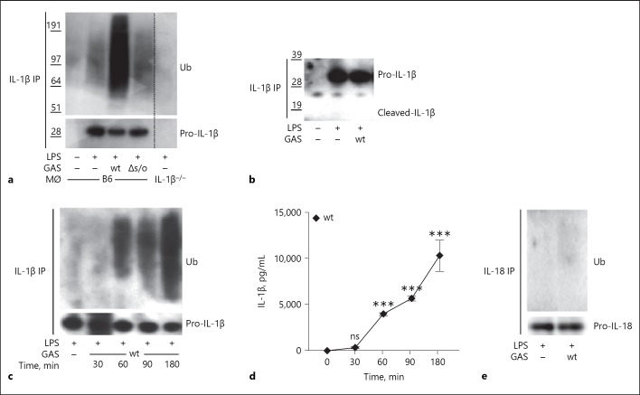
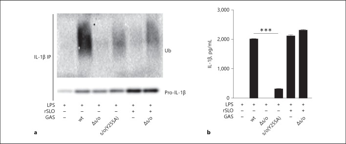
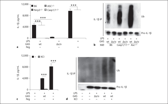
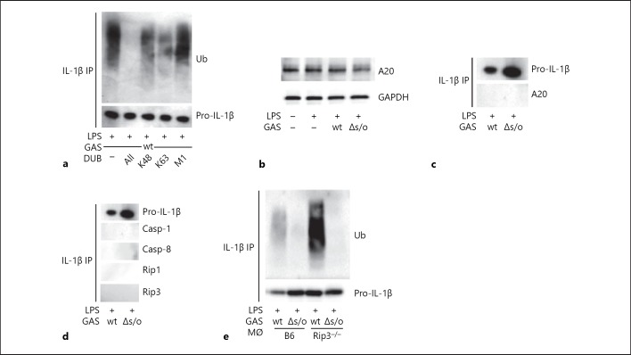
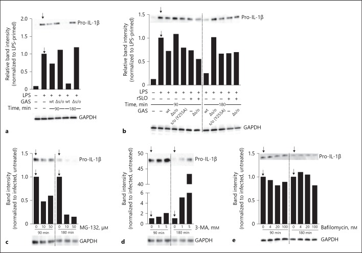
Group A Streptococcus (GAS) is a common and versatile human pathogen causing a variety of diseases. One of the many virulence factors of GAS is the secreted pore-forming cytotoxin streptolysin O (SLO), which has been ascribed multiple properties, including inflammasome activation leading to release of the potent inflammatory cytokine IL-1β from infected macrophages. IL-1β is synthesized as an inactive pro-form, which is activated intracellularly through proteolytic cleavage. Here, we use a macrophage infection model to show that SLO specifically induces ubiquitination and degradation of pro-IL-1β. Ubiquitination was dependent on SLO being released from the infecting bacterium, and pore formation by SLO was required but not sufficient for the induction of ubiquitination. Our data provide evidence for a novel SLO-mediated mechanism of immune regulation, emphasizing the importance of this pore-forming toxin in bacterial virulence and pathogenesis.

Keywords: Group A Streptococcus; Streptolysin O; Ubiquitin, IL-1β.


Conflict of interest statement

The authors declare that they have no conflicts of interest to disclose.

Figures

Fig. 1
Fig. 1
Group A Streptococcus (GAS) expressing streptolysin O (SLO) induces ubiquitination of pro-IL-1β in macrophages. a B6 macrophages were primed with lipopolysaccharide (LPS) or primed and infected with GAS as indicated and cell lysates were subjected to IL-1β immunoprecipitation (IP) and immunoblot analysis for ubiquitin (Ub) and (pro-)IL-1β. Primed IL-1β−/− macrophages were used as a control for specificity. b IL-1β IP and immunoblot analysis of lysates from LPS-primed or primed and infected macrophages, as indicated. c Macrophages were primed with LPS or primed and infected with wild type (wt) GAS for the indicated times. IL-1β was precipitated from cell lysates and analyzed for Ub and (pro-)IL-1β by immunoblot. d LPS-primed macrophages were infected by wt GAS and levels of released IL-1β were assessed by ELISA. e IL-18 was precipitated from cell lysates of LPS-primed or primed and infected macrophages, as indicated, followed by immunoblot analysis for Ub and (pro-)IL-18. Blots show 1 representative of 2 or 3 independent experiments. Graph shows means plus SD for triplicate samples and is representative of 2 independent experiments.
Fig. 2
Fig. 2
Pro-IL-1β ubiquitination is dependent on bacterial secretion of and pore formation by streptolysin O (SLO). LPS-primed B6 macrophages were infected with wild type (wt), Δslo or slo(Y255A) Group A Streptococcus (GAS) or stimulated with 10 µg/mL recombinant SLO (rSLO) for 90 min in the presence or absence of Δslo bacteria, as indicated. a IL-1β precipitates from cell lysates were assayed for ubiquitin (Ub) and (pro-)IL-1β by immunoblot. b Supernatants were analyzed for IL-1β by ELISA. Blot shows 1 representative of at least 3 independent experiments. Graph shows means plus SD for triplicate samples and is representative of 3 independent experiments.
Fig. 3
Fig. 3
Streptococcus-induced ubiquitination of pro-IL-1β occurs independently of SLO-mediated inflammasome signaling. a Primed macrophages of different genotypes were infected with wild type (wt) or Δslo Group A Streptococcus (GAS), as indicated. The Nlrp3 inflammasome-activating toxin Nigericin (Nig) was used as a control. Supernatants were assessed for IL-1β by ELISA. b Different genotype macrophages were primed and infected with wt or Δslo GAS. IL-1β was precipitated from cell lysates and ubiquitin (Ub) and (pro-)IL-1β were assessed by immunoblot. c B6 macrophages were primed or primed and infected or treated with Nig, as indicated, in the presence or absence of KCl and IL-1β in the supernatant was assessed by ELISA. d B6 macrophages were primed or primed and infected, as indicated, in the presence or absence of KCl and IL-1β was precipitated from cell lysates and ubiquitin (Ub) and (pro-)IL-1β were assessed by immunoblot. Graphs show means plus SD for triplicate samples and are representatives of at least 3 independent experiments. Blots show 1 representative of 3 independent experiments.
Fig. 4
Fig. 4
Pro-IL-1β ubiquitination after Group A Streptococcus (GAS) infection is of heterogeneous linkage specificity. a lipopolysaccharide (LPS)-primed macrophages were infected with wild type (wt) GAS followed by precipitation of IL-1β. Precipitates were subjected to deubiquitinase (DUB) proteolysis removing ubiquitin (Ub) of all linkages (“All”) or specifically K48, K63 or M1 linkages, as indicated, and ubiquitin and (pro-)IL-1β were assessed by immunoblot. b Immunoblot for A20 in lysates of untreated, primed or primed, and infected (wt or Δslo GAS) macrophages, as indicated. c Immunoblot for A20 in IL-1β precipitates from macrophages primed and infected with wt or Δslo GAS. d IL-1β was precipitated from whole cell lysates from macrophages after priming and infection with wt or Δslo GAS and analyzed for co-precipitation of the indicated proteins. e B6 and receptor-interacting kinase 3 (Rip3)−/− macrophages were primed and infected with wt or Δslo GAS. Precipitates of IL-1β from cell lysates were assayed for ubiquitin (Ub) and (pro-)IL-1β. Blots show 1 representative of 2 or 3 independent experiments.
Fig. 5
Fig. 5
Pro-IL-1β is degraded upon streptolysin O (SLO) proficient Group A Streptococcus (GAS) infection. a Macrophages deficient for the inflammasome linker protein ASC were left untreated, primed or primed, and infected with wild type (wt) or Δslo Group A Streptococcus (GAS) for the designated time points, as indicated, and (pro-)IL-1β levels in whole cell lysates were evaluated by immunoblot. b Macrophages deficient for ASC were left untreated, primed or primed, and infected with wt, Δslo, or slo(Y255A) GAS in the presence or absence of 10 µg/mL recombinant SLO (rSLO) for the designated time points, as indicated, and pro-IL-1β levels in whole cell lysates were evaluated by immunoblot. Lipopolysaccharide (LPS)-primed ASC deficient macrophages were infected with wt GAS for the indicated times in the presence of specific inhibitors as follows; c MG-132 (proteasome), d 3-MA (autophagy, PI3K inhibitor) or e Bafilomycin (autophagy, V-ATPase inhibitor). Band intensities were quantified based on whole protein content of cell lysates loaded onto gels followed by normalization to infected cells (without inhibitor) at each time point (indicated by arrows). The housekeeping protein GAPDH was used as a visual loading control but not as a basis for quantification. Figure shows representative blots of at least 3 independent experiments.

References

    1. Lin YH, Machner MP. Exploitation of the host cell ubiquitin machinery by microbial effector proteins. J Cell Sci. 2017 Jun;130((12)):1985–96. - PMC - PubMed
    1. Malynn BA, Ma A. Ubiquitin makes its mark on immune regulation. Immunity. 2010 Dec;33((6)):843–52. - PMC - PubMed
    1. Shaid S, Brandts CH, Serve H, Dikic I. Ubiquitination and selective autophagy. Cell Death Differ. 2013 Jan;20((1)):21–30. - PMC - PubMed
    1. Hafner-Bratkovič I, Pelegrín P. Ion homeostasis and ion channels in NLRP3 inflammasome activation and regulation. Curr Opin Immunol. 2018 Jun;52:8–17. - PubMed
    1. Shi J, Zhao Y, Wang K, Shi X, Wang Y, Huang H, et al. Cleavage of GSDMD by inflammatory caspases determines pyroptotic cell death. Nature. 2015 Oct;526((7575)):660–5. - PubMed

Publication types