. 2019 Mar 18;20(6):1363.
doi: 10.3390/ijms20061363.

Curcumin Analogue C1 Promotes Hex and Gal Recruitment to the Plasma Membrane via mTORC1-Independent TFEB Activation

Affiliations

Curcumin Analogue C1 Promotes Hex and Gal Recruitment to the Plasma Membrane via mTORC1-Independent TFEB Activation

Alessandro Magini et al. Int J Mol Sci. .

Abstract

The monocarbonyl analogue of curcumin (1E,4E)-1,5-Bis(2-methoxyphenyl)penta-1,4-dien-3-one (C1) has been used as a specific activator of the master gene transcription factor EB (TFEB) to correlate the activation of this nuclear factor with the increased activity of lysosomal glycohydrolases and their recruitment to the cell surface. The presence of active lysosomal glycohydrolases associated with the lipid microdomains has been extensively demonstrated, and their role in glycosphingolipid (GSL) remodeling in both physiological and pathological conditions, such as neurodegenerative disorders, has been suggested. Here, we demonstrate that Jurkat cell stimulation elicits TFEB nuclear translocation and an increase of both the expression of hexosaminidase subunit beta (HEXB), hexosaminidase subunit alpha (HEXA), and galactosidase beta 1 (GLB1) genes, and the recruitment of β-hexosaminidase (Hex, EC 3.2.1.52) and β-galactosidase (Gal, EC 3.2.1.23) on lipid microdomains. Treatment of Jurkat cells with the curcumin analogue C1 also resulted in an increase of both lysosomal glycohydrolase activity and their targeting to the cell surface. Similar effects of C1 on lysosomal glycohydrolase expression and their recruitment to lipid microdomains was observed by treating the SH-SY5Y neuroblastoma cell line; the effects of C1 treatment were abolished by TFEB silencing. Together, these results clearly demonstrate the existence of a direct link between TFEB nuclear translocation and the transport of Hex and Gal from lysosomes to the plasma membrane.

Keywords: TFEB; curcumin; curcumin analogue C1; lysosomal glycohydrolases; plasma membrane-associated glycohydrolases.


Conflict of interest statement

The authors declare no conflict of interest.

Figures

Figure 1
Figure 1
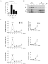
Phytohaemagglutinin (PHA)-stimulation of Jurkat cells induces transcription factor EB (TFEB) nuclear translocation and exocytosis. (A) Immunoblot analysis of TFEB in cytosolic and nuclear fractions from resting (Rest) and PHA-stimulated (PHA) Jurkat cells. Cytosolic TFEB level was normalized over β-actin, whereas nuclear TFEB level was normalized over H3. Values are the mean ± SEM of three independent experiments. ** p < 0.01 and *** p < 0.001 (PHA-stimulated vs. resting cells). (B) Horseradish peroxidase (HRP) enzyme activity in culture medium from resting and PHA-stimulated cells. Values are the mean ± SEM of three independent experiments. *** p < 0.001 (PHA-stimulated vs. resting cells).
Figure 2
Figure 2
Hex and Gal glycohydrolases increase their targeting to lipid microdomains after cell stimulation. (A) Gene expression analysis by Q-PCR of HEXB, HEXA, and GLB1 genes in resting and PHA-stimulated Jurkat cells. The ACTB gene was used as the endogenous control. The values are expressed as Relative Quantity (RQ). The mean ± SEM of three independent experiments is reported. *** p < 0.001 (PHA-stimulated vs. resting cells). Lipid microdomains were isolated from resting and PHA-stimulated Jurkat cells (1 × 10⁸) by a discontinuous sucrose-density gradient. (B) Fractions were collected from the top to the bottom of the tube and were analyzed by immunoblotting for flot-2 and lck (#, p56lck; ##, p60lck). Representative Western blotting of five independent experiments is reported. (C) Distribution of Total Hex, Hex A, and Gal enzymatic activities is expressed as total mU (tot. mU) in each fraction. Values are the mean ± SEM of five independent experiments. *** p < 0.001 (PHA-stimulated vs. resting cells). LM, lipid microdomain fractions; H, high-density fractions; Rest, resting cells; PHA, PHA-stimulated cells.
Figure 3
Figure 3
Hex and Gal are localized on external leaflet microdomains of the plasma membrane. Jurkat cells were treated with EZ-Link Sulfo-NHS-LC-Biotin to label cell surface proteins. After lipid microdomain purification, the biotinylated proteins contained in light-density fraction 3 were purified by avidin affinity chromatography. (A) Aliquots of concentrated and solubilized fraction 3 lipid microdomains (LM3), flow-through (F), and eluate (E) were analyzed by Dot blotting using HRP-conjugated streptavidin (Strep-HRP). (B) Total Hex, Hex A, and Gal enzymatic activities found in LM3, F, and E for resting (-) and PHA-stimulated (+) cells were expressed as total mU (tot. mU). The mean ± SEM of three independent experiments is reported. *** p < 0.001 (PHA-stimulated vs. resting cells).
Figure 4
Figure 4
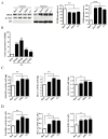
Curcumin analogue C1 promotes the recruitment of Hex and Gal on lipid microdomains by TFEB nuclear translocation in Jurkat cells. Jurkat cells were treated with PHA (24 h; 1 mg/mL), the curcumin analogue C1 (C1; 24 h, 1 μM), and torin 1 (Tor-1; 2 h, 0,1 μM,). Resting cells (Rest) were used as the negative control; torin 1 treated cells were used as the TFEB activation control. (A) Immunoblot analysis of cytosolic and nuclear protein fractions. TFEB level was normalized over β-actin and H3 in cytosolic and nuclear fractions, respectively. Values are the mean ± SEM of three independent experiments. *** p < 0.001 (treated vs. resting cells). (B) Gene expression analysis by Q-PCR of TFEB, HEXB, HEXA, and GLB1 genes on curcumin analogue C1 (C1, 1 µM)-treated Jurkat cells. The ACTB gene was used as the endogenous control. Values are expressed as Relative Quantity (RQ). The mean ± SEM of three independent experiments is reported. ** p < 0.01 and *** p < 0.001 (C1 vs. resting cells). (C,D) Hex and Gal activities were assayed by using fluorogenic substrates in cell extract (mU/mg) and in the flot-2-enrhiched fractions 2–4 (tot mU), respectively. Values are the mean ± SEM of five independent experiments. * p < 0.1 and ** p < 0.01 (treated vs. resting cells).
Figure 5
Figure 5
Curcumin analogue C1 promotes mechanistic target of rapamycin complex 1 (mTORC1)-independent TFEB nuclear translocation. SH-SY5Y cells were treated for 24 h with either curcumin (Cur, 5 µM) or the curcumin analogue C1 (C1, 1 µM). (A) Immunoblot analysis of the nuclear fractions from SH-SY5Y cells. TFEB level was normalized over H3. Starved cells were used as the positive control. Values are the mean ± SEM of three independent experiments. ** p < 0.01 and *** p < 0.001 (treated or starved vs. untreated cells, CTRL). (B) Immunofluorescence analysis of TFEB subcellular distribution on untreated (CTRL) and SH-SH5Y cells treated with torin 1 (Tor1), curcumin (Cur), or the curcumin analogue C1 (C1). Torin 1 was used as the positive control. Magnification, 40×; scale bar: 50 µm. (C) Immunoblot analysis of SH-SY5Y cell extracts. P-S6 (S235/236) was normalized over total S6, and LC3B-II was normalized over LC3B-I. β-Actin was used as the immunoblotting loading control. Values are the mean ± SEM of three independent experiments. ** p < 0.01 and *** p < 0.001 (treated or starved vs. untreated cells, CTRL).
Figure 6
Figure 6
Curcumin analogue C1 promotes the expression of Hex and Gal. (A) Gene expression analysis by Q-PCR of TFEB, HEXB, HEXA, and GLB1 genes on curcumin analogue C1 (C1, 1 µM) treated SH-SY5Y cells. The ACTB gene was used as the endogenous control. Values are expressed as Relative Quantity (RQ). The mean ± SEM of three independent experiments is reported. *** p < 0.001 (treated vs. untreated cells, CTRL). (B) SH-SY5Y cells were starved or treated for 24 h with either curcumin (Cur, 5 µM) or the curcumin analogue C1 (C1, 1 µM). Hex and Gal specific activities (mU/mg) were assayed by using fluorogenic substrates. Values are the mean ± SEM of five independent experiments. ** p < 0.01 and *** p < 0.001 (treated or starved vs. untreated cells, CTRL).
Figure 7
Figure 7
Curcumin analogue C1 promotes the recruitment of Hex and Gal on lipid microdomains. Lipid microdomains were isolated from starved (Starv), curcumin (Cur, 5 μM). and curcumin analogue C1 (C1, 1 μM) 24 h-treated and untreated (CTRL) SH-SY5Y cells. (A) Collected fractions were analyzed by Dot blotting for the lipid microdomain maker GM1 by using the cholera toxin B subunit. Representative Dot blotting of five independent experiments is reported. (B) Collected fractions were also analyzed by immunoblotting for the lipid microdomain maker flotillin 2 (Flot-2). Representative immunoblotting of five independent experiments is reported. (C) Total Hex, Hex A, and Gal enzymatic activities in the GM1-enrhiched fractions 2–4 are reported as total mU (tot. mU). Values are the mean ± SEM of five independent experiments. *** p < 0.001 (treated or starved vs. untreated cells, CTRL). LM, lipid microdomain fractions; H, high-density fractions.
Figure 8
Figure 8
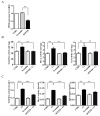
Curcumin analogue C1 failed to increase Hex and Gal activity and their recruitment on lipid microdomains in TFEB knock-down cells. SH-SY5Y cells were transfected with shRNA for TFEB (shTFEB) or scrambled shRNA (Scramble) as the control. (A) Gene expression analysis by Q-PCR of the TFEB gene in SH-SY5Y (CTRL), Scramble, and shTFEB cells. The values are expressed as Relative Quantity (RQ). The mean ± SEM of three independent experiments is reported. ** p < 0.01 (Scramble vs. shTFEB). (B,C) SH-SY5Y (C1) and shTFEB (shTFEB + C1) cells were treated for 24 h with the curcumin analogue C1 (C1, 1 µM); untreated SH-SY5Y (CTRL) and untreated scramble (Scramble) cells are reported as controls. Hex and Gal activities were assayed by using fluorogenic substrates in cell extract (mU/mg) and in the flot-2-enrhiched fractions 2–4 (tot mU), respectively. Values are the mean ± SEM of five independent experiments. ** p < 0.01 and *** p < 0.001 (treated vs. untreated SH-SY5Y cells or treated shTFEB vs. treated SH-SY5Y cells).

Similar articles

Cited by

References

    1. Sardiello M., Palmieri M., Di Ronza A., Medina D.L., Valenza M., Gennarino V.A., Di Malta C., Donaudy F., Embrione V., Polishchuk R.S., et al. A gene network regulating lysosomal biogenesis and function. Science. 2009;325:473–477. doi: 10.1126/science.1174447. - DOI - PubMed
    1. Settembre C., Di Malta C., Polito V.A., Garcia Arencibia M., Vetrini F., Erdin S., Erdin S.U., Huynh T., Medina D., Colella P., et al. TFEB links autophagy to lysosomal biogenesis. Science. 2011;332:1429–1433. doi: 10.1126/science.1204592. - DOI - PMC - PubMed
    1. Reddy A., Caler E.V., Andrews N.W. Plasma membrane repair is mediated by Ca(2+) regulated exocytosis of lysosomes. Cell. 2001;106:157–169. doi: 10.1016/S0092-8674(01)00421-4. - DOI - PubMed
    1. Encarnação M., Espada L., Escrevente C., Mateus D., Ramalho J., Michelet X., Santarino I., Hsu V.W., Brenner M.B., Barral D.C., et al. A Rab3a-dependent complex essential for lysosome positioning and plasma membrane repair. J. Cell Biol. 2016;213:631–640. doi: 10.1083/jcb.201511093. - DOI - PMC - PubMed
    1. Giatromanolaki A., Kalamida D., Sivridis E., Karagounis I.V., Gatter K.C., Harris A.L., Koukourakis M.I. Increased expression of transcription factor EB (TFEB) is associated with autophagy, migratory phenotype and poor prognosis in non-small cell lung cancer. Lung Cancer. 2015;90:98–105. doi: 10.1016/j.lungcan.2015.07.008. - DOI - PubMed

MeSH terms

LinkOut - more resources