. 2019 Apr;50(4):978-988.
doi: 10.1161/STROKEAHA.118.021590.

Mitophagy Reduces Oxidative Stress Via Keap1 (Kelch-Like Epichlorohydrin-Associated Protein 1)/Nrf2 (Nuclear Factor-E2-Related Factor 2)/PHB2 (Prohibitin 2) Pathway After Subarachnoid Hemorrhage in Rats

Affiliations

Mitophagy Reduces Oxidative Stress Via Keap1 (Kelch-Like Epichlorohydrin-Associated Protein 1)/Nrf2 (Nuclear Factor-E2-Related Factor 2)/PHB2 (Prohibitin 2) Pathway After Subarachnoid Hemorrhage in Rats

Tongyu Zhang et al. Stroke. 2019 Apr.

Erratum in

Abstract

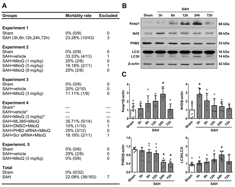
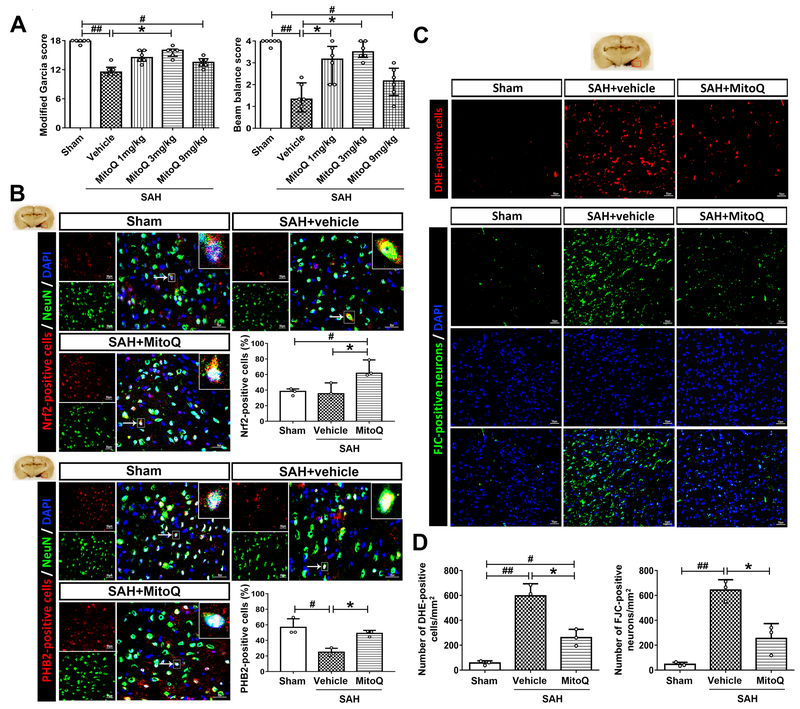
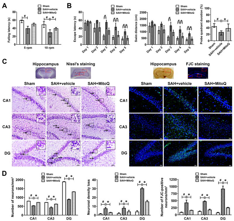
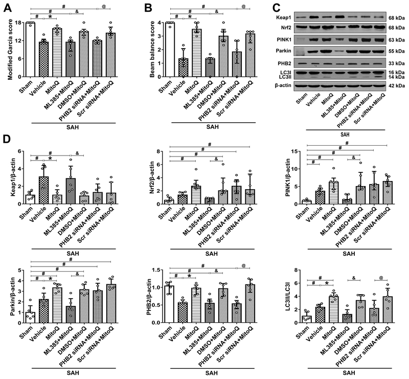
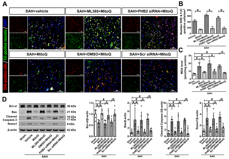
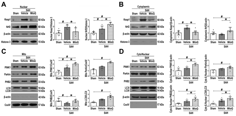
Background and Purpose- Mitoquinone has been reported as a mitochondria-targeting antioxidant to promote mitophagy in various chronic diseases. Here, our aim was to study the role of mitoquinone in mitophagy activation and oxidative stress-induced neuronal death reduction after subarachnoid hemorrhage (SAH) in rats. Methods- Endovascular perforation was used for SAH model of male Sprague-Dawley rats. Exogenous mitoquinone was injected intraperitoneally 1 hour after SAH. ML385, an inhibitor of Nrf2 (nuclear factor-E2-related factor 2), was given intracerebroventricularly 24 hours before SAH. Small interfering RNA for PHB2 (prohibitin 2) was injected intracerebroventricularly 48 hours before SAH. Nuclear, mitochondrial, and cytoplasmic fractions were gathered using nucleus and mitochondria isolation kits. SAH grade evaluation, short- and long- term neurological function tests, oxidative stress, and apoptosis measurements were performed. Pathway related proteins were investigated with Western blot and immunofluorescence staining. Results- Expression of Keap1 (Kelch-like epichlorohydrin-associated protein 1, 2.84× at 24 hours), Nrf2 (2.78× at 3 hours), and LC3II (light chain 3-II; 1.94× at 24 hours) increased, whereas PHB2 (0.46× at 24 hours) decreased after SAH compared with sham group. Mitoquinone treatment attenuated oxidative stress and neuronal death, both short-term and long-term. Administration of mitoquinone resulted in a decrease in expression of Keap1 (0.33×), Romo1 (reactive oxygen species modulator 1; 0.24×), Bax (B-cell lymphoma-2 associated X protein; 0.31×), Cleaved Caspase-3 (0.29×) and an increase in Nrf2 (2.13×), Bcl-xl (B-cell lymphoma-extra large; 1.67×), PINK1 (phosphatase and tensin-induced kinase 1; 1.67×), Parkin (1.49×), PHB2 (1.60×), and LC3II (1.67×) proteins compared with SAH+vehicle group. ML385 abolished the treatment effects of mitoquinone on behavior and protein levels. PHB2 small interfering RNA reversed the outcomes of mitoquinone administration through reduction in protein expressions downstream of PHB2. Conclusions- Mitoquinone inhibited oxidative stress-related neuronal death by activating mitophagy via Keap1/Nrf2/PHB2 pathway after SAH. Mitoquinone may serve as a potential treatment to relieve brain injury after SAH.

Keywords: Kelch-like epichlorohydrin-associated protein 1; mitoquinone; nuclear factor E2-related factor 2; prohibitin 2; rats; subarachnoid hemorrhage.


Conflict of interest statement

Conflict of Interest

There is no conflict of interest.

Figures

Figure 1.
Figure 1.. Mortality rate and expression changes of Kelch-like ECH-associated protein (Keap1), NF-E2-related factor 2 (Nrf2), Prohibitin 2 (PHB2) and LC3 after subarachnoid hemorrhage (SAH).
(A) The number of mortality and excluded rats of each group. * means shared groups with Experiment 2. (B) Representative Western blot images and (C) quantitative analysis of Keap1, Nrf2, PHB2 and LC3 time course expression. n=6 per group. Data of LC3 time course expression was expressed as the medians with interquartile range using Kruskal–Wallis test followed by the Dunn’s post hoc test. Other data were expressed as the means ± SD using one-way analysis of variance (ANOVA) followed by the Tukey post hoc test. #P < 0.05 vs. Sham group.
Figure 2.
Figure 2.. Mitoquinone (MitoQ) attenuated short-term neurological deficits, oxidative stress and neuronal death and increased the expression of Nrf2 and PHB2 after subarachnoid hemorrhage (SAH).
(A) Modified Garcia and beam balance scores. n=6 per group. (B) Double immunofluorescence staining and quantifications for Nrf2 (red) and PHB2 (red) in neuron (NeuN, green) of Sham, SAH+vehicle and SAH+MitoQ groups. Nuclei are stained with DAPI (blue). Top panel indicates the location of staining (small red box). Arrows indicate a cell chosen for 10 times magnification in right upper corner of merged panels. n=3 per group. (C) DHE and FJC staining in three groups above. (D) The quantifications of DHE and FJC staining. n=3 per group. Scale bar = 50μm. Data of Modified Garcia and beam balance scores were expressed as the medians with interquartile range using Kruskal–Wallis test followed by the Dunn’s post hoc test. Other data were expressed as the means ± SD using one-way analysis of variance (ANOVA) followed by the Tukey post hoc test. #P < 0.05 vs. Sham group; ##P < 0.01 vs. Sham group; *P < 0.05 vs. SAH+vehicle group.
Figure 3.
Figure 3.. Mitoquinone (MitoQ) attenuated long-term neurological deficits and hippocampus injury after subarachnoid hemorrhage (SAH).
(A) Rotarod test of 5 RPM and 10 RPM on the 21th day. n=8 per group. (B) Water maze test related escape latency and swim distance and probe quadrant duration for 21–25 days. n=8 per group. (C) Representative images and neuronal quantifications of Nissl’s and FJC staining for hippocampus in CA1, CA3 and DG regions. (D) Quantitative analysis of Nissl’s and FJC staining. n=8 per group. Arrows indicate shrunken pyramidal or died neurons. Scale bar = 200μm (general) and 50 μm (regions). Data of long-term neurological behavior tests were expressed as the medians with interquartile range using Kruskal–Wallis test followed by the Dunn’s post hoc test. Other data were expressed as the means ± SD using one-way analysis of variance (ANOVA) followed by the Tukey post hoc test. #P < 0.05 vs. Sham group; *P < 0.05 vs. SAH+vehicle group.
Figure 4.
Figure 4.. Blokade of Nrf2 and PHB2 abolished mitoquinone (MitoQ) treatment effects on the neurological function and mitophagy 24 h after subarachnoid hemorrhage (SAH).
(A) Modified Garcia score. (B) Beam balance score. (C) Representative Western bolt images of Keap1, Nrf2, PINK1, Parkin, PHB2 and LC3. (D) Quantitative analyses of those proteins above. n=6 per group. Data of Modified Garcia scores, beam balance scores and Nrf2, PINK1, PHB2 protein expressions were expressed as the medians with interquartile range using Kruskal–Wallis test followed by the Dunn’s post hoc test. Other data were expressed as the means ± SD using one-way analysis of variance (ANOVA) followed by the Tukey post hoc test. #P < 0.05 vs. Sham group; *P < 0.05 vs. SAH+vehicle group; &P< 0.05 vs. SAH+ML385+MitoQ group; @P< 0.05 vs. SAH+PHB2 siRNA+MitoQ group.
Figure 5.
Figure 5.. Blokade of Nrf2 and PHB2 reversed the effects of mitoquinone (MitoQ) on the oxidative stress-induced neuronal death 24 h after subarachnoid hemorrhage (SAH).
(A) Representative images and (B) quantitative analyses of Double staining for DHE and FJC. (C) MDA levels. (D) Representative Western blot images and quantitative analyses of Bcl-xl, Bax, Cleaved Caspase-3 and Romo1. Data of Cleaved Caspase-3 protein expression was expressed as the medians with interquartile range using Kruskal–Wallis test followed by the Dunn’s post hoc test. Other data were expressed as the means ± SD using one-way analysis of variance (ANOVA) followed by the Tukey post hoc test. #P <0.05 vs. Sham group; *P < 0.05 vs. SAH+vehicle group. &P< 0.05 vs. SAH+ML385+MitoQ group; @P< 0.05 vs. SAH+PHB2 siRNA+MitoQ group.
Figure 6.
Figure 6.. The effects of Mitoquinone (MitoQ) on nuclear Keap1/Nrf2 and mitochondrial mitophagy-associated proteins after subarachnoid hemorrhage (SAH).
Representative Western blot images and quantitative analyses of Nrf2 and Keap1 in (A) nuclear and in (B) cytoplasmic fractions. Representative Western blot images and quantitative analyses of PINK1, Parkin, PHB2 and LC3 in (C) mitochondrial and in (D) cytoplasmic (except mitochondria) plus nuclear fractions. Data of Keap1 and Nrf2 cytoplasmic expressions; Parkin, PHB2 and LC3 mitochondrial expressions; Parkin and LC3 cytoplasmic (except mitochondria) plus nuclear expressions were expressed as the medians with interquartile range using Kruskal–Wallis test followed by the Dunn’s post hoc test. Other data were expressed as the means ± SD using one-way analysis of variance (ANOVA) followed by the Tukey post hoc test. #P < 0.05 vs. Sham group; *P< 0.05 vs. SAH+vehicle group.

Similar articles

Cited by

References

    1. Cahill J, Calvert JW, Zhang JH. Mechanisms of early brain injury after subarachnoid hemorrhage. J. Cereb. Blood Flow Metab 2006;26:1341–1353 - PubMed
    1. Han Y, Zhang T, Su J, Zhao Y, Chenchen W, Li X. Apigenin attenuates oxidative stress and neuronal apoptosis in early brain injury following subarachnoid hemorrhage. J. Clin. Neurosci 2017;40:157–162 - PubMed
    1. Chen S, Wu H, Tang J, Zhang J, Zhang JH. Neurovascular events after subarachnoid hemorrhage: Focusing on subcellular organelles. Acta Neurochir. Suppl 2015;120:39–46 - PMC - PubMed
    1. Jing CH, Wang L, Liu PP, Wu C, Ruan D, Chen G. Autophagy activation is associated with neuroprotection against apoptosis via a mitochondrial pathway in a rat model of subarachnoid hemorrhage. Neuroscience. 2012;213:144–153 - PubMed
    1. McManus MJ, Murphy MP, Franklin JL. The mitochondria-targeted antioxidant mitoq prevents loss of spatial memory retention and early neuropathology in a transgenic mouse model of alzheimer’s disease. J. Neurosci 2011;31:15703–15715 - PMC - PubMed

Publication types

MeSH terms