. 2019 Mar 19;38(1):132.
doi: 10.1186/s13046-019-1129-8.

Pancreatic cancer-initiating cell exosome message transfer into noncancer-initiating cells: the importance of CD44v6 in reprogramming

Affiliations

Pancreatic cancer-initiating cell exosome message transfer into noncancer-initiating cells: the importance of CD44v6 in reprogramming

Zhe Wang et al. J Exp Clin Cancer Res. .

Erratum in

Retraction in

Abstract

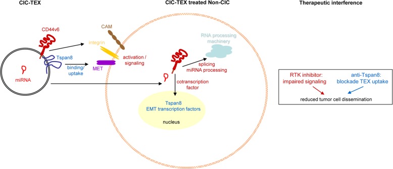
Background: Cancer-initiating cell (CIC) exosomes (CIC-TEX) are suggested reprogramming Non-CIC. Mode of message transfer and engagement of CIC-markers being disputed, we elaborated the impact of CD44v6 and Tspan8 on the response of Non-CIC.

Methods: Non-metastasizing CD44v6- and Tspan8-knockdown (kd) pancreatic cancer cells served as Non-CIC. CIC-TEX coculture-induced changes were evaluated by deep-sequencing and functional assays. Tumor progression was surveyed during in vivo CIC-TEX treatment.

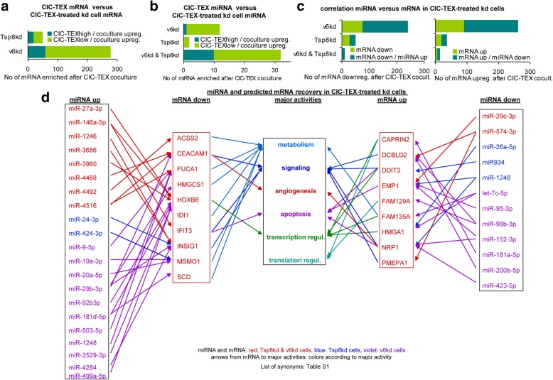
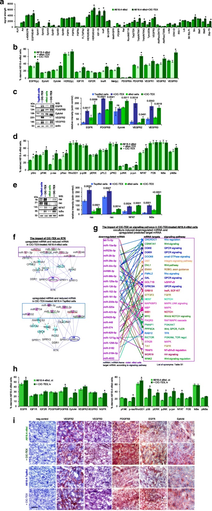
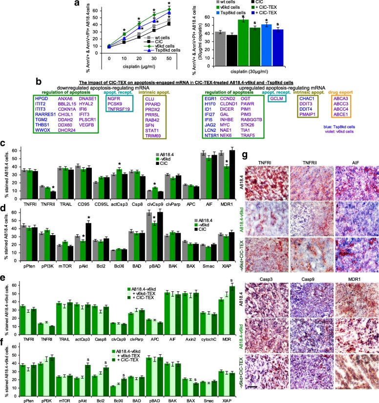
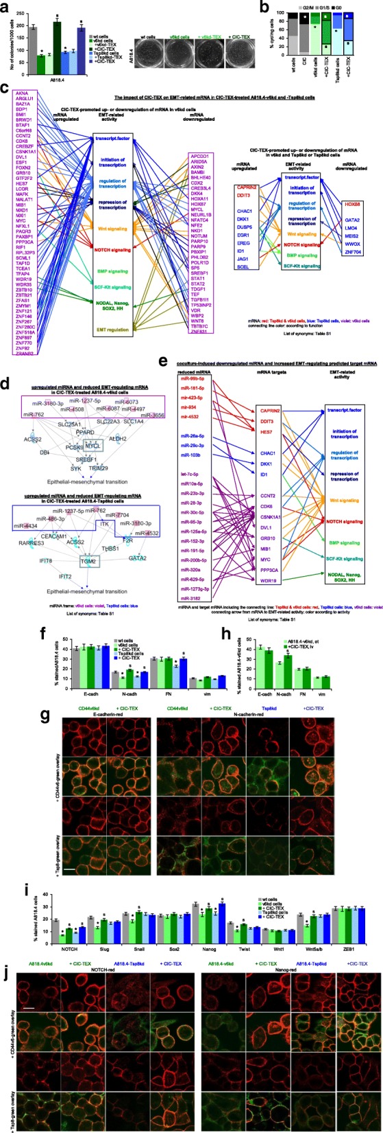
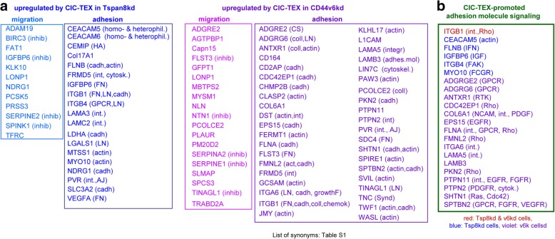
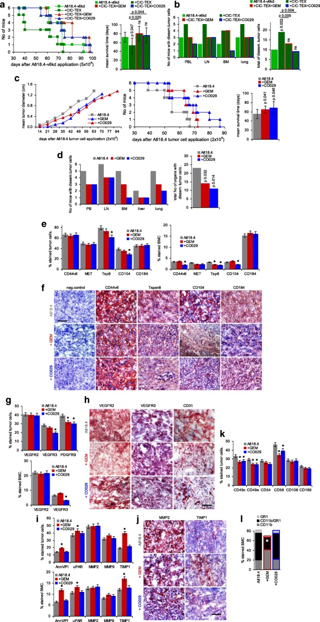
Results: Deep-sequencing of CIC-TEX-cocultured CD44v6kd-Non-CIC revealed pronounced mRNA changes in signaling, transport, transcription and translation; altered miRNA affected metabolism, signaling and transcription. CIC-TEX coculture-induced changes in Tspan8kd-Non-CIC mostly relied on CIC-TEX-Tspan8 being required for targeting. CIC-TEX transfer supported apoptosis resistance and significantly promoted epithelial mesenchymal transition, migration, invasion and (lymph)angiogenesis of the kd Non-CIC in vitro and in vivo, deep-sequencing allowing individual mRNA and miRNA assignment to altered functions. Importantly, CIC-TEX act as a hub, initiated by CD44v6-dependent RTK, GPCR and integrin activation and involving CD44v6-assisted transcription and RNA processing. Accordingly, a kinase inhibitor hampered CIC-TEX-fostered tumor progression, which was backed by an anti-Tspan8 blockade of CIC-TEX binding.

Conclusions: This in depth report on the in vitro and in vivo impact of CIC-TEX on CD44v6kd and Tspan8kd Non-CIC unravels hub CIC-TEX activity, highlighting a prominent contribution of the CIC-markers CD44v6 to signaling cascade activation, transcription, translation and miRNA processing in Non-CIC and of Tspan8 to CIC-TEX targeting. Blocking CIC-TEX binding/uptake and uptake-initiated target cell activation significantly mitigated the deleterious CIC-TEX impact on CD44v6kd and Tspan8kd Non-CIC.

Keywords: CD44v6; Exosome biogenesis; Exosome message transfer; Non-cancer stem cell reprogramming; Pancreatic cancer stem cells; Tspan8.


Conflict of interest statement

Authors’ information

The manuscript was edited for proper English language, grammar, punctuation, spelling, and overall style by one or more of the highly qualified native English speaking editors at Wiley Editing Services.

Ethics approval

Animal experiments were approved by the Government of Baden-Wuerttemberg, Germany. Animals were housed under specific pathogen-free conditions at the central animal facility of the University of Heidelberg (IBF) (Interfakultäre Biomedizinische Forschungseinrichtung). Yearly reports on animal experiments were delivered to the Government of Baden-Wuerttemberg, Germany, according to the rules of the European Union Directive 2010/63/EU for animal experiments.

Consent for publication

not applicable.

Competing interests

The authors declare that they have no competing interests.

Publisher’s Note

Springer Nature remains neutral with regard to jurisdictional claims in published maps and institutional affiliations.

Figures

Fig. 1
Fig. 1
The impact of CIC-TEX on CD44v6kd and Tspan8kd tumor growth and progression. a-c Nude mice received GFP-transfected A818.4 CIC on the left upper back and/or A818.4-v6kd cells on the right upper back. a Mean tumor diameter ± SD (3mice/group); b,c mean percent GFP+, GFP + marker+ and marker+ (v6, Tsp8, EpC) cells was evaluated at autopsy (flow-cytometry); a-c significant differences between mice receiving only CD44v6kd cells and mice receiving a contralateral injection of CIC-GFP: s. d,e Nude mice received an ot injection of 1 × 106 wt or v6kd or Tsp8kd cells and weekly 200 μg/mouse CIC-TEX, iv; d survival time and mean survival time of 6 and 5, respectively, mice/group; p-values comparing wt versus kd tumor and kd tumor depending on CIC-TEX application are indicated. e The indicated organs, collected at autopsy, were dispersed and maintained in culture to observe tumor cell outgrowth; p-values (Kruskal-Wallis after Bonferroni-Holm correction) for the total No of organs with disseminated tumor cells are indicated comparing wt versus kd TB mice and CIC-TEX-treated kd TB mice. After reaching a mean tumor diameter of ~ 0 .5cm, CIC assist the growth of v6kd Non-CIC, likely via delivery of TEX. The shortened survival time and the increase in disseminated tumor cells of v6kd TB mice receiving CIC-TEX supports this assumption
Fig. 2
Fig. 2
Correlation between CIC-TEX and CIC-TEX-induced changes in mRNA and miRNA profiles. a Numbers of ≥2-fold enriched mRNA (signal strength ≥1000) in CIC-TEX-treated v6kd and Tsp8kd cells sorted according to ≥2-fold higher versus comparable or lower recovery in CIC-TEX than kd cells; b numbers of miRNA ≥1.5-fold upregulated in CIC-TEX-treated v6kd and Tsp8kd cells sorted according to ≥1.5-fold higher versus comparable or lower recovery in CIC-TEX than kd cells; c Correlation between ≥1.5-fold up- or downregulated mRNA in CIC-TEX treated v6kd and Tsp8kd cells sorted according to reverse miRNA recovery in CIC-TEX-treated cells; d major activities (IPA-based Reactome analysis) of ≥2-fold up- or downregulated mRNA that are predicted targets of inversely recovered miRNA (miRNA database, target scan database) in both CIC-TEX-treated v6kd and Tsp8kd cell (List of synonyms: Additional file 1: Table S1). mRNA and miRNA recovery are more strongly affected by CIC-TEX treatment in CD44v6kd than Tsp8kd cells. However, at the mRNA and the miRNA level coculture-induced changes rarely correlate with the CIC-TEX content. Instead, there is a strong correlation between coculture-induced up- or downregulated miRNA and reversely down- or upregulated mRNA in CIC-TEX-treated kd cells, indicating that not the CIC-TEX content, but CIC-TEX-induced target cell activation is dominating
Fig. 3
Fig. 3
CIC-TEX-initiated changes in RTK and downstream signaling molecules in CD44v6kd and Tspan8kd cells. a Signaling array of A818.4-v6kd cells cultured for 72 h with/without CIC-TEX. The relative signal strength was evaluated by ImageJ; significant differences by coculture with CIC-TEX: *. Flow-cytometry and WB analysis of (b,c) RTK expression in kd-TEX- or CIC-TEX-treated kd cells and (d,e) major pathway-engaged cytosolic signaling molecules; b,d mean % stained cells±SD (3 assays), significant differences by coculture with TEX: *; c,e representative examples and relative signal strength±SD of 3 independent experiments including p-values for kd cells compared to CIC-TEX-treated kd cells; f pathways from miRNA to RTK (IPA-based STRING analysis after predicted target mRNA selection by microrna.org and targetscan.org) for ≥2-fold upregulated miRNA (framed) and ≥ 2-fold reduced mRNA recovery in CIC-TEX-treated v6kd or Tsp8kd cells compared to untreated kd cells; g IPA-based STRING analysis after predicted target mRNA selection by microrna.org and targetscan.org for ≥2-fold reduced miRNA in CIC-TEX-treated compared to untreated v6kd cells and of ≥2-fold upregulated predicted mRNA targets that are engaged in signal transduction. h Flow-cytometry analysis of ex vivo harvested intrapancreatic A818.4-v6kd cells from nude mice with/without weekly iv CIC-TEX treatment; mean % stained cells±SD (3 tumors), significant differences by CIC-TEX treatment: s; i Representative immunohistology examples of A818.4-v6kd and -Tsp8kd shock-frozen tumor sections with/without CIC-TEX treatment stained with the indicated antibodies (scale bar: 100 μm). (List of synonyms: Additional file 1: Table S1). CIC-TEX treatment strongly affects RTK expression and downstream signaling molecules in vitro and in vivo. Changes in the recovery of mRNA engaged in signal transduction (Additional file 1: Figure S2d, S2e) are accompanied at a noteworthy frequency by reversely altered miRNA expression in CIC-TEX-treated kd, predominantly v6kd cells
Fig. 4
Fig. 4
CIC-TEX-promoted apoptosis-resistance in CD44v6kd and/or Tspan8kd cells. a Flow-cytometry of cells cultured for 48 h in the presence of cisplatin; mean % AnnexinV+ and AnnexinV+/PI+ cells±SD (triplicates); significant differences of wt cells versus CIC or kd cells: *; significant differences by CIC-TEX treatment: s. b mRNA that expression differed by ≥2-fold after CIC-TEX treatment in Tsp8kd (blue) or v6kd (violet) cells were sorted by KEGG analysis according to the engagement in distinct apoptotic processes. c-f Flow-cytometry analysis of apoptosis-related receptor and cytoplasmic signaling molecules in A818.4, −v6kd and CIC-enriched cells and in v6kd cells cocultured with v6kd- or CIC-TEX; mean % stained cells±SD (3 assays), significant differences between wt cells, v6kd cells and CIC: *, significant differences by coculture of v6kd cells with TEX: s. g Representative immunohistology examples of A818.4 and -v6kd shock-frozen tumor sections from untreated or CIC-TEX-treated mice stained with the indicated antibodies (scale bar: 100 μm). (List of synonyms: Additional file 1: Table S1). Slightly reduced apoptosis resistance of v6kd and Tsp8kd tumor cells becomes mitigated by CIC-TEX treatment. The impact of CIC-TEX on apoptosis resistance is mostly restricted to the regulation of molecules engaged in the intrinsic pathway of apoptosis induction. An exception is the increased expression of drug transporters in CIC-TEX-treated v6kd cells
Fig. 5
Fig. 5
The impact of CIC-TEX on EMT gene expression. a Tumor cells were seeded in soft agar containing 30 μg/ml TEX, where indicated; mean No of colonies±SD (5 replicates) and representative examples after 3wk of culture; b cell cycle progression (flow-cytometry, PI staining) of wt, CIC and kd cells cultured with/without CIC-TEX; mean % of cells (5 replicates) in G0, G1/S and G2/M; c IPA-based Reactome analysis of transcription factor-, stem cell-, EMT-, transcription-, and EMT-regulating genes that mRNA level is ≥2-fold up- or downregulated in CIC-TEX-treated v6kd and Tsp8kd cells (red), Tsp8kd cells (blue) or CD44v6kd cells (violet). d,e Reactome analysis after IPA coordination of miRNA with predicted mRNA targets (miRNA and targetscan databases) of (d) > 2-fold upregulated miRNA (framed) and (e) downregulated miRNA in CIC-TEX-treated kd cells affecting EMT-related genes in kd cells; in (d) mRNA pathways from upregulated miRNA towards EMT are included; for downregulated miRNA (e) only direct predicted mRNA targets are shown (color code as in c). f Flow-cytometry of EMT markers in A818.4 and kd cells with/without CIC-TEX-treatment (72 h); g confocal microscopy of kd cells with/without CIC-TEX-treatment stained for E- or N-cadherin and counterstained for v6 or Tsp8 (scale bar: 10 μm); h Flow-cytometry of ex vivo analyzed EMT markers in dispersed intrapancreatic v6kd tumors from mice with/without CIC-TEX-treatment; (i) Flow-cytometry of EMT-related transcription factors in A818.4 and kd cells with/without CIC-TEX-treatment (72 h); j Confocal microscopy of kd cells with/without CIC-TEX-treatment stained for EMT-related transcription factors NOTCH and Nanog and counterstained with anti-v6 or anti-Tsp8 (scale bar: 10 μm); f,h,i mean % stained cells±SD (3 assays/tumors); a,b,f,h,i significant differences between wt and kd cells: *, significant differences by CIC-TEX-treatment: s. (List of synonyms: Additional file 1: Table S1). CIC-TEX partly rescue impaired anchorage-independent growth and accelerate kd cell cycle progression. DS, confirmed at the protein level, unraveled a strong impact of CIC-TEX on EMT-related transcription factors mostly in v6kd cells at the mRNA and miRNA level, the latter being particularly engaged in Wnt and NOTCH signaling
Fig. 6
Fig. 6
CIC-TEX engagment in protease, adhesion molecule and related signaling regulation in CD44v6kd and Tspan8kd cells. a CIC-TEX coculture-induced ≥1.5-fold upregulated RNA expression was analyzed by KEGG for engagement in proteolysis and adhesion. The analysis was restricted to plasma membrane or plasma membrane attached proteins; proteases and inhibitors (indicated in brackets) are listed for migration; for adhesion molecules the predominant ligands are included in brackets. b Assignment of major adhesion / motility promoting signaling molecules (STRING analysis) that expression was ≥1.5-fold upregulated by CIC-TEX coculture; major targets are indicated in brackets. (Abbreviations: AJ: adherens junction, cadh: cadherin, chemok: chemokine, coll: collagen, CS: chondroitin sulphate, cytosk: cytoskeleton, FN: fibronectin, GPCR: G-protein coupled receptor, growthF: growth factors, HA: hyaluronan, inhib: inhibitor, int: integrin, LN: laminin, Synd: syndecan, List of synonyms: Additional file 1: Table S1). CIC-TEX affect proteases and more frequently adhesion molecules in v6kd and Tsp8kd cells. CIC-TEX often promote protease inhibitor upregulation. Besides matrix binding molecules, the linkage to the actin cytoskeleton and binding to cadherin is dominating. Altered adhesion molecule-promoted signaling frequently proceeds directly via activated integrins, RTK and GPCR
Fig. 7
Fig. 7
Binding and RTK inhibition for blocking CIC-TEX activity as adjuvant PaCa therapy. Mice received an ot (A818.4-v6kd) or a sc (A818.4) tumor cell injection and weekly the RTK inhibitor GEM (iv) or anti-Tsp8 (CO029) (iv). a Survival time and mean survival time of v6kd-TB mice (intrapancreatic) treated with CIC-TEX (2x/wk) and GEM or CO029 (1x/wk). b Recovery of disseminated tumor cells in cultures of dispersed organs at autopsy. c Tumor growth rate, survival time and mean survival time of sc A818.4-TB mice, treated with GEM or CO029; d disseminated tumor cell recovery in cultures of dispersed organs at autopsy; a-d p-values for the mean survival time and the numbers of organs containing dispersed tumor cells (after Bonferroni-Holm correction) of GEM- or CO029-treated compared to untreated v6kd-TB and CIC-TEX-treated-TB or wt-TB are indicated. e,g,i,k,l Flow-cytometry of dispersed tumor cells and BMC of untreated and GEM- or CO029-treated A818.4-TB mice evaluating CIC, angiogenic, apoptosis, proteolysis, adhesion (only tumor cells) marker expression and MDSC (CD11b + Gr1+) (only BMC); mean % stained cells±SD (3 mice), significant differences by GEM- or CO029-treatment: *. f,h,j Immunohistology of A818.4 shock-frozen tumor sections from untreated, GEM- or CO029-treated mice stained for CIC markers, VEGFR2, VEGFR3, CD31, MMP2 and TIMP1 (scale bar: 100 μm). GEM and CO029 weakly affect tumor growth and strongly tumor cell dissemination. Ex vivo analysis indicates that GEM and CO029 act independently. GEM primarily affects tumor cell apoptosis, proteases and MDSC expansion, CO029 treatment is accompanied by a reduction in (Tsp8-associated) CIC markers and compromises (lymph)angiogenesis
Fig. 8
Fig. 8
Summary on the importance of CD44v6 and Tspan8 on CIC-TEX-promoted Non-CIC reprogramming and potential therapeutic interference. CIC-TEX promote a shift towards stem cell features in v6kd and Tsp8kd Non-CIC, CIC-TEX binding/transfer acting as a hub. The v6 importance relies on initiating activation of signal transduction and contributing to mRNA processing. Tspan8 is central in CIC-TEX communication. The complementing v6 and Tsp8 activities open new adjuvant therapeutic options by blocking CIC-TEX binding/uptake and by interfering with CIC-TEX-promoted Non-CIC activation. (CAM: cell adhesion molecules, EMT: epithelial mesenchymal transition, RTK: receptor tyrosine kinases)

References

    1. Siegel R, Naishadham D, Jemal A. Cancer statistics 2013. CA Cancer J Clin. 2013;63:11–30. doi: 10.3322/caac.21166. - DOI - PubMed
    1. Del Chiaro M, Segersvärd R, Löhr M, Verbeke C. Early detection and prevention of pancreatic cancer: is it really possible today? World J Gastroenterol. 2014;20:12118–12131. doi: 10.3748/wjg.v20.i34.12118. - DOI - PMC - PubMed
    1. Ajani JA, Song S, Hochster HS, Steinberg IB. Cancer stem cells: the promise and the potential. Semin Oncol. 2015;42(Suppl 1):S3–17. doi: 10.1053/j.seminoncol.2015.01.001. - DOI - PubMed
    1. Heiler S, Wang Z, Zöller M. Pancreatic cancer stem cell markers and exosomes - the incentive push. World J Gastroenterol. 2016;22:5971–6007. doi: 10.3748/wjg.v22.i26.5971. - DOI - PMC - PubMed
    1. Mateescu B, Kowal EJ, van Balkom BW, Bartel S, Bhattacharyya SN, Buzás EI, et al. Obstacles and opportunities in the functional analysis of extracellular vesicle RNA - an ISEV position paper. J Extracell Vesicles. 2017;6:1286095. doi: 10.1080/20013078.2017.1286095. - DOI - PMC - PubMed

Publication types

MeSH terms