. 2019 Mar 19;7(1):79.
doi: 10.1186/s40425-019-0550-z.

Interleukin-1 alpha increases anti-tumor efficacy of cetuximab in head and neck squamous cell carcinoma

Affiliations

Interleukin-1 alpha increases anti-tumor efficacy of cetuximab in head and neck squamous cell carcinoma

Madelyn Espinosa-Cotton et al. J Immunother Cancer. .

Abstract

Background: Despite the high prevalence of epidermal growth factor receptor (EGFR) overexpression in head and neck squamous cell carcinomas (HNSCCs), incorporation of the EGFR inhibitor cetuximab into the clinical management of HNSCC has not led to significant changes in long-term survival outcomes. Therefore, the identification of novel therapeutic approaches to enhance the clinical efficacy of cetuximab could lead to improved long-term survival for HNSCC patients. Our previous work suggests that EGFR inhibition activates the interleukin-1 (IL-1) pathway via tumor release of IL-1 alpha (IL-1α), although the clinical implications of activating this pathway are unclear in the context of cetuximab therapy. Given the role of IL-1 signaling in anti-tumor immune response, we hypothesized that increases in IL-1α levels would enhance tumor response to cetuximab.

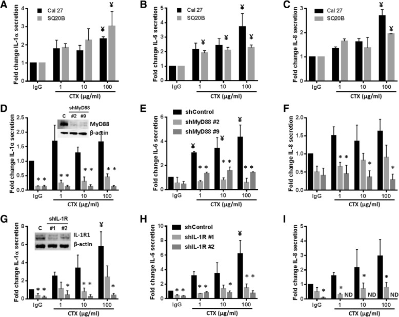
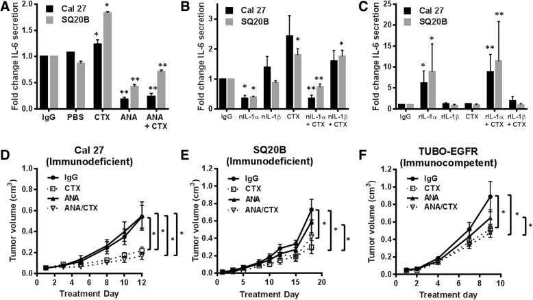
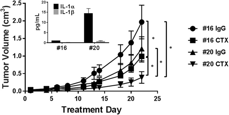
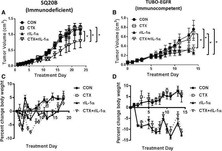
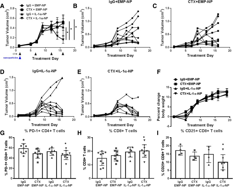
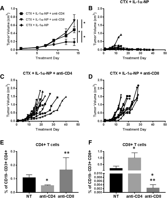
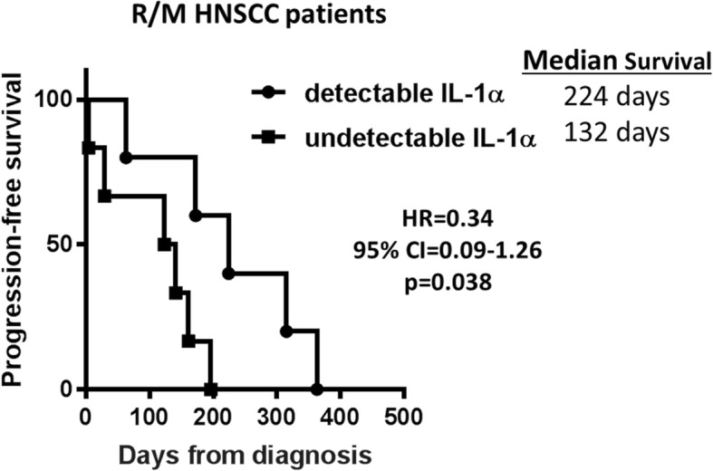
Methods: Parental and stable myeloid differentiation primary response gene 88 (MyD88) and IL-1 receptor 1 (IL-1R1) knockdown HNSCC cell lines, an IL-1R antagonist (IL-1RA), neutralizing antibodies to IL-1α and IL-1β, and recombinant IL-1α and IL-1β were used to determine cytokine production (using ELISA) in response to cetuximab in vitro. IL-1 pathway modulation in mouse models was accomplished by administration of IL-1RA, stable overexpression of IL-1α in SQ20B cells, administration of rIL-1α, and administration of a polyanhydride nanoparticle formulation of IL-1α. CD4+ and CD8+ T cell-depleting antibodies were used to understand the contribution of T cell-dependent anti-tumor immune responses. Baseline serum levels of IL-1α were measured using ELISA from HNSCC patients treated with cetuximab-based therapy and analyzed for association with progression free survival (PFS).

Results: Cetuximab induced pro-inflammatory cytokine secretion from HNSCC cells in vitro which was mediated by an IL-1α/IL-1R1/MyD88-dependent signaling pathway. IL-1 signaling blockade did not affect the anti-tumor efficacy of cetuximab, while increased IL-1α expression using polyanhydride nanoparticles in combination with cetuximab safely and effectively induced a T cell-dependent anti-tumor immune response. Detectable baseline serum levels of IL-1α were associated with a favorable PFS in cetuximab-based therapy-treated HNSCC patients compared to HNSCC patients with undetectable levels.

Conclusions: Altogether, these results suggest that IL-1α in combination with cetuximab can induce a T cell-dependent anti-tumor immune response and may represent a novel immunotherapeutic strategy for EGFR-positive HNSCCs.

Keywords: Anakinra; Biomarker; Cetuximab; Cytokines; EGFR; HNSCC; Interleukin-1; Nanoparticle.


Conflict of interest statement

Ethics approval and consent to participate

Not applicable.

Consent for publication

Not applicable.

Competing interests

The authors declare that they have no competing interests.

Publisher’s Note

Springer Nature remains neutral with regard to jurisdictional claims in published maps and institutional affiliations.

Figures

Fig. 1
Fig. 1
Cetuximab induces secretion of pro-inflammatory cytokines via IL-1R/MyD88 signaling. Cal 27 and SQ20B HNSCC cells (a-c), SQ20B cells derived from MyD88 stable knockout clones (shMyD88 #2, shMyD88 #9) and control cells (shControl) (d-f), and SQ20B cells derived from IL-1R1 stable knockout clones (shIL-1R #1, shIL-1R #2) and control cells (shControl) (g-i) were treated with 1–100 μg/mL cetuximab (CTX) or 100 μg/mL IgG for 48 H. media was collected and ELISAs were performed to measure IL-1α (a, d, g), IL-6 (b,e,h), and IL-8 (c,f,i). Cells were analyzed for expression of MyD88 (D inset) and IL-1R1 (G inset) by Western blot and β-actin was used as a control. Error bars = SEM. N = 3. ¥:p < 0.05 vs. respective IgG treatment; *p < 0.05 vs. respective shControl cell line. ND = not detected
Fig. 2
Fig. 2
IL-1 blockade does not improve the anti-tumor efficacy of cetuximab. a Cal 27 and SQ20B HNSCC cells were pretreated with 10 μg/mL anakinra (ANA) for 4 h with or without 100 μg/mL cetuximab (CTX) for 48 h. IgG and PBS were used as controls. b Cal 27 and SQ20B HNSCC cells were pretreated with 1 μg/mL nIL-1αab or 1 μg/mL nIL-1βab for 4 h with or without 100 μg/mL CTX for 48 h. IgG was used as a control. c Cal 27 and SQ20B HNSCC cells were pretreated with 0.5 ng/mL human rIL-1α or 0.5 ng/mL human rIL-1β for 4 h with or without 100 μg/mL CTX for 48 h. IgG was used as a control. Media was collected and ELISAs were performed to measure IL-6 secretion. N = 3–4. *: p < 0.05 vs. IgG; **: p < 0.05 vs CTX and IgG. d-f: Athymic nu/nu mice (n = 12 [n = 6 male/n = 6 female]) bearing Cal 27 (d) and SQ20B (e) tumors and BALB/c mice (n = 10 [n = 5 male/n = 5 female]) bearing TUBO-EGFR tumors (f) were treated with CTX (2 mg/kg [8 mg/kg for TUBO-EGFR tumors]) twice weekly and anakinra (10 mg/kg daily) i.p. for two weeks. Tumors were measured three times/week. Tumor growth curves shown were stopped after a mouse in any treatment group reached euthanasia criteria. Error bars = SEM. *:p < 0.05
Fig. 3
Fig. 3
IL-1α overexpression enhances the anti-tumor efficacy of cetuximab. Female athymic nu/nu mice bearing IL-1α overexpressing (#20) or control (#16) SQ20B tumors were treated with cetuximab (CTX, 2 mg/kg, twice/week) or IgG for 3 weeks. Overexpression was confirmed by ELISA (inset). Tumors were measured three times weekly. Tumor growth curves shown were stopped after a mouse in any treatment group reached euthanasia criteria. Error bars = SEM. N = 4–5 mice/treatment group. *: p < 0.05
Fig. 4
Fig. 4
Tumor response to recombinant IL-1α differs between immunodeficient and immunocompetent mouse models. Athymic nu/nu mice (n = 10 [n = 5 male/n = 5 female]) bearing SQ20B tumors (a, c) or BALB/c mice (n = 10 [n = 5 male/n = 5 female]) bearing TUBO-EGFR tumors (b, d) were treated with cetuximab (CTX, 2 mg/kg [8 mg/kg for TUBO-EGFR tumors], twice/week) with or without 0.6 μg human (a, c) or murine (b, d) recombinant IL-1α (rIL-1α) for 2 weeks. IgG and H2O were used as controls. IL-α was given at least half an hour prior to CTX or IgG administration, and again 24 h later totaling 4 doses of CTX and IgG, and 8 doses of IL-1α and H2O. Tumor growth (a, b) and mouse weights (C,D) were measured 3–5 times per week. Tumor growth curves shown were stopped after a mouse in any treatment group reached euthanasia criteria. Error bars = SEM. *: p < 0.05
Fig. 5
Fig. 5
Nanoparticle delivery of IL-1α demonstrates anti-tumor efficacy. a Female BALB/c mice (n = 9–10 mice/treatment group) bearing TUBO-EGFR tumors were treated with cetuximab (CTX, 8 mg/kg, twice/week) for 2 weeks with or without a single administration of IL-1α nanoparticles (IL-1α-NPs (0.5 mg NPs containing 7.5 μg IL-1α)) on the first day of treatment. IgG and empty nanoparticles (EMP-NP) were used as controls. Tumor volumes were measured three times per week. Tumor growth curves shown were stopped after a mouse in any treatment group reached euthanasia criteria. *:p < 0.05. Error bars = SEM. b-e: Shown are spaghetti plots for each individual mouse in each treatment group shown in A. F: Mouse weights were measured three times per week. g-i: Spleens were harvested after therapy and PD-1 + CD4+ T cells (g), CD8+ T cells (h) and CD25 + CD8+ T cells (i) were analyzed using flow cytometry. Error bars = SDM. N = 4–9 per group. *: p < 0.05 vs IgG
Fig. 6
Fig. 6
The anti-tumor effects of cetuximab+IL-1α-NP are T cell dependent. Female BALB/c mice (n = 9–10 mice/treatment group) bearing TUBO-EGFR tumors were treated with cetuximab (CTX, 8 mg/kg twice/week) in combination with a single i.p. dose on treatment day 1 of IL-1α-NPs (0.5 mg NPs containing 7.5 μg IL-1α) (CTX + IL-1α-NP (a,b) with or without anti-CD4 (100 μg (clone GK1.5)) (a,c) or anti-CD8 (300 μg (clone 53–6.7)) (a,d) 1 and 3 days prior to tumor inoculation, and every 3–4 days after tumor inoculation. Treatment duration was 3 weeks. Tumor volumes were measured three times per week. Tumor growth curves shown were stopped after a mouse in any treatment group reached euthanasia criteria. Error bars = SEM. *:p < 0.05. b-d: Shown are spaghetti plots for each individual mouse in each treatment group shown in a. Tumors from female BALB/c mice bearing TUBO-EGFR tumors (n = 3–4) were treated as described in A and harvested after 2 weeks of therapy for validation of CD4+ T cell (E) and CD8+ T cell (F) depletion by flow cytometry. *:p < 0.05 vs NT, **:p < 0.05 vs anti-CD4. Error bars = SDM
Fig. 7
Fig. 7
High serum IL-1α predicts progression free survival (PFS) in HNSCC patients treated with cetuximab-containing therapy. Baseline serum samples from 11 recurrent and/or metastatic (R/M) HNSCC patients scheduled for cetuximab-based chemotherapy (i.e. carboplatin, cisplatin, 5-FU, paclitaxel) at the University of Iowa Hospitals and Clinics Holden Comprehensive Cancer Center were collected. Serum IL-1α levels were measured by ELISA and patients were divided into two groups: detectable (n = 5) and undetectable (n = 6) IL-1α levels. Kaplan Meier survival curves were plotted for PFS for both groups. HR: hazard ratio; CI: confidence interval

Similar articles

Cited by

References

    1. Burris HA, 3rd, Taylor CW, Jones SF, Koch KM, Versola MJ, Arya N, et al. A phase I and pharmacokinetic study of oral lapatinib administered once or twice daily in patients with solid malignancies. Clin Cancer Res. 2009;15:6702–6708. doi: 10.1158/1078-0432.CCR-09-0369. - DOI - PMC - PubMed
    1. Cohen EE, Kane MA, List MA, Brockstein BE, Mehrotra B, Huo D, et al. Phase II trial of gefitinib 250 mg daily in patients with recurrent and/or metastatic squamous cell carcinoma of the head and neck. Clin Cancer Res. 2005;11:8418–8424. doi: 10.1158/1078-0432.CCR-05-1247. - DOI - PubMed
    1. Cohen EE, Rosen F, Stadler WM, Recant W, Stenson K, Huo D, et al. Phase II trial of ZD1839 in recurrent or metastatic squamous cell carcinoma of the head and neck. J Clin Oncol. 2003;21:1980–1987. doi: 10.1200/JCO.2003.10.051. - DOI - PubMed
    1. Soulieres D, Senzer NN, Vokes EE, Hidalgo M, Agarwala SS, Siu LL. Multicenter phase II study of erlotinib, an oral epidermal growth factor receptor tyrosine kinase inhibitor, in patients with recurrent or metastatic squamous cell cancer of the head and neck. J Clin Oncol. 2004;22:77–85. doi: 10.1200/JCO.2004.06.075. - DOI - PubMed
    1. Vermorken JB, Mesia R, Rivera F, Remenar E, Kawecki A, Rottey S, et al. Platinum-based chemotherapy plus cetuximab in head and neck cancer. N Engl J Med. 2008;359:1116–1127. doi: 10.1056/NEJMoa0802656. - DOI - PubMed

Publication types

MeSH terms