. 2019 May;60(5):995-1004.
doi: 10.1194/jlr.M092494. Epub 2019 Mar 19.

AdipoR1 and AdipoR2 maintain membrane fluidity in most human cell types and independently of adiponectin

Affiliations

AdipoR1 and AdipoR2 maintain membrane fluidity in most human cell types and independently of adiponectin

Mario Ruiz et al. J Lipid Res. 2019 May.

Abstract

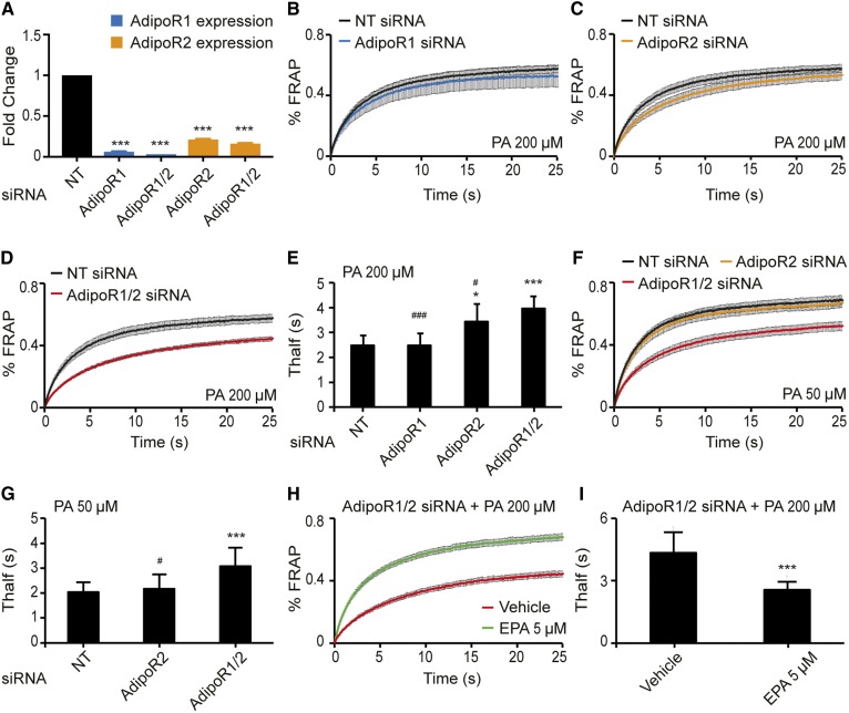
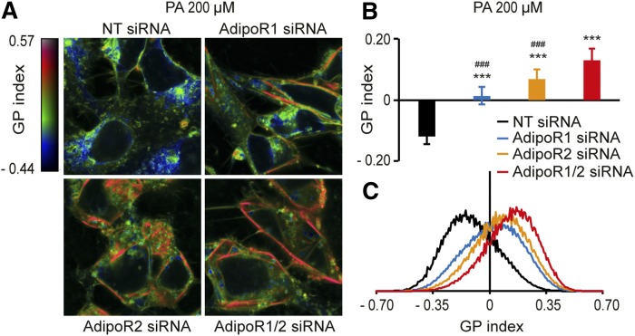
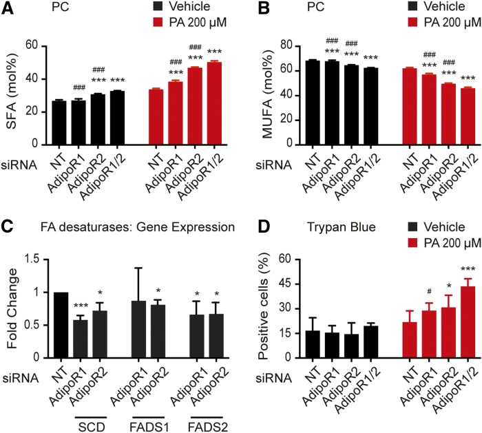
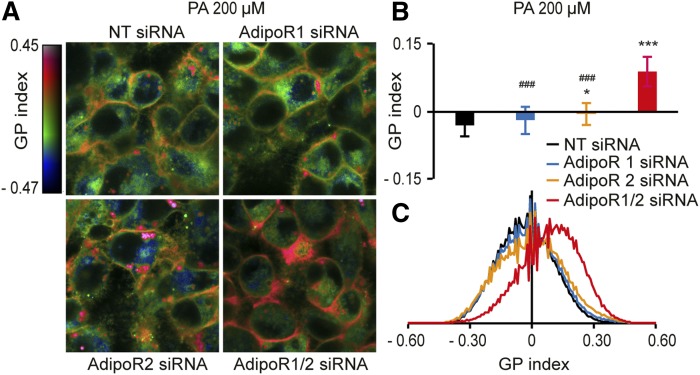
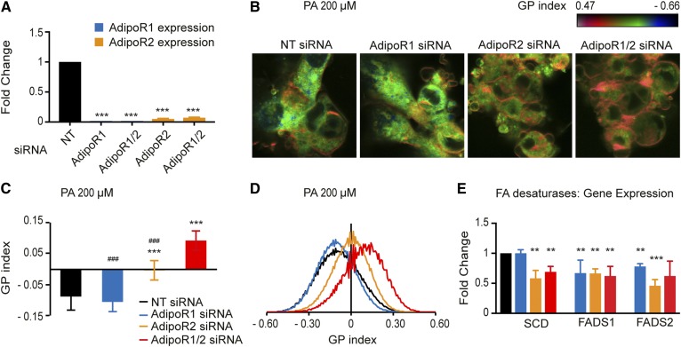
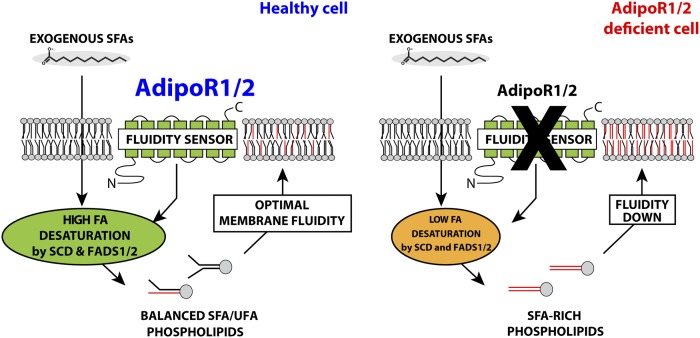
The FA composition of phospholipids must be tightly regulated to maintain optimal cell membrane properties and compensate for a highly variable supply of dietary FAs. Previous studies have shown that AdipoR2 and its homologue PAQR-2 are important regulators of phospholipid FA composition in HEK293 cells and Caenorhabditiselegans, respectively. Here we show that both AdipoR1 and AdipoR2 are essential for sustaining desaturase expression and high levels of unsaturated FAs in membrane phospholipids of many human cell types, including primary human umbilical vein endothelial cells, and for preventing membrane rigidification in cells challenged with exogenous palmitate, a saturated FA. Three independent methods confirm the role of the AdipoRs as regulators of membrane composition and fluidity: fluorescence recovery after photobleaching, measurements of Laurdan dye generalized polarization, and mass spectrometry to determine the FA composition of phospholipids. Furthermore, we show that the AdipoRs can prevent lipotoxicity in the complete absence of adiponectin, their putative ligand. We propose that the primary cellular function of AdipoR1 and AdipoR2 is to maintain membrane fluidity in most human cell types and that adiponectin is not required for this function.

Keywords: desaturases; fatty acids; lipotoxicity; metabolism; palmitate; phospholipids; plasma membrane; receptors.


Conflict of interest statement

There are no competing interests.

Figures

Fig. 1.
Fig. 1.
FRAP analysis showing that AdipoR1 and AdipoR2 redundantly maintain membrane fluidity in HEK293 cells. A: qPCR results showing the efficiency of the knockdown using NT, AdipoR1, and/or AdipoR2 siRNA. The expression levels are normalized to the NT value. B–D: FRAP results in HEK293 cells challenged with 200 μM PA and treated with NT, AdipoR1, AdipoR2, or AdipoR1/2 siRNA (n = 6–14). E: Average Thalf values (the time by which half of the maximum fluorescence recovery is reached) from multiple experiments as in panels B–D. F: FRAP results in HEK293 cells challenged with 50 μM PA and treated with NT, AdipoR2, or AdipoR1/2 siRNA (n = 10–13). G: Average Thalf values from panel F. H, I: FRAP results in HEK293 cells challenged with 200 μM PA and treated with either vehicle (DMSO) or 5 μM EPA (n = 10). Error bars show the SDs in histograms and SEMs in FRAP panels. *P < 0.05 and ***P < 0.001 compared with the control treatment. #P < 0.05 and ###P < 0.001 compared with the AdipoR1/2 siRNA treatment. NT, nontarget.
Fig. 2.
Fig. 2.
The Laurdan dye method confirms that AdipoR1 and AdipoR2 are required to maintain membrane fluidity in HEK293 cells. A: Pseudocolor images showing the Laurdan dye GP index at each pixel position in HEK293 cells challenged with 200 μM PA and treated with NT, AdipoR1, and/or AdipoR2 siRNA. Note the pronounced rigidification of the plasma membrane in the AdipoR1/2 siRNA-treated cells. B: Average GP index from several images as in panel A (n = 10–15). C: Distribution of the GP index values in representative images for each treatment. Error bars show the SDs. ***P < 0.001 compared with the control treatment. ###P < 0.001 compared with the AdipoR1/2 siRNA treatment. GP, generalized polarization; NT, nontarget.
Fig. 3.
Fig. 3.
The AdipoRs are required to maintain MUFA levels in PCs, sustain desaturase gene expression, and prevent lipotoxicity by PA. A, B: SFA and MUFA abundance (mol.%) in the PCs of HEK293 cells cultivated in the presence of either vehicle (DMSO) or 200 μM PA and treated with NT, AdipoR1, and/or AdipoR2 siRNA (n = 3). C: qPCR results showing the expression of three desaturases in HEK293 cells following knockdown using NT, AdipoR1, and/or AdipoR2 siRNA. The expression levels are normalized to the NT value. D: Percentage of dead HEK293 cells (trypan blue-positive) following cultivation in the presence of either vehicle (DMSO) or 200 μM PA and treated with NT, AdipoR1, and/or AdipoR2 siRNA. Error bars show the SDs. *P < 0.05 and ***P < 0.001 compared with the control treatment. #P < 0.05 and ###P < 0.001 compared with the AdipoR1/2 siRNA treatment. NT, nontarget.
Fig. 4.
Fig. 4.
The Laurdan dye method shows that AdipoR1 and AdipoR2 are required to maintain membrane fluidity in HepG2 cells. A: Pseudocolor images showing the Laurdan dye GP index at each pixel position in HepG2 cells challenged with 200 μM PA and treated with NT, AdipoR1, and/or AdipoR2 siRNA. Note the pronounced rigidification of the plasma membrane in the AdipoR1/2 siRNA-treated cells. B: Average GP index from several images as in panel A (n = 15). C: Distribution of the GP index values in representative images for each treatment. Error bars show the SDs. *P < 0.05 and ***P < 0.001 compared with the control treatment. #P < 0.05 and ###P < 0.001 compared with the AdipoR1/2 siRNA treatment. GP, generalized polarization; NT, nontarget.
Fig. 5.
Fig. 5.
The AdipoRs maintain membrane fluidity and desaturase expression in primary human cells (HUVECs). A: qPCR results showing the efficiency of the knockdown in HUVECs using NT, AdipoR1, and/or AdipoR2 siRNA. The expression levels are normalized to the NT value. B: Pseudocolor images showing the Laurdan dye GP index at each pixel position in HUVECs challenged with 200 μM PA and treated with NT, AdipoR1, and/or AdipoR2 siRNA. Note the pronounced rigidification of the plasma membrane in the AdipoR1/2 siRNA-treated cells. C: Average GP index from several images as in panel A (n = 10–15). D: Distribution of the GP index values in representative images for each treatment. E: qPCR results showing the expression of three desaturases in HEK293 cells following knockdown using NT, AdipoR1, and/or AdipoR2 siRNA. The expression levels are normalized to the NT value. Error bars show the SDs. **P < 0.01 and ***P < 0.001 compared with the control treatment. ###P < 0.001 compared with the AdipoR1/2 siRNA treatment. GP, generalized polarization; NT, nontarget.
Fig. 6.
Fig. 6.
Model for the role of the AdipoRs in membrane homeostasis. In healthy cells challenged with exogenous SFAs, the AdipoRs sense membrane rigidification and signals to promote desaturase enzyme expression, resulting in increased incorporation of fluidizing UFAs into phospholipids. AdipoR-deficient cells fail to promote desaturase activity to compensate for the exogenously provided SFAs that become incorporated into phospholipids, leading to membrane rigidification and hence lipotoxicity.

References

    1. Yamauchi T., Kamon J., Ito Y., Tsuchida A., Yokomizo T., Kita S., Sugiyama T., Miyagishi M., Hara K., Tsunoda M., et al. . 2003. Cloning of adiponectin receptors that mediate antidiabetic metabolic effects. Nature. 423: 762–769. - PubMed
    1. Yamauchi T., Nio Y., Maki T., Kobayashi M., Takazawa T., Iwabu M., Okada-Iwabu M., Kawamoto S., Kubota N., Kubota T., et al. . 2007. Targeted disruption of AdipoR1 and AdipoR2 causes abrogation of adiponectin binding and metabolic actions. Nat. Med. 13: 332–339. - PubMed
    1. Kubota N., Yano W., Kubota T., Yamauchi T., Itoh S., Kumagai H., Kozono H., Takamoto I., Okamoto S., Shiuchi T., et al. . 2007. Adiponectin stimulates AMP-activated protein kinase in the hypothalamus and increases food intake. Cell Metab. 6: 55–68. - PubMed
    1. Iwabu M., Yamauchi T., Okada-Iwabu M., Sato K., Nakagawa T., Funata M., Yamaguchi M., Namiki S., Nakayama R., Tabata M., et al. . 2010. Adiponectin and AdipoR1 regulate PGC-1alpha and mitochondria by Ca(2+) and AMPK/SIRT1. Nature. 464: 1313–1319. - PubMed
    1. Bjursell M., Ahnmark A., Bohlooly-Y M., William-Olsson L., Rhedin M., Peng X-R., Ploj K., Gerdin A-K., Arnerup G., Elmgren A., et al. . 2007. Opposing effects of adiponectin receptors 1 and 2 on energy metabolism. Diabetes. 56: 583–593. - PubMed

Publication types