. 2019 Apr 5;146(7):dev175570.
doi: 10.1242/dev.175570.

The Hunchback temporal transcription factor determines motor neuron axon and dendrite targeting in Drosophila

Affiliations

The Hunchback temporal transcription factor determines motor neuron axon and dendrite targeting in Drosophila

Austin Q Seroka et al. Development. .

Abstract

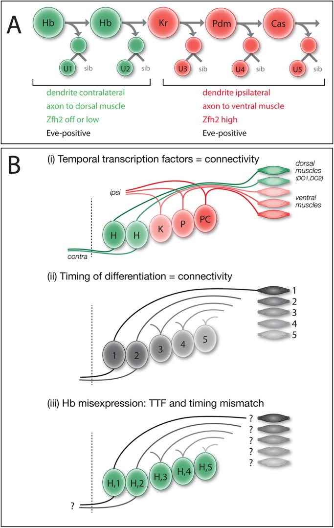
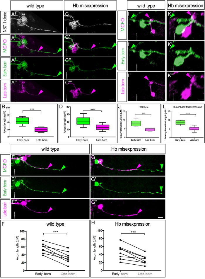
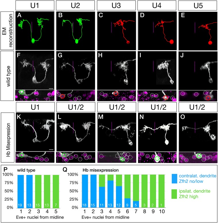
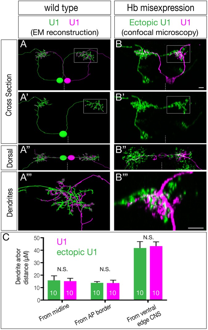
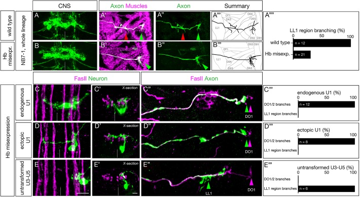
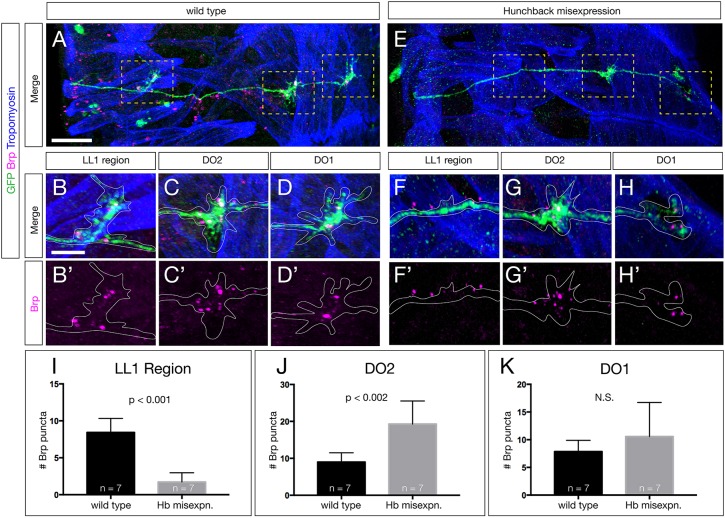
The generation of neuronal diversity is essential for circuit formation and behavior. Morphological differences in sequentially born neurons could be due to intrinsic molecular identity specified by temporal transcription factors (henceforth called intrinsic temporal identity) or due to changing extrinsic cues. Here, we have used the Drosophila NB7-1 lineage to address this issue. NB7-1 generates the U1-U5 motor neurons sequentially; each has a distinct intrinsic temporal identity due to inheritance of different temporal transcription factors at its time of birth. We show that the U1-U5 neurons project axons sequentially, followed by sequential dendrite extension. We misexpressed the earliest temporal transcription factor, Hunchback, to create 'ectopic' U1 neurons with an early intrinsic temporal identity but later birth-order. These ectopic U1 neurons have axon muscle targeting and dendrite neuropil targeting that are consistent with U1 intrinsic temporal identity, rather than with their time of birth or differentiation. We conclude that intrinsic temporal identity plays a major role in establishing both motor axon muscle targeting and dendritic arbor targeting, which are required for proper motor circuit development.

Keywords: Dendrite morphology; Heterochronic; Hunchback; Larval locomotion; Motor circuits; Motor neuron; Neural circuits; Temporal identity; Temporal transcription factors.


Conflict of interest statement

Competing interestsThe authors declare no competing or financial interests.

Figures

Fig. 1.
Fig. 1.
Models: intrinsic temporal identity or time of differentiation determines U1-U5 motor neuron morphology. (A) NB7-1 (top ) sequentially expresses the temporal transcription factors Hb, Kr, Pdm and Cas. The U1-U2 neurons (bottom) born during the Hb window have an ‘early-born’ identity (green) characterized by contralateral dendrites, an axon projection to dorsal body wall muscles DO1 and DO2, and little or no nuclear Zfh2. The U3-U5 neurons born after the Hb window have a ‘late-born’ identity (red) characterized by ipsilateral dendrites, an axon projection to more ventral muscles DA3/LL1 and high nuclear Zfh2. All U1-U5 neurons have nuclear Eve. (B) Models for specification of U1-U5 axon and dendrite targeting. (i) Intrinsic temporal identity could determine axon and dendrite targeting. (ii) Neuronal time of differentiation could determine axon and dendrite targeting. (iii) Misexpression of Hb can generate late-differentiating neurons with an early intrinsic temporal identity; this mismatch reveals which mechanism is more important for axon and dendrite targeting.
Fig. 2.
Fig. 2.
The U1-U5 motor neurons extend axons and dendrites sequentially. (A-H) Axon outgrowth timing in early- and late-born neurons of the NB7-1 lineage in stage 13-15 embryos. (A-B) Wild-type multicellular MCFO labeling (NB7-1-Gal4KZ UAS-MCFO). Analysis was restricted to lineages in which early-born medially located neurons were stochastically labeled in one MCFO color, and in which the later-born laterally located neurons were stochastically labeled in a different MCFO color. (A) All labeled cells. (A′-A‴) Early-born medial neurons (green) project out of the CNS ahead of later-born lateral neurons (magenta). (B) Quantification of axon length as a representation of timing of axon outgrowth; early-born neurons project further (earlier) than late-born neurons (***P<0.001). (C-D) Hb misexpression (NB7-1-Gal4KZ UAS-hb UAS-MCFO) multicellular MCFO labeling. (C) All labeled cells. (C′-C‴) Early-born medial neurons (green) project out of the CNS ahead of later-born lateral neurons (magenta). (D) Quantification of axon length as a representation of timing of axon outgrowth; early-born neurons project further (earlier) than late-born neurons (***P<0.001). (E-F) Wild-type single-neuron MCFO labeling (NB7-1-Gal4KZ UAS-MCFO). (E) A single early-born medial neuron (green) always projects out of the CNS ahead of a single later-born lateral neuron (magenta). (F) Quantification of axon length as a representation of timing of axon outgrowth; early-born neurons project further (earlier) than late-born neurons (***P<0.001). (G-H) Hb misexpression (NB7-1-Gal4KZ UAS-hb UAS-MCFO) single-neuron MCFO. (G-G″) A single early-born medial neuron (green) always projects out of the CNS ahead of a single later-born lateral neuron (magenta). Quantification of axon length as a representation of timing of axon outgrowth; early-born neurons project further (earlier) than late-born neurons (***P<0.001). (I-L) Dendrite outgrowth timing in early- and late-born neurons of the NB7-1 lineage in stage 13-15 embryos. (I-I″) Wild-type single-neuron MCFO labeling (NB7-1-Gal4KZ UAS-MCFO). A single early-born medial neuron (green) extends a dendrite before a single later-born lateral neuron (magenta). (J) Quantification of axon length as a representation of the timing of axon outgrowth; early-born neurons project further (earlier) than late-born neurons (***P<0.001). (K-K″) Hb misexpression (NB7-1-Gal4KZ UAS-hb UAS-MCFO) single neuron MCFO labeling. A single early-born medial neuron (green) extends a dendrite before a single later-born lateral neuron (magenta). (L) Quantification of axon length as a representation of timing of axon outgrowth; early-born neurons project further (earlier) than late-born neurons (P<0.001). Arrowheads indicate the early-born axon (green) and late-born axon (magenta); dashed line indicates the midline. Scale bars: 5 μm.
Fig. 3.
Fig. 3.
Late-born neurons with early intrinsic temporal identity have ‘early’ dendrite morphology. (A-E) U1-U5 neuronal morphology determined by EM reconstruction in the first instar larval CNS. Early-born U1-U2 neurons (green) have a bipolar morphology with a contralateral dendrite arbor (left of dashed midline), whereas later-born U3-U5 neurons (red) have a monopolar morphology and ipsilateral dendritic arbors. Neuronal birth-order is determined by mediolateral position (U1 most medial/earliest, U5 most lateral/latest). (F-J) Wild-type U1-U5 single neuronal morphology by MCFO in L1 larvae (NB7-1-Gal4KZ UAS-MCFO). Neurons are shown from left to right based on birth-order, determined by their position within the five Eve+ neurons (inset). Scale bar: 5 μm. (K-O) Hb misexpression U1-U5 single neuronal morphology by MCFO in L1 larvae (NB7-1-Gal4KZ UAS-MCFO). Neurons are shown from left to right based on birth-order, determined by their position within the Eve+ neurons (inset). The later-born neurons (‘ectopic U1’) have acquired a contralateral dendrite, more consistent with their early intrinsic temporal identity than their late time of differentiation. Scale bar: 5 μm. (P,Q) Quantification. In wild type, early-born U1-U2 neurons have low/no nuclear Zfh2, a marker for their early intrinsic temporal identity, and contralateral dendrites; later-born neurons have high Zfh2 and no contralateral projection. In Hb misexpression embryos, all neurons with low/no Zfh2 have a contralateral dendrite, even when they have a late-born time of differentiation (>3 Eve+ nuclei from the midline). The number of neurons scored is shown within each bar.
Fig. 4.
Fig. 4.
Ectopic U1 dendrites target the normal U1 neuropil domain. (A-A‴) Wild-type bilateral U1 neurons (green, magenta) assayed in the EM reconstruction of the L1 larval CNS. A U1 neuron (magenta) targets its contralateral dendrite to the same neuropil volume as the ipsilateral dendrite of the contralateral U1 neuron (green; boxed region in A,A′, shown enlarged in A″). (B-B‴) Hb misexpression (NB7-1-Gal4KZ UAS-hb UAS-MCFO) assayed by MCFO labeling in L1 larvae, showing an endogenous U1 (magenta; defined by its medial cell body position, bipolar morphology and contralateral projection) and an ectopic U1 neuron (green; defined by its lateral cell body position, monopolar morphology and contralateral projection). The endogenous and ectopic U1 neurons target the same dorsal neuropil domain (boxed in B,B′ shown enlarged in B″). Dashed line indicates the midline; all views are cross-sections; dorsal is upwards except A″ and B″ (dorsal views, anterior upwards). Scale bars: 5 μm. (C) Quantification. Endogenous and ectopic U1 dendrites are the same distance from the midline, anterior-posterior (AP) border and ventral edge of the CNS. n=10 for U1 and ectopic U1.
Fig. 5.
Fig. 5.
Ectopic U1 axons project to dorsal muscles and lack ventral muscle targets. (A-B‴) Wild-type (NB7-1-Gal4KZ UAS-GFP) and Hb-misexpression (NB7-1-Gal4KZ UAS-hb UAS-GFP) L1 larvae stained for U motor neurons (green) and muscles (magenta). (A-A‴) Wild type: U motor neurons project axons to dorsal muscles (DO1/DO3) and more ventral muscles in the LL1/DA3 region. (B-B‴) Hb misexpression: U motor neurons project only to dorsal muscle targets (magenta, shown in B′) consistent with ectopic U1-U2 identity at the expense of U3-U5 neuronal identity. Arrowheads indicate U1-U2 muscle targets (green) and U3-U5 muscle targets (red). (A″″) Quantification. (C-C″,D-D″,E-E″) Hb misexpression L1 larvae (NB7-1-Gal4KZ UAS-hb UAS-MCFO) showing MCFO-labeled single neurons. (C-C″) The endogenous U1 motor neuron (green; closest to midline). (C) Dorsal view showing medial cell body position, contralateral dendrites and ipsilateral axon (Zfh2 negative; not shown). (C′) Cross-sectional view of the same U1 neuron. (C″) Dorsal view of the body wall showing the U1 axon (green arrowhead) projecting to the most dorsal extent of the FasII+ motor neurons (magenta arrowhead). (C‴) Quantification. (D-D″) An ectopic U1 motor neuron (green). (D) Dorsal view showing lateral cell body position, contralateral dendrites and ipsilateral axon (Zfh2 negative; not shown). (D′) Cross-sectional view of the same neuron; the contralateral dendrite has a dorsal origin and there is a lack of bipolar morphology. (D″) Dorsal view of body wall showing the ectopic U1 axon (green arrowhead) projecting to the most dorsal extent of the FasII+ motor neurons (magenta arrowhead). (D‴) Quantification. (E-E″) A late-born laterally positioned U3, U4 or U5 motor neuron that was not transformed (based on being Zfh2+; not shown). (E) Dorsal view showing far lateral cell body position, ipsilateral dendrites and ipsilateral axon. (E′) Cross-sectional view of the same neuron. (E″) Dorsal view of the body wall showing the U3-U5 axon (green arrowhead) projecting to a more ventral region along the FasII+ motor neurons (magenta arrowhead). (E‴) Quantification.
Fig. 6.
Fig. 6.
Ectopic U1 axons shift synaptic input from ventral to dorsal muscle targets. (A-D′) Wild-type L1 larva stained for all U motor neurons in the NB7-1 lineage (GFP, green), Brp+ puncta (magenta) and body wall muscles (Tropomyosin, blue). The U motor neurons have Brp+ puncta contacting muscles around LL1 (B,B′), the DO2 muscle (C,C′) and the DO1 muscle (D,D′). (E-H′) Hb-misexpression L1 larva (NB7-1-Gal4KZ UAS-hb) stained for all U motor neurons in the NB7-1 lineage (GFP, green), Brp+ puncta (magenta) and body wall muscles (Tropomyosin, blue). There are reduced Brp+ puncta around LL1 (F,F′), increased Brp+ puncta on the DO2 muscle (G,G′) and a similar number of Brp+ puncta on the DO1 muscle (H,H′). Areas outlined in A and E are shown at higher magnification in B-D′ and F-H′, respectively. (I-K) Quantification. Scale bars: 15 μm in A,E; 5 μm in B-H′.

Similar articles

Cited by

References

    1. Barsh G. R., Isabella A. J. and Moens C. B. (2017). Vagus motor neuron topographic map determined by parallel mechanisms of hox5 expression and time of axon initiation. Curr. Biol. 27, 3812-3825.e3813. 10.1016/j.cub.2017.11.022 - DOI - PMC - PubMed
    1. Bertet C., Li X., Erclik T., Cavey M., Wells B. and Desplan C. (2014). Temporal patterning of neuroblasts controls Notch-mediated cell survival through regulation of Hid or Reaper. Cell 158, 1173-1186. 10.1016/j.cell.2014.07.045 - DOI - PMC - PubMed
    1. Chang F.-L. F., Steedman J. G. and Lund R. D. (1986). The lamination and connectivity of embryonic cerebral cortex transplanted into newborn rat cortex. J. Comp. Neurol. 244, 401-411. 10.1002/cne.902440311 - DOI - PubMed
    1. Doe C. Q. (2017). Temporal patterning in the Drosophila CNS. Annu. Rev. Cell Dev. Biol. 33, 219-240. 10.1146/annurev-cellbio-111315-125210 - DOI - PubMed
    1. Doe C. Q., Bastiani M. J. and Goodman C. S. (1986). Guidance of neuronal growth cones in the grasshopper embryo. IV. Temporal delay experiments. J. Neurosci. 6, 3552-3563. 10.1523/JNEUROSCI.06-12-03552.1986 - DOI - PMC - PubMed

Publication types

MeSH terms

LinkOut - more resources