. 2019 Mar 19;10(2):e02417-18.
doi: 10.1128/mBio.02417-18.

Yet More Evidence of Collusion: a New Viral Defense System Encoded by Gordonia Phage CarolAnn

Affiliations

Yet More Evidence of Collusion: a New Viral Defense System Encoded by Gordonia Phage CarolAnn

Matthew T Montgomery et al. mBio. .

Abstract

Temperate phages play important roles in the physiology of their bacterial hosts and establish a lysogenic relationship with the host through which prophage-expressed genes confer new phenotypes. A key phenotype is prophage-mediated defense against heterotypic viral attack, in which temperate phages collude with their bacterial host to prevent other phages from attacking, sometimes with exquisite specificity. Such defense systems have been described in Pseudomonas and Mycobacterium phages but are likely widespread throughout the microbial community. Here, we describe a novel prophage-mediated defense system encoded by Gordonia phage CarolAnn, which defends against infection by unrelated phages grouped in cluster CZ. CarolAnn genes 43 and 44 are coexpressed with the repressor and are necessary and sufficient to confer defense against phage Kita and its close relatives. Kita and these relatives are targeted through Kita gene 53, a gene that is of unknown function but which is the location of defense escape mutations that overcome CarolAnn defense. Expression of Kita gene 53 is toxic to Gordonia terrae in the presence of CarolAnn genes 43 and 44, suggesting that defense may be mediated by an abortive infection type of mechanism. CarolAnn genes 43 and 44 are distant relatives of mycobacteriophage Sbash genes 31 and 30, respectively, which also confer viral defense but use a different targeting system.IMPORTANCE Prophage-mediated viral defense systems play a key role in microbial dynamics, as lysogeny is established relatively efficiently, and prophage-expressed genes can strongly inhibit lytic infection of other, unrelated phages. Demonstrating such defense systems in Gordonia terrae suggests that these systems are widespread and that there are a multitude of different systems with different specificities for the attacking phages.

Keywords: Gordonia; bacteriophage; viral defense.


Figures

FIG 1
FIG 1
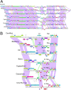
Comparisons of cluster CV Gordonia phages. (A) The genome maps of cluster CV phages CarolAnn, Blueberry, Utz, Barco, CaptainKirk2, Guacamole, and UmaThurman (top to bottom) are shown with spectrum-colored shading between each genome reflecting nucleotide sequence similarity (with violet representing greatest similarity and red the least similarity above a threshold E value of 10−4; white areas are below the threshold value). Phages shown correspond to the lysogens shown in Table 1. (B) An expanded view of the central parts of these genomes displayed as described in the panel A legend. The locations of the integrase (Int) and immunity repressor (Rep) genes are indicated. Genes are shown as colored boxes above and below genome rulers. Gene numbers are shown within the boxes, and the assigned Pham numbers (45) are shown above the boxes, with the number of Pham members shown in parentheses. Gene boxes are colored according to their Pham assignment.
FIG 2
FIG 2
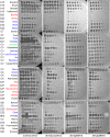
Plating efficiencies of Gordonia phages and defense patterns. Ten-fold serial dilutions of the phages indicated on the left were plated onto lawns of G. terrae 3612, a CarolAnn lysogen [3612(CarolAnn)], 3612pMM15, and 3612pMM16 (see Fig. 3). Phage names shown in red correspond to those that have markedly reduced plating efficiencies on the CarolAnn lysogen as well as the pMM15 and pMM16 strains. Phage names in blue correspond to those that plate with reduced efficiencies on the CarolAnn lysogen but not on the pMM15 and pMM16 strains. CarolAnn, shown in green, is subject to repressor-mediated immunity on both the lysogen and pMM15 but not on the pMM16 strain (see Table 1).
FIG 3
FIG 3
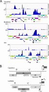
Transcriptomic patterns of cluster CV lysogens. (A) RNA was isolated from G. terrae 3612 lysogens of CarolAnn, Blueberry, and Utz and analyzed by RNAseq. The central portions of the genome are shown; the full genome profiles are in Fig. S1 to S3. The forward and reverse strand reads are shown scaled to the map of the phage genome indicated at the bottom. Arrows below the map indicate the transcribed regions. Gene boxes are colored according to their Pham assignment; colors reflect sequence similarity. Integrase (Int) and repressor genes (Rep) are indicated. (B) Comparison of CarolAnn and lambda genomes. Segments of the two phage genomes are aligned to show similarities between CarolAnn genes 43 to 46 and lambda exclusion genes rexA, rexB, cI, and cro. The positions of lambda promoters PR and PRM are shown as well as those of putative CarolAnn promoters. The locations of a DUF4747 conserved domain in CarolAnn gp44 and lambda RexA are indicated. CarolAnn gp46 is likely a cro-like protein. Genome coordinates (indicated in kilobase pairs) are shown centrally.
FIG 4
FIG 4
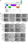
CarolAnn genes 43 and 44 confer defense against phage infection. (A) The organization of CarolAnn genes 41 to 46 is shown at the top, with arrows indicating the positions of putative promoters and the direction of transcription. Genes are represented as boxes, with the gene names within the box; the Phamily designation and the number of Pham members (in parentheses) are shown below each box. The segments of CarolAnn DNA present in plasmids pMM15, pMM16, pMM18, and pMM59 are shown as black lines. Plasmids pMM55 and pMM56 contain the same CarolAnn genes as plasmids pMM16 and pMM59, respectively, but the genes are inserted into Tet-ON vector pCCK39 (Table S2). (B) Lysates of phages Bialota, BatStarr, Nymphadora, Yeezy, CarolAnn, KatherineG, OneUp, and Kita were serially diluted 10-fold and plated onto lawns of G. terrae strains. Plasmid pMM16 confers defense against all of the phages except CarolAnn. (C) Dilutions of the same phages were plated onto lawns of G. terrae 3612 on solid medium either lacking (Uninduced) or containing (Induced) ATc inducer.
FIG 5
FIG 5
Transcriptional profile of phage Kita infection. RNA was isolated at early (30 min) and late (120 min) time points after infection of G. terrae 3612 or a CarolAnn lysogen [G. terrae 3612(CarolAnn)]—shown in red and aqua, respectively—with phage Kita; RNAseq reads were mapped to the forward and reverse strands as indicated. Arrows at the bottom indicate the regions transcribed early and late, with functional notation and with the position of Kita gene 53 shown.
FIG 6
FIG 6
Isolation and mapping of defense escape mutants. (A) Defense escape mutants of Kita efficiently escape CarolAnn 43/44-mediated defense. (B) Locations of defense escape mutant substitutions in Kita gp53 (and its homologues) are shown in black type, and the positions of nontoxic mutants are shown in red type. Of 24 independent defense escape mutants isolated, 6 introduce either termination codons or single amino acid substitutions (see Table 2). The M1A, and E31* mutants are DEMs of Kita, the G19D substitution was isolated independently in Kita and Nymphadora, the V67F substitution was identified in a Nymphadora DEM, and V51A and W80* are both DEM derivatives of Yeezy. The translation initiation codon shown for Kita 53 is at coordinate 36992 and corresponds to Kita 53-short (see the text and Fig. 7).
FIG 7
FIG 7
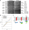
Kita gp53 is toxic in combination with CarolAnn gp43/gp44. (A) Plasmids pCCK38 (vector), pMM53 (Kita 53-long), pMM63 (Kita 53-short), pMM60 (Nymphadora 53), and pMM61 (Nymphadora 53 with DEM mutation) were electroporated into G. terrae strains carrying no integrated plasmid (‘-‘), or with integrated plasmid pMH94 (vector) or pMM16 (with CarolAnn genes 43 and 44), as indicated at the left; key plasmid features are indicated on the right. Liquid cultures of the plasmids were serially diluted 10-fold (right to left) and spotted onto solid media in the absence (‘Uninduced’) or presence (‘Induced’) of Atc inducer. (B) Growth inhibition by expression of Kita gp53 and Nymphadora gp53. Overnight cultures of G. terrae strains expressing CarolAnn genes 43 and 44 (plasmid pMM16) and either vector (pCCK38) or plasmids expressing Kita 53 (pMM63), Nymphadora 53 (pMM60), or the DEM 2C mutant of Nymphadora 53 (pM61) were subcultured into liquid medium at time zero and grown at 37°C. ATc inducer was added to each culture after 4 h (vertical arrow), and cell density (OD600) was measured every 2 h. (C) A model for CarolAnn 43/44-mediated defense. CarolAnn gp43 is proposed to be membrane located but inactive as an ion channel until infection with phage Kita (or relatives). During early lytic growth of Kita, gp53 acts either directly or indirectly through CarolAnn gp44 to activate the gp43 ion channel, leading to loss of membrane potential and of intracellular ATP, interruption of macromolecular synthesis, and loss of cell viability.
FIG 8
FIG 8
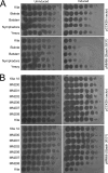
Sbash genes 30 and 31 confer defense against Gordonia phages. (A) Gordonia phages (as indicated on left) were serially diluted 10-fold and spotted onto lawns of Gordonia strains (as indicated on the right) carrying either plasmid vector pCCK39 or plasmid pMM66 with inducible Sbash genes 30 and 31. Uninduced, solid media with no ATc inducer; Induced, solid media with ATc. Under conditions of induction, Sbash genes 30 and 31 confer defense patterns with respect to these phages that are similar to those seen with a strain expressing CarolAnn genes 43 and 44 (see Fig. 2). (B) Isolates of defense escape mutants against CarolAnn 43/44 defense also escape Sbash 30/31 defense. Ten-fold serial dilutions of seven DEMs (see Table 2) and wild-type Kita (bottom) were spotted on lawns of Gordonia strains as described above.

Similar articles

Cited by

References

    1. Stern A, Sorek R. 2011. The phage-host arms race: shaping the evolution of microbes. Bioessays 33:43–51. doi:10.1002/bies.201000071. - DOI - PMC - PubMed
    1. Bondy-Denomy J, Pawluk A, Maxwell KL, Davidson AR. 2013. Bacteriophage genes that inactivate the CRISPR/Cas bacterial immune system. Nature 493:429–432. doi:10.1038/nature11723. - DOI - PMC - PubMed
    1. Tock MR, Dryden DT. 2005. The biology of restriction and anti-restriction. Curr Opin Microbiol 8:466–472. doi:10.1016/j.mib.2005.06.003. - DOI - PubMed
    1. Doron S, Melamed S, Ofir G, Leavitt A, Lopatina A, Keren M, Amitai G, Sorek R. 2018. Systematic discovery of antiphage defense systems in the microbial pangenome. Science 359:eaar4120. doi:10.1126/science.aar4120. - DOI - PMC - PubMed
    1. Goldfarb T, Sberro H, Weinstock E, Cohen O, Doron S, Charpak-Amikam Y, Afik S, Ofir G, Sorek R. 2015. BREX is a novel phage resistance system widespread in microbial genomes. EMBO J 34:169–183. doi:10.15252/embj.201489455. - DOI - PMC - PubMed

Publication types