

Low oxygen post conditioning prevents thalamic secondary neuronal loss caused by excitotoxicity after cortical stroke
- PMID: 30890719
- PMCID: PMC6425023
- DOI: 10.1038/s41598-019-39493-8
Low oxygen post conditioning prevents thalamic secondary neuronal loss caused by excitotoxicity after cortical stroke
Abstract
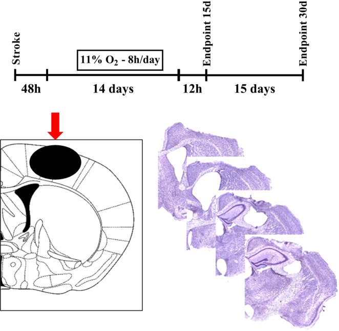
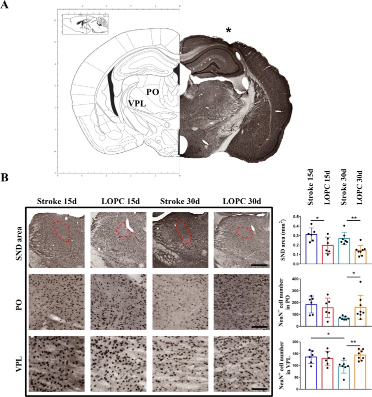
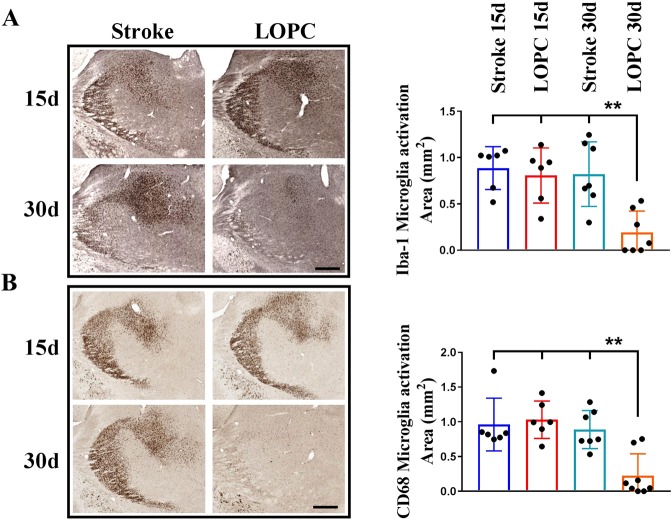
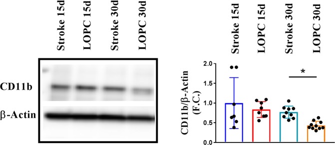
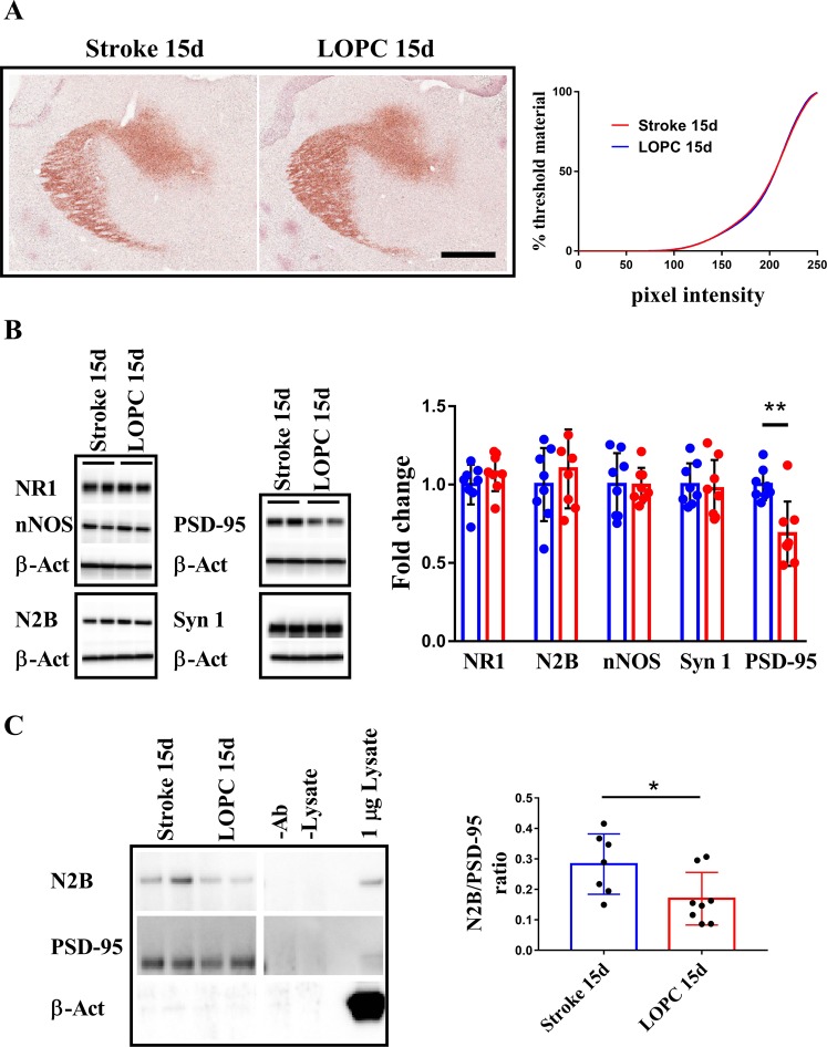
In the current study, we were interested in investigating whether Low oxygen post-conditioning (LOPC) was capable of limiting the severity of stroke-induced secondary neurodegeneration (SND). To investigate the effect of LOPC we exposed adult male C57/BL6 mice to photothrombotic occlusion (PTO) of the motor and somatosensory cortex. This is known to induce progressive neurodegeneration in the thalamus within two weeks of infarction. Two days after PTO induction mice were randomly assigned to one of four groups: (i) LOPC-15 day exposure group; (ii) a LOPC 15 day exposure followed by a 15 day exposure to normal atmosphere; (iii) normal atmosphere for 15 days and (iv) normal atmosphere for 30 days (n = 20/group). We observed that LOPC reduced the extent of neuronal loss, as indicated by assessment of both area of loss and NeuN+ cell counts, within the thalamus. Additionally, we identified that LOPC reduced microglial activity and decreased activity within the excitotoxic signalling pathway of the NMDAR axis. Together, these findings suggest that LOPC limits neuronal death caused by excitotoxicity in sites of secondary damage and promotes neuronal survival. In conclusion, this work supports the potential of utilising LOPC to intervene in the sub-acute phase post-stroke to restrict the severity of SND.
Conflict of interest statement
The authors declare no competing interests.
Figures
Similar articles
-
Exploring How Low Oxygen Post Conditioning Improves Stroke-Induced Cognitive Impairment: A Consideration of Amyloid-Beta Loading and Other Mechanisms.Front Neurol. 2021 Mar 24;12:585189. doi: 10.3389/fneur.2021.585189. eCollection 2021. Front Neurol. 2021. PMID: 33841293 Free PMC article.
-
Chronic stress exacerbates neuronal loss associated with secondary neurodegeneration and suppresses microglial-like cells following focal motor cortex ischemia in the mouse.Brain Behav Immun. 2015 Aug;48:57-67. doi: 10.1016/j.bbi.2015.02.014. Epub 2015 Mar 4. Brain Behav Immun. 2015. PMID: 25749481
-
Age-dependent Disturbances of Neuronal and Glial Protein Expression Profiles in Areas of Secondary Neurodegeneration Post-stroke.Neuroscience. 2018 Nov 21;393:185-195. doi: 10.1016/j.neuroscience.2018.07.034. Epub 2018 Jul 29. Neuroscience. 2018. PMID: 30059704
-
Neuronal Loss after Stroke Due to Microglial Phagocytosis of Stressed Neurons.Int J Mol Sci. 2021 Dec 14;22(24):13442. doi: 10.3390/ijms222413442. Int J Mol Sci. 2021. PMID: 34948237 Free PMC article. Review.
-
NMDARs in Cell Survival and Death: Implications in Stroke Pathogenesis and Treatment.Trends Mol Med. 2020 Jun;26(6):533-551. doi: 10.1016/j.molmed.2020.03.001. Epub 2020 Apr 4. Trends Mol Med. 2020. PMID: 32470382 Review.
Cited by
-
Venous stroke-a stroke subtype that should not be ignored.Front Neurol. 2022 Oct 6;13:1019671. doi: 10.3389/fneur.2022.1019671. eCollection 2022. Front Neurol. 2022. PMID: 36277910 Free PMC article. Review.
-
Inflammatory Responses in the Secondary Thalamic Injury After Cortical Ischemic Stroke.Front Neurol. 2020 Apr 7;11:236. doi: 10.3389/fneur.2020.00236. eCollection 2020. Front Neurol. 2020. PMID: 32318016 Free PMC article. Review.
-
Exploring How Low Oxygen Post Conditioning Improves Stroke-Induced Cognitive Impairment: A Consideration of Amyloid-Beta Loading and Other Mechanisms.Front Neurol. 2021 Mar 24;12:585189. doi: 10.3389/fneur.2021.585189. eCollection 2021. Front Neurol. 2021. PMID: 33841293 Free PMC article.
-
Determining the effect of aging, recovery time, and post-stroke memantine treatment on delayed thalamic gliosis after cortical infarct.Sci Rep. 2021 Jun 15;11(1):12613. doi: 10.1038/s41598-021-91998-3. Sci Rep. 2021. PMID: 34131204 Free PMC article.
-
Single-cell analysis identifies Ifi27l2a as a gene regulator of microglial inflammation in the context of aging and stroke in mice.Nat Commun. 2025 Feb 14;16(1):1639. doi: 10.1038/s41467-025-56847-1. Nat Commun. 2025. PMID: 39953063 Free PMC article.
References
MeSH terms
Substances
LinkOut - more resources
Full Text Sources
Medical
